Pyruvate dehydrogenase (PDH) is a giant enzyme complex (three enzymes, ~60 subunits) that converts pyruvate to acetyl-CoA. It is the bridge between glycolysis (cytoplasm) and the TCA cycle (mitochondrial matrix). Without PDH, the pyruvate produced by glycolysis could not be fully oxidized - so the whole aerobic pipeline depends on this single committed step. Pyruvate from glycolysis enters the mitochondrion, PDH strips a CO2 and adds a CoA, and the resulting acetyl-CoA is handed off to the TCA cycle (Krebs / citric acid cycle) for complete oxidation.
Pyruvate dehydrogenase: the one-way bridge
Pathway map
1
Scroll sideways to see the whole map.
Junction metabolite Irreversible NADH made Turns the complex on
Why it only runs one wayThe reaction is an oxidative decarboxylation: a carbon leaves as CO₂ and is gone. That makes it irreversible, and it is the single reason fatty acids can never become glucose. Every carbon that crosses this bridge has left the sugar pool for good.
The five cofactorsTender Loving Care For Nancy: TPP, Lipoic acid, CoA, FAD, NAD⁺. Four of the five are vitamins, which is why thiamine deficiency stalls this step and causes the lactate build-up seen in beriberi and Wernicke-Korsakoff. α-ketoglutarate dehydrogenase uses exactly the same five.
How it is switchedTwo layers. Allosterically, the products (acetyl-CoA, NADH) and a full energy tank (ATP) slow it, while ADP, NAD⁺, CoA, and Ca²⁺ speed it up. On top of that, PDH kinase phosphorylates and switches it off, and PDH phosphatase (stimulated by insulin and by Ca²⁺ in contracting muscle) switches it back on.
One reaction, three enzymes, five cofactors, and no way back. PDH is not part of glycolysis and not part of the TCA cycle; it is the gate between them, and it is where the cell commits carbohydrate carbon to being burned rather than stored as sugar.
The Reaction
Pyruvate + CoA + NAD+→Acetyl-CoA + CO2+NADH
This is an oxidative decarboxylation. Pyruvate loses one carbon as CO2 and gains a CoA to become a 2-carbon acetyl-CoA. NADH is produced. The reaction is irreversible and commits carbon to the TCA cycle.
Five Required Coenzymes
PDH is one of three mitochondrial complexes that share the same five coenzymes:
TPP (thiamine pyrophosphate, from vitamin B1).
Lipoic acid (a covalent cofactor on E2).
CoA (coenzyme A, from vitamin B5 - pantothenate).
FAD (riboflavin, B2).
NAD+ (niacin, B3).
Alpha-ketoglutarate dehydrogenase (in the TCA cycle) and branched-chain alpha-ketoacid dehydrogenase (for branched amino acid catabolism) use the same five coenzymes.
Regulation
PDH is shut off when the cell has enough energy:
Inhibited by: ATP, NADH, acetyl-CoA (product inhibition), and by PDH kinase-mediated phosphorylation.
Activated by: ADP, NAD+, pyruvate, and dephosphorylation by PDH phosphatase.
Insulin activates PDH (favors oxidation of glucose). Glucagon inactivates PDH (in the liver).
What are the products of the pyruvate dehydrogenase reaction per pyruvate?
Click to reveal answer
Per pyruvate: 1 acetyl-CoA, 1 CO2, 1 NADH. The reaction is irreversible and commits carbon to the TCA cycle. Per glucose (2 pyruvates), 2 acetyl-CoA, 2 CO2, 2 NADH are produced by PDH.
Which vitamin is required as TPP for pyruvate dehydrogenase activity, and what happens with its deficiency?
Click to reveal answer
Vitamin B1 (thiamine) is required as thiamine pyrophosphate (TPP). Deficiency impairs PDH, alpha-ketoglutarate dehydrogenase, and other TPP-dependent enzymes. Clinical picture: beriberi (wet with heart failure, dry with neuropathy) and Wernicke-Korsakoff syndrome in chronic alcoholics (encephalopathy, memory loss). Pyruvate accumulates and is converted to lactate, causing lactic acidosis.
Why is the PDH reaction irreversible?
Click to reveal answer
PDH couples decarboxylation (CO2 released as gas, lost to the environment) with oxidation and thioester formation. The reaction has a large negative ΔG and the CO2 physically leaves the system. This is one of the defining features of metabolism - once acetyl-CoA is made from pyruvate, the cell cannot regenerate pyruvate from acetyl-CoA. That is why humans cannot convert fatty acids (which produce only acetyl-CoA) to glucose.
The citric acid cycle - also written as the TCA cycle (tricarboxylic acid cycle) or the Krebs cycle - completes the oxidation of acetyl-CoA. All three names refer to the same 8-step cycle. Each turn burns off two carbons as CO2 and harvests electrons as NADH and FADH2 for the electron transport chain. It happens in the mitochondrial matrix.
The TCA cycle’s main job is not ATP production (it makes only 1 GTP per turn). Its main job is to strip electrons from fuel and load them onto NADH and FADH2, which then carry the electrons to the ETC where the bulk of ATP is made. Think of the TCA cycle as an electron-stripping machine, and the ETC as the actual ATP factory that runs on those electrons.
Any fuel that can be converted to acetyl-CoA enters here - glucose (via glycolysis → PDH), fatty acids (via beta-oxidation, Chapter 11), and many amino acids (Chapter 11). So the TCA cycle is the shared convergence point of essentially all catabolism.
The citric acid cycle
Pathway map
1
Scroll sideways to see the whole map.
Oxaloacetate: the gatekeeper NADH FADH₂ CO₂ released
Ways inPyruvate carboxylase tops up oxaloacetate when the cycle runs low (anaplerosis). Glutamate enters at α-ketoglutarate, and odd-chain fatty acids plus valine, isoleucine, and methionine enter at succinyl-CoA.
Ways outCitrate leaves for fatty acid and cholesterol synthesis. Malate and oxaloacetate leave for gluconeogenesis. Succinyl-CoA leaves for heme synthesis. Draining any of them slows the cycle unless OAA is replaced.
Why it needs O₂No step in the cycle uses oxygen directly, but every turn dumps NADH and FADH₂ that only the electron transport chain can re-oxidize. Without O₂, NAD⁺ runs out and the cycle stops within seconds.
The cycle is a roundabout, not a road. Acetyl-CoA joins oxaloacetate at the top, two carbons leave as CO₂ on the right, and the four-carbon skeleton is rebuilt on the way back round. The carbons that leave as CO₂ are not the two that just arrived, which is why the cycle can never make net glucose from fat.
Succinate dehydrogenase (TCA step 6) is the same protein as Complex II of the electron transport chain. It is the only TCA enzyme embedded in the inner mitochondrial membrane. Electrons from succinate oxidation go directly into CoQ (bypassing Complex I), which is why FADH2 yields less ATP than NADH (enters the ETC one step later).
What is the yield of one turn of the TCA cycle?
Click to reveal answer
Per turn (per acetyl-CoA): 3 NADH + 1 FADH2 + 1 GTP + 2 CO2. Per glucose (2 turns): 6 NADH + 2 FADH2 + 2 GTP + 4 CO2. The NADH and FADH2 carry electrons to the ETC for additional ATP production via oxidative phosphorylation.
Why does succinate dehydrogenase produce FADH2 instead of NADH like most TCA dehydrogenases?
Click to reveal answer
Succinate → fumarate is a simple alkene formation (oxidation of C-C single bond to double bond) with a smaller free energy change than the oxidation of a hydroxyl to a ketone. FAD is a better fit for lower-energy oxidations. Succinate dehydrogenase is also Complex II of the ETC - the enzyme is in the inner membrane rather than in the matrix, and FADH2 electrons feed CoQ directly.
Why does the TCA cycle not require oxygen directly yet only operates when oxygen is present?
Click to reveal answer
No TCA reaction uses O2 as a substrate. But the cycle requires NAD+ and FAD as electron acceptors. These are regenerated by the electron transport chain, which uses O2 as the final electron acceptor. Without oxygen, NAD+ and FAD remain reduced as NADH and FADH2, which would back up the cycle. So the TCA cycle is aerobic in the sense that it depends on oxygen indirectly, via the ETC.
The TCA cycle is regulated primarily by energy state (ATP/ADP and NADH/NAD+ ratios). When the cell has enough energy, the cycle slows. When energy is low or demand rises (exercise, muscle contraction with Ca2+ spike), the cycle speeds up.
Three Regulated Steps
Three enzymes are the main control points:
Enzyme
Step
Inhibited by
Activated by
Citrate synthase
Step 1 (OAA + Acetyl-CoA → Citrate)
ATP, NADH, citrate, succinyl-CoA
High substrates
Isocitrate dehydrogenase
Step 3 (Isocitrate → α-KG + NADH + CO2)
ATP, NADH
ADP, Ca2+
α-KG dehydrogenase
Step 4 (α-KG → Succinyl-CoA + NADH + CO2)
NADH, succinyl-CoA
Ca2+
Why These Three
Step 1 is the commitment point. Controlling it controls whether acetyl-CoA enters the cycle.
Step 3 is the rate-limiting step of the TCA cycle proper.
Step 4 is another major NAD+-using step.
All three produce CO2 or commit carbon further down the cycle. Regulating them controls net flux.
Which TCA enzyme is the rate-limiting step, and what are its main regulators?
Click to reveal answer
Isocitrate dehydrogenase. It is inhibited by ATP and NADH (high energy shuts down the cycle), and activated by ADP and Ca2+ (low energy or contraction speeds it up). This makes it the main throttle of TCA flux.
Why does calcium activate TCA cycle enzymes during muscle contraction?
Click to reveal answer
Ca2+ rises in the cytoplasm during contraction and also enters the mitochondrial matrix. There Ca2+ activates isocitrate dehydrogenase and α-ketoglutarate dehydrogenase, speeding the production of NADH. More NADH means more ETC activity and more ATP - exactly when the muscle is burning ATP rapidly. Ca2+ effectively couples contraction demand to ATP supply.
Why does a high NADH/NAD+ ratio inhibit multiple TCA enzymes?
Click to reveal answer
NADH is a product of several TCA reactions. When the ETC cannot keep up (low oxygen or high ATP), NADH accumulates. High NADH product-inhibits isocitrate dehydrogenase and α-KG dehydrogenase and also inhibits PDH via phosphorylation. This prevents wasteful fuel oxidation when the ETC cannot process the electrons.
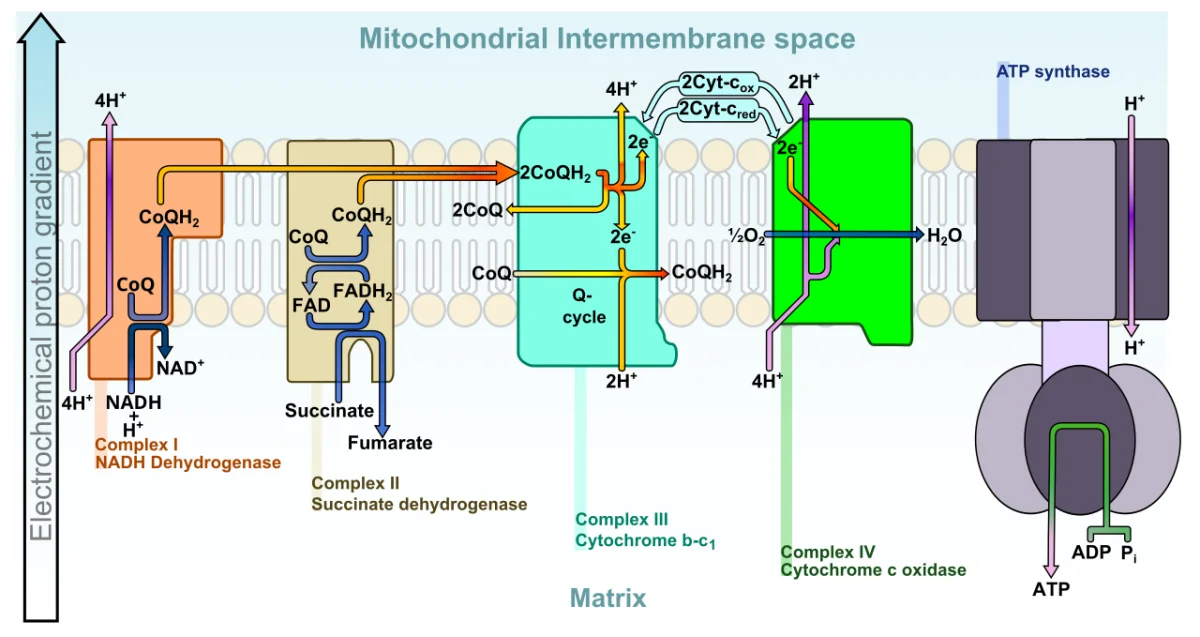
The electron transport chain (ETC) is where the NADH and FADH2 produced by glycolysis, PDH, the TCA cycle, and beta-oxidation finally cash in. The ETC is a series of protein complexes embedded in the inner mitochondrial membrane. NADH donates electrons at Complex I; FADH2 donates at Complex II. The electrons pass through the complexes, releasing energy used to pump protons from the matrix to the intermembrane space. Oxygen accepts the electrons at the end, forming water.
Put simply: the upstream pathways exist to produce NADH and FADH2 for the ETC. The ETC exists to build the proton gradient. ATP synthase (next section) uses the proton gradient to make ATP. Three linked machines, one goal.
The electron transport chain and chemiosmosis
Pathway map
1
Scroll sideways to see the whole map.
Electron path Proton (H⁺) path Inner membrane Where inhibitors bind
Where the electrons come fromEvery NADH and FADH₂ made in glycolysis, PDH, the TCA cycle, and β-oxidation ends up here. This is the only place their energy becomes ATP, which is why every pathway upstream stalls the moment oxygen runs out.
Why FADH₂ is worth lessFADH₂ hands its electrons to coenzyme Q, skipping Complex I. Four fewer protons are pumped per pair of electrons, so FADH₂ yields about 1.5 ATP against NADH's 2.5.
Inhibitors vs uncouplersInhibitors stop electron flow, so the gradient collapses, ATP synthesis stops, and O₂ consumption stops with it. Uncouplers (2,4-DNP, thermogenin in brown fat) let protons leak back without passing through ATP synthase: electrons keep flowing and O₂ is still consumed, but the energy comes out as heat instead of ATP.
Electrons go across, protons go around. Complexes I, III, and IV use the energy of electron transfer to push protons into the intermembrane space; ATP synthase lets them fall back into the matrix and captures the energy as ATP. Complex II is the only complex that pumps nothing, which is the whole reason FADH₂ is worth less than NADH.
The Four Complexes
| Complex | Role | Proton pumping |
|---------|------|---------------|
| I (NADH dehydrogenase) | Accepts electrons from NADH; transfers to CoQ | Pumps 4 H+ |
| II (Succinate dehydrogenase) | Accepts electrons from FADH2 (succinate); transfers to CoQ | Does NOT pump protons |
| III (Cytochrome bc1) | Transfers electrons from CoQ to cytochrome c | Pumps 4 H+ |
| IV (Cytochrome c oxidase) | Transfers electrons from cytochrome c to O2, forming water | Pumps 2 H+ |
Mobile carriers:
CoQ (ubiquinone): lipid-soluble carrier that shuttles electrons from I and II to III within the membrane.
Cytochrome c: small peripheral protein that shuttles electrons from III to IV in the intermembrane space.
Why NADH > FADH2 in ATP Yield
NADH enters at Complex I. Complex I + III + IV all pump protons = 10 H+ total per NADH. FADH2 enters at Complex II (which does not pump), so only Complexes III and IV pump = 6 H+ total per FADH2. Fewer protons pumped means less ATP made.
Standard approximations:
Per NADH: ~2.5 ATP.
Per FADH2: ~1.5 ATP.
Final Electron Acceptor
Oxygen at Complex IV. Four electrons + 4 H+ + O2 → 2 H2O. Without oxygen, electrons cannot leave the chain, and the entire chain backs up. NADH cannot be recycled back to NAD+, so the TCA cycle and pyruvate dehydrogenase stop. Only glycolysis can continue (anaerobically), relying on lactate fermentation to recycle NAD+.
Oxidative stress: the cost of breathing
Pathway map
1
Scroll sideways to see the whole map.
Inner mitochondrial membrane Damaging species The enzymes that defuse it NADPH, from the pentose phosphate pathway
Why the pentose phosphate pathway matters hereGlutathione is the cell's reusable antioxidant, but it only works in its reduced form. Glutathione reductase resets it and the reducing power comes from NADPH, which for most cells means the pentose phosphate pathway. That is the whole reason a pathway that makes no ATP is still essential.
G6PD deficiency in one lineA red blood cell has no mitochondria and no nucleus, so the pentose phosphate pathway is its only source of NADPH. Lose glucose-6-phosphate dehydrogenase and glutathione cannot be reset, so an oxidative challenge (fava beans, sulfa drugs, infection) denatures hemoglobin into Heinz bodies, and the spleen bites them out, leaving bite cells and hemolysis.
The same organelle kills the cellCytochrome c is a normal carrier between complexes III and IV. If the mitochondrion is damaged badly enough it leaks into the cytosol, where it assembles the apoptosome and activates caspase 9 and then caspase 3. BCL-2 holds the membrane shut and BAX opens it, which is why BCL-2 behaves as an oncogene when overexpressed: the cell simply refuses to die.
Oxygen is a superb electron acceptor and a dangerous one. A small fraction of electrons escape complexes I and III early and make superoxide. Everything on the top row exists to defuse that, everything on the left is what happens when it is not defused, and the bottom row is the cell's decision to give up.
Why does NADH yield more ATP than FADH2 per electron pair?
Click to reveal answer
NADH enters the ETC at Complex I, so electrons pass through I, III, and IV - three proton-pumping complexes. FADH2 enters at Complex II (succinate dehydrogenase), which does not pump protons. FADH2 electrons therefore only pass through III and IV. Fewer protons pumped means less ATP made per FADH2 (~1.5) compared to NADH (~2.5).
What is the role of oxygen in the electron transport chain?
Click to reveal answer
Oxygen is the final electron acceptor at Complex IV. Four electrons combine with O2 and 4 H+ to form 2 water molecules. Without oxygen, electrons cannot leave the chain, all the carriers become reduced, NADH cannot be recycled back to NAD+, and the TCA cycle halts. The entire aerobic energy system depends on O2 accepting electrons at the end.
What do CoQ and cytochrome c do in the ETC?
Click to reveal answer
Both are mobile electron carriers. CoQ (ubiquinone) is lipid-soluble and shuttles electrons from Complex I and Complex II to Complex III within the membrane. Cytochrome c is a peripheral membrane protein in the intermembrane space that shuttles electrons from Complex III to Complex IV. Without these carriers, the large complexes could not pass electrons between themselves.
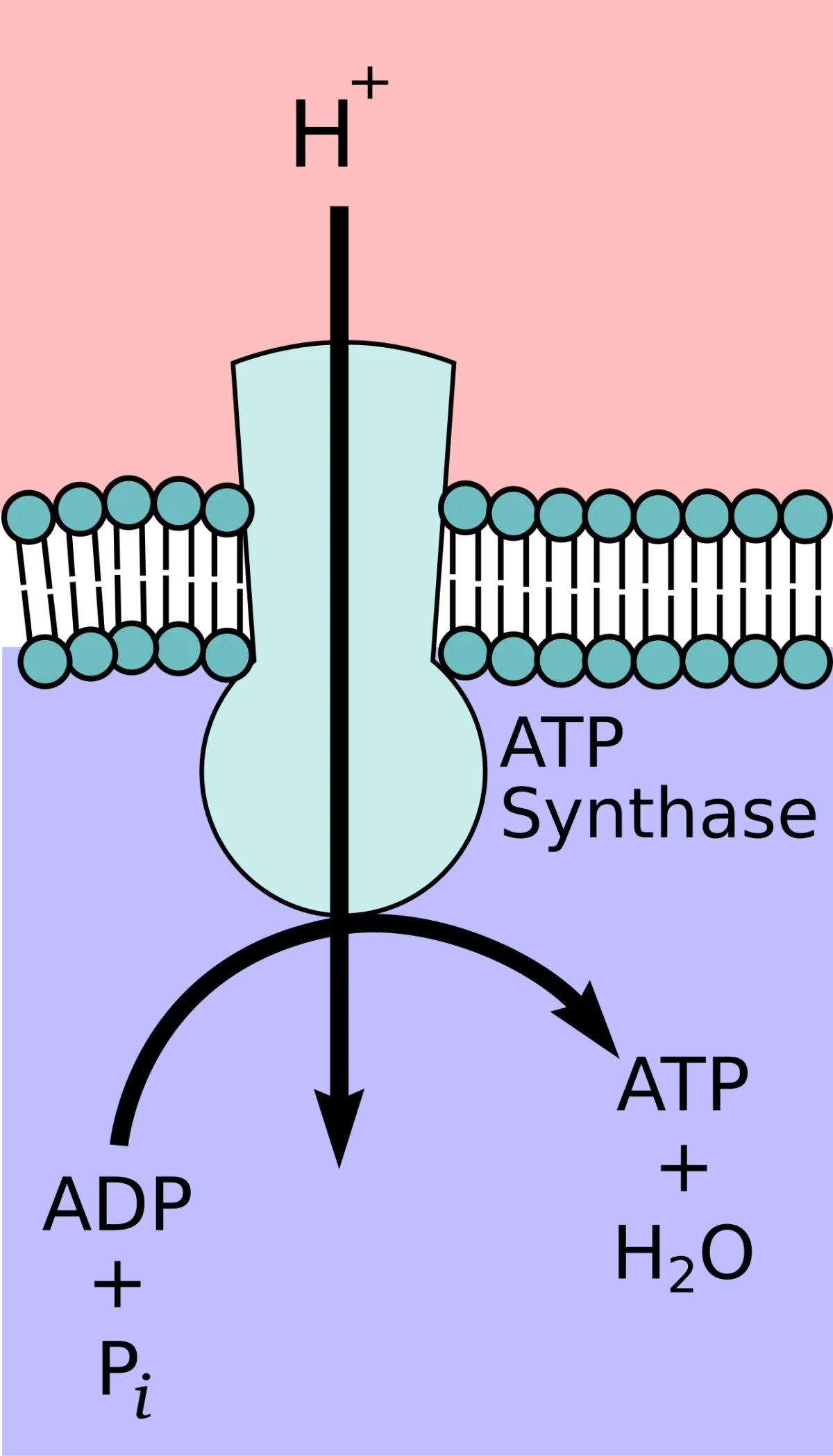
The electron transport chain builds a proton gradient. Protons flow back through ATP synthase, and that flow spins the enzyme like a water mill, producing ATP. This is chemiosmosis, a Nobel-winning insight by Peter Mitchell (1961). This step is the payoff for everything upstream: the purpose of glycolysis, PDH, the TCA cycle, and the ETC is to build the gradient that ATP synthase is about to convert into ATP.
The Proton Motive Force
As the ETC pumps H+ from the matrix to the intermembrane space, two gradients are established:
A chemical gradient: [H+] is higher outside than inside the matrix.
An electrical gradient: the intermembrane space becomes more positive.
Together these make the proton motive force (PMF). The PMF stores energy equivalent to the redox cascade that built it. Mitochondria use the PMF to drive ATP synthesis, thermogenesis, and some transport.
Oxidative phosphorylation in full. The ETC builds the proton motive force. ATP synthase uses the flow of protons back into the matrix to spin its rotor and synthesize ATP. Coupling these two processes is the essence of aerobic ATP production. Credit: Wikimedia Commons, CC BY-SA
ATP Synthase Structure
ATP synthase has two parts:
F0 (in membrane): a rotor. Protons flowing through it turn the central stalk. Named “F-zero” because it is inhibited by oligomycin.
F1 (in matrix): a stationary head with three catalytic sites. As F0 turns the stalk, F1’s catalytic sites cycle through three conformations (open, loose, tight), producing ATP from ADP + Pi with each rotation.
ATP synthase. F0 (membrane-embedded rotor) and F1 (matrix-facing ATP-forming head). Proton flow through F0 spins the central stalk, driving conformational changes in F1 that synthesize ATP. Credit: Wikimedia Commons, CC BY-SAThe rotary mechanism of ATP synthesis. Protons flowing through F0 rotate the stalk. The three F1 catalytic sites cycle through open (O), loose (L), and tight (T) conformations, producing ATP with each rotation. Per full rotation: 3 ATP. Credit: Wikimedia Commons, CC BY-SA
The Rotary Mechanism
Each full rotation of the F1 head produces 3 ATP. The number of protons required per rotation depends on the species (~8-14); in humans, roughly 4 H+ per ATP synthesized. This is why NADH (~10 H+ pumped) yields ~2.5 ATP, and FADH2 (~6 H+) yields ~1.5 ATP.
Coupling
ATP synthase normally couples proton flow directly to ATP synthesis. If you block proton pumping (an ETC inhibitor) or block proton return (an ATP synthase inhibitor like oligomycin), both shut down. If you put a “hole” in the membrane that lets protons leak back without going through ATP synthase, the PMF dissipates as heat without making ATP - see the next section on uncouplers.
What is the proton motive force, and what builds it?
Click to reveal answer
The proton motive force (PMF) is the combined chemical and electrical gradient of protons across the inner mitochondrial membrane, higher outside the matrix. It is built by the electron transport chain (Complexes I, III, IV), which uses the energy from electron flow to pump protons from matrix to intermembrane space. The PMF stores the energy harvested by the ETC.
How does ATP synthase produce ATP from the proton gradient?
Click to reveal answer
Protons flow through the F0 membrane rotor, causing it to rotate. This rotation turns a central stalk that drives conformational changes in the F1 catalytic head. The three F1 catalytic sites cycle through open, loose, and tight conformations, binding ADP + Pi in one and releasing ATP in the next. Each full rotation of F1 produces about 3 ATP.
What happens to ATP production if you punch a hole in the inner mitochondrial membrane that lets protons leak?
Click to reveal answer
The proton gradient dissipates - protons leak back into the matrix without passing through ATP synthase, so no ATP is made. The energy originally stored in the gradient is released as heat. The ETC keeps running (in fact, speeds up because matrix NADH is still processed), but no ATP is produced. This is uncoupling, the basis of thermogenesis in brown fat and the mechanism of DNP.
Blocking any step of the ETC shuts down aerobic ATP production because electrons cannot flow and the proton gradient cannot be maintained. Four classic inhibitors - one per complex plus one for ATP synthase.
Inhibitor
Target
Effect
Rotenone
Complex I
Blocks NADH’s entry point. FADH2 can still deliver to Complex II.
Antimycin A
Complex III
Blocks electron transfer from CoQ to cytochrome c
Cyanide (CN-), Carbon monoxide (CO)
Complex IV
Blocks electron transfer to O2
Oligomycin
ATP synthase (F0)
Blocks proton flow through ATP synthase, stopping ATP production directly
What Happens Downstream
Block any complex and everything upstream of it backs up. Block Complex IV and electrons cannot reach O2; all previous complexes become reduced; NADH cannot be oxidized; TCA cycle stops. Block ATP synthase and protons accumulate outside, increasing PMF, eventually stopping the ETC because no more protons can be pumped against the gradient.
Cyanide Poisoning
Cyanide binds Fe3+ in cytochrome c oxidase (Complex IV), preventing electron transfer to O2. Cellular respiration halts within seconds. All tissues suffer, but the brain and heart (high aerobic demand) are fastest to fail. Paradoxically, blood O2 stays high because cells cannot use it - “histotoxic hypoxia.” Antidote: sodium nitrite + sodium thiosulfate converts cyanide to non-toxic thiocyanate, or hydroxocobalamin (vitamin B12 precursor) binds cyanide.
How does cyanide cause cell death?
Click to reveal answer
Cyanide binds Complex IV (cytochrome c oxidase), preventing electron transfer to O2. The electron transport chain halts. Protons cannot be pumped, ATP synthase stops, NADH accumulates, the TCA cycle halts. Cells die within minutes from ATP depletion. Oxygen is available but unusable - histotoxic hypoxia.
An investigator adds rotenone to isolated mitochondria. O2 consumption drops. When succinate is added, O2 consumption recovers. Explain.
Click to reveal answer
Rotenone blocks Complex I, so NADH cannot deliver electrons. Succinate is oxidized by Complex II (succinate dehydrogenase) to produce FADH2, whose electrons enter the ETC at CoQ (downstream of Complex I). Succinate thus bypasses the rotenone block, and electron flow resumes through Complex III, IV, and O2 consumption continues.
Why does blocking ATP synthase eventually stop the ETC too?
Click to reveal answer
Without ATP synthase to consume protons, protons accumulate in the intermembrane space and the proton motive force grows. Eventually the ETC cannot pump additional protons against the steep gradient - it is thermodynamically blocked. Electrons back up, reduced carriers pile up, and electron flow stops. This is why coupled ETC and ATP synthase are both essential for aerobic respiration.
An uncoupler dissipates the proton gradient as heat instead of ATP. It does this by letting protons leak back across the inner mitochondrial membrane without passing through ATP synthase. The ETC keeps running (in fact, runs faster because NADH is consumed faster), but no ATP is made; the energy becomes heat.
Why Uncoupling Exists Biologically
Newborns and hibernating animals use controlled uncoupling to generate body heat. Brown adipose tissue has mitochondria rich in thermogenin (uncoupling protein 1, UCP1). When activated (e.g., by cold via norepinephrine and beta-3 adrenergic receptors), UCP1 creates a channel that lets protons leak back to the matrix. The heat warms the infant or bear.
2,4-Dinitrophenol (DNP)
DNP is a synthetic lipid-soluble weak acid. Once in the intermembrane space it picks up a proton, diffuses through the membrane to the matrix, and drops the proton - effectively carrying protons across without going through ATP synthase.
DNP was briefly used as a weight loss drug in the 1930s. It works - uncoupling burns enormous amounts of fat as heat - but the window between effective and fatal is narrow. Fatal hyperthermia was common, and it was banned. Illicit use persists, with periodic deaths.
Aspirin Overdose
Salicylate (from aspirin) is also a weak lipid-soluble acid and uncouples at high doses. Aspirin toxicity features hyperthermia, tachypnea (trying to blow off CO2 as the body tries to compensate for lactic acidosis), and metabolic acidosis. This is why aspirin overdose is a serious poisoning.
How does 2,4-dinitrophenol (DNP) act as an uncoupler?
Click to reveal answer
DNP is a lipid-soluble weak acid. In the intermembrane space (acidic) it picks up a proton; it diffuses through the membrane to the matrix (alkaline) and releases the proton. In effect, it shuttles protons across the membrane without going through ATP synthase. The proton motive force dissipates as heat; ATP production drops; ETC runs faster trying to rebuild the gradient; the body overheats.
What is thermogenin, and what does it do?
Click to reveal answer
Thermogenin (uncoupling protein 1, UCP1) is an inner-mitochondrial-membrane protein in brown adipose tissue that creates a proton channel, allowing protons to flow back from the intermembrane space to the matrix without making ATP. The energy is released as heat. UCP1 is activated by norepinephrine binding beta-3 adrenergic receptors during cold exposure. It keeps newborns and hibernating animals warm.
Why does aspirin overdose cause hyperthermia?
Click to reveal answer
Salicylate (from aspirin) is a lipid-soluble weak acid that can shuttle protons across the inner mitochondrial membrane, similarly to DNP. At high doses it uncouples oxidative phosphorylation, dissipating the proton gradient as heat. This causes hyperthermia, along with metabolic acidosis (lactic acid builds up from incomplete oxidation) and tachypnea (respiratory compensation).
Adding up all the ATP produced from one glucose in aerobic respiration gives about 30-32 ATP. The range exists because of the NADH shuttle used to get cytoplasmic NADH into the mitochondrial matrix.
Where the ATP per glucose actually comes from
The ledger
1
Scroll sideways to see the whole map.
Made directly (substrate-level) From NADH, at ≈ 2.5 ATP each From FADH₂, at ≈ 1.5 ATP each
Why 30 or 32Glycolysis makes its 2 NADH in the cytosol, and NADH cannot cross the inner membrane. The malate-aspartate shuttle delivers their electrons as mitochondrial NADH (2.5 ATP each); the glycerol-3-phosphate shuttle delivers them to FADH₂ instead (1.5 ATP each). Two electrons, two prices, and a 2 ATP difference in the final total.
Why the numbers are not wholeATP synthase needs roughly 4 protons per ATP, and Complexes I, III, and IV pump 10 protons per NADH but only 6 per FADH₂. Nothing divides evenly, which is why modern textbooks quote 2.5 and 1.5 rather than the older 3 and 2.
The point of the chartOnly 4 of the 32 are made directly by an enzyme handing a phosphate to ADP. Everything else is made by the electron transport chain from carriers the other pathways filled. Metabolism spends most of its effort collecting electrons, not making ATP.
Glycolysis gets the fame and contributes about a sixteenth of the yield. Its real job is not to make ATP but to make NADH and pyruvate, which is why anaerobic tissue running the same pathway to lactate gets 2 ATP where aerobic tissue gets 30 or more from the same glucose.
Step by Step Per Glucose
1
Glycolysis (cytoplasm)
Net: 2 ATP directly + 2 NADH (cytoplasmic, shuttle-dependent)
5 or 7 ATP
2
PDH (2× pyruvate → acetyl-CoA)
2 NADH (mitochondrial) × 2.5 ATP each
5 ATP
3
TCA cycle (2 turns)
6 NADH × 2.5 + 2 FADH₂ × 1.5 + 2 GTP = 15 + 3 + 2
20 ATP
Total per glucose: ~30-32 ATP. The 2 ATP range comes from which shuttle delivers cytoplasmic NADH to the matrix.
The “wiggle room” is the 2 glycolysis-produced cytoplasmic NADH, which cannot directly enter the mitochondrial matrix. They are brought in by one of two shuttle systems.
The Two Shuttles
Malate-Aspartate Shuttle (heart, liver, kidney)
Cytoplasmic NADH reduces OAA to malate. Malate crosses the inner membrane, is re-oxidized to OAA, regenerating NADH inside the matrix. The matrix NADH then enters the ETC at Complex I, yielding 2.5 ATP per original NADH.
Cytoplasmic NADH reduces DHAP to glycerol-3-phosphate. G3P transfers electrons to FAD on the outer face of the inner membrane, producing FADH2. The FADH2 electrons enter the ETC at CoQ (bypassing Complex I), yielding only 1.5 ATP.
Why does the total ATP yield from glucose oxidation range from 30 to 32?
Click to reveal answer
The 2 NADH produced by glycolysis in the cytoplasm must reach the mitochondrial matrix via a shuttle system. The malate-aspartate shuttle (heart, liver, kidney) delivers them as matrix NADH (2.5 ATP each, contributing 5 ATP total). The glycerol-3-phosphate shuttle (brain, skeletal muscle) delivers them as FADH2 (1.5 ATP each, contributing 3 ATP total). The 2 ATP difference explains the 30 vs. 32 range.
How many ATP come from the TCA cycle per glucose?
Click to reveal answer
20 ATP equivalents. Per glucose, two turns of the TCA cycle produce 6 NADH (15 ATP via ETC) + 2 FADH2 (3 ATP) + 2 GTP (directly counted as ATP equivalents) = 20 ATP. Plus the 5 ATP from the 2 PDH NADH that fed the cycle.
Why is aerobic respiration about 15x more efficient than anaerobic glycolysis?
Click to reveal answer
Glycolysis alone yields 2 ATP per glucose (anaerobic, pyruvate converted to lactate to regenerate NAD+). Aerobic respiration adds the TCA cycle and oxidative phosphorylation, producing about 28-30 more ATP for a total of 30-32. Oxidative phosphorylation extracts most of the chemical energy from glucose by coupling electron flow to proton pumping and ATP synthesis; glycolysis alone captures only a tiny fraction of the available energy.
Glycolysis, PDH, TCA, and the ETC are not isolated pathways. They are tightly coordinated so that fuel oxidation matches ATP demand. Understanding how they interconnect is how the MCAT asks you to think about metabolism as a whole.
Glucose-6-phosphateThe first fork. G6P can be stored as glycogen, burned through glycolysis, or diverted to the pentose phosphate pathway for NADPH and ribose-5-P. It cannot leave the cell: only liver and kidney have glucose-6-phosphatase to turn it back into free glucose.
PyruvateThe last cytosolic fork. With oxygen it crosses into the mitochondrion for PDH; without oxygen it becomes lactate; in muscle it is transaminated to alanine; and in the fasting liver it is carboxylated to oxaloacetate to start gluconeogenesis.
Acetyl-CoAThe great convergence: carbohydrate, fat, and ketogenic amino acids all arrive here. It burns in the TCA cycle, leaves as citrate for fatty acid and cholesterol synthesis, or condenses into ketone bodies. It can never become glucose, which is why fat is not a gluconeogenic fuel.
OxaloacetateGatekeeper of the TCA cycle and the doorway out of it. Acetyl-CoA cannot enter the cycle without OAA, so when OAA is drained for gluconeogenesis during fasting, acetyl-CoA backs up and is shunted into ketone bodies. OAA also trades with the urea cycle as aspartate and fumarate.
Read it top to bottom for catabolism, bottom to top for anabolism. Fuels enter at the top, carbon funnels through four junction metabolites, and the electron carriers cash out at the electron transport chain. The four navy pills are the only molecules you need to reason from: know what enters and leaves each one and you can rebuild the rest of the map from memory.
Each arrow is a regulatory checkpoint. The overall rate is set by the slowest committed step of the entire chain.
Respiratory Control
In isolated mitochondria, the rate of O2 consumption depends on ADP availability. When ADP is low (cell has plenty of ATP), ATP synthase cannot run; the PMF builds up; the ETC cannot pump more protons; NADH accumulates; TCA and PDH slow; glycolysis slows. When ADP rises (ATP being consumed), the whole chain speeds up in synchrony.
This is respiratory control: the rate of fuel oxidation = the rate of ATP consumption. The cell never oxidizes fuel faster than it uses ATP.
The Pasteur Effect
Louis Pasteur observed that yeast glucose consumption slows dramatically when oxygen is added. Anaerobic glycolysis burns glucose fast (2 ATP per glucose). Aerobic respiration is 15x more efficient (30-32 ATP per glucose), so less glucose is needed. Adding O2 raises ATP, which inhibits PFK-1, slowing glycolysis. The Pasteur effect is the textbook name for this regulatory coupling.
Cancer and the Warburg Effect
Many cancer cells show the opposite - they rely heavily on glycolysis even in the presence of O2 (aerobic glycolysis), producing lactate as their main end product. This is the Warburg effect. Reasons are complex but likely include mitochondrial dysfunction, enhanced biosynthesis, and rapid growth requirements. PET scans exploit the Warburg effect: radioactive glucose analogs (FDG) accumulate in cancer cells because of their high glucose uptake.
What is respiratory control?
Click to reveal answer
The coupling of fuel oxidation rate to ATP consumption rate. ADP availability ultimately sets the rate - when ADP is low (ATP high), ATP synthase cannot run, the proton gradient backs up, the ETC slows, NADH accumulates, TCA and PDH slow, and glycolysis slows. When ADP rises (ATP being used), the entire chain speeds up in synchrony. The cell never oxidizes fuel faster than it needs ATP.
What is the Pasteur effect?
Click to reveal answer
When oxygen is added to a culture, glucose consumption slows dramatically. Because aerobic respiration yields ~15x more ATP per glucose than anaerobic glycolysis, far less glucose is needed to meet the same ATP demand. The added ATP inhibits PFK-1, slowing glycolysis. Observed by Pasteur in fermenting yeast, this is a textbook example of metabolic regulation coupling.
What is the Warburg effect, and why is it exploited in PET imaging?
Click to reveal answer
Many cancer cells rely heavily on aerobic glycolysis (converting pyruvate to lactate even with O2 present) instead of oxidative phosphorylation. The Warburg effect results in high glucose uptake by tumors. PET scans use fluorodeoxyglucose (FDG), a radioactive glucose analog. Tumors take up FDG at elevated rates and become visible on PET, enabling non-invasive cancer imaging.