Metabolism is every chemical reaction happening in a cell. It splits into two directions: catabolism breaks complex molecules into simpler ones and harvests energy, and anabolism builds complex molecules from simpler ones using energy.
Glucose-6-phosphateThe first fork. G6P can be stored as glycogen, burned through glycolysis, or diverted to the pentose phosphate pathway for NADPH and ribose-5-P. It cannot leave the cell: only liver and kidney have glucose-6-phosphatase to turn it back into free glucose.
PyruvateThe last cytosolic fork. With oxygen it crosses into the mitochondrion for PDH; without oxygen it becomes lactate; in muscle it is transaminated to alanine; and in the fasting liver it is carboxylated to oxaloacetate to start gluconeogenesis.
Acetyl-CoAThe great convergence: carbohydrate, fat, and ketogenic amino acids all arrive here. It burns in the TCA cycle, leaves as citrate for fatty acid and cholesterol synthesis, or condenses into ketone bodies. It can never become glucose, which is why fat is not a gluconeogenic fuel.
OxaloacetateGatekeeper of the TCA cycle and the doorway out of it. Acetyl-CoA cannot enter the cycle without OAA, so when OAA is drained for gluconeogenesis during fasting, acetyl-CoA backs up and is shunted into ketone bodies. OAA also trades with the urea cycle as aspartate and fumarate.
Read it top to bottom for catabolism, bottom to top for anabolism. Fuels enter at the top, carbon funnels through four junction metabolites, and the electron carriers cash out at the electron transport chain. The four navy pills are the only molecules you need to reason from: know what enters and leaves each one and you can rebuild the rest of the map from memory.
For this chapter and the next two, keep this simple map in your head for catabolism:
NADH and FADH2 → electron transport chain → proton gradient.
Proton gradient → ATP synthase → ATP.
Fatty acids and amino acids plug into the same pipeline at acetyl-CoA (or a TCA intermediate). The TCA cycle’s main job is not ATP - it is making NADH and FADH2 to feed the ETC. Think of the TCA cycle as the electron loader and the ETC as the ATP maker.
From food to fuel: digestion and absorption
Pathway map
1
Scroll sideways to see the whole map.
Fuel that reaches the master map Lumen and enterocyte Rate-setting or clinically tested step Intermediate
Only fat skips the liver firstSugars and amino acids go straight into the portal vein, so the liver sees them before anyone else and can buffer the load. Dietary fat leaves in chylomicrons through the lymphatics and enters the bloodstream at the thoracic duct, which means muscle and adipose get first refusal on it. That is why a fatty meal raises plasma triglyceride for hours while a sugary one is cleared much faster.
The two lipases students confusePancreatic lipase works in the gut lumen on food you have just eaten. Lipoprotein lipase sits on the capillary wall of muscle and adipose and unloads triglyceride out of circulating chylomicrons and VLDL. Hormone-sensitive lipase is different again: it is inside the fat cell and releases stored fat during fasting, which is the arrow that feeds β-oxidation.
Lactose intolerance in one lineLactase is a brush border enzyme and the first one to be lost with age. Undigested lactose stays in the lumen, pulls in water osmotically, and is fermented by colonic bacteria into gas and short-chain acids. Nothing is wrong with absorption itself, which is why the symptoms are bloating and diarrhea rather than malnutrition.
Three fuels, three routes, one destination. Carbohydrate and protein are broken to their monomers and go to the liver by the portal vein. Fat is emulsified, cut, ferried in micelles, rebuilt inside the enterocyte, and shipped out in chylomicrons through the lymph. Everything here arrives at the top of the master map as glucose, fatty acids, or amino acids.
Catabolism vs. Anabolism
| Process | Direction | Energy | Examples |
|---------|-----------|--------|----------|
| Catabolism | Large → small | Releases energy (captured as ATP, NADH, FADH2) | Glycolysis, beta-oxidation, amino acid catabolism |
| Anabolism | Small → large | Consumes energy (ATP, NADPH) | Gluconeogenesis, fatty acid synthesis, protein synthesis |
Acetyl-CoA - The Central Hub
All major fuels converge to acetyl-CoA, which enters the TCA cycle:
Some amino acids → directly or via pyruvate → acetyl-CoA.
Energy Carriers
Cells store catabolic energy in three main molecules:
ATP: immediate energy currency. Hydrolysis of ATP to ADP releases ~7.3 kcal/mol.
NADH and FADH2: electron carriers. Deliver electrons to the ETC, which makes ATP.
NADPH: like NADH but used for biosynthetic reductions (fatty acid synthesis, antioxidant regeneration) and made by the pentose phosphate pathway.
What is the difference between catabolism and anabolism?
Click to reveal answer
Catabolism breaks down complex molecules to simpler ones and releases energy (captured as ATP, NADH, FADH2). Anabolism builds complex molecules from simpler ones and consumes energy (ATP, NADPH). Cells run both simultaneously and regulate the balance based on energy needs and hormonal signals.
How do glucose, fatty acids, and amino acids converge metabolically?
Click to reveal answer
All three ultimately produce acetyl-CoA, which enters the TCA cycle. Glucose does so via glycolysis → pyruvate → pyruvate dehydrogenase. Fatty acids via beta-oxidation. Amino acids enter at various points depending on their carbon skeleton, but many converge on acetyl-CoA. Acetyl-CoA is the central junction of catabolism.
Why do cells maintain separate NADH and NADPH pools?
Click to reveal answer
NADH is primarily oxidized back to NAD+ by the electron transport chain to make ATP (catabolism). NADPH is used as a reducing agent in biosynthetic reactions (fatty acid synthesis, cholesterol synthesis) and in antioxidant systems (reducing glutathione). Keeping them separate lets the cell independently control catabolic energy production and anabolic/reductive reactions.
The first half of glycolysis consumes 2 ATP to prepare glucose for splitting and cleaves the resulting 6-carbon molecule into two 3-carbon molecules.
Glycolysis: glucose to pyruvate in ten steps
Pathway map
1
Scroll sideways to see the whole map.
Irreversible (the 3 regulated steps) ATP made NADH made Junction metabolite
Glucose-6-POff-ramps to glycogen synthesis and to the pentose phosphate pathway. This is why hexokinase traps glucose in the cell: G6P carries a charge and cannot leave through GLUT transporters.
DHAPWhere the glycerol backbone of fat joins and leaves. Glycerol from lipolysis enters here, which is the only part of a triglyceride that can become glucose.
PyruvateThe exit into fermentation (lactate), transamination (alanine), or the mitochondrion for PDH and the TCA cycle.
The only three steps you can regulate are the three you cannot reverse. Hexokinase, PFK-1, and pyruvate kinase release far too much free energy to run backwards, so gluconeogenesis has to route around all three. Everything below triose phosphate isomerase happens twice per glucose, which is where the doubled yields come from.
The Five Investment Steps
1
Glucose → G6P
Hexokinase · liver uses glucokinase regulated
-1 ATP
2
G6P → F6P
Phosphoglucose isomerase · aldose to ketose
–
3
F6P → F1,6BP
PFK-1 rate-limiting
-1 ATP
4
F1,6BP → DHAP + G3P
Aldolase · splits 6C into two 3C
–
5
DHAP ⇌ G3P
Triose phosphate isomerase · both feed the payoff phase
–
Investment cost: -2 ATP per glucose. End state: 2 G3P, ready for the payoff phase.
Key Points
Step 1 (hexokinase): glucose enters the cell via GLUT transporters, then is phosphorylated. Once phosphorylated, glucose cannot leave the cell - it is committed. Hexokinase has a low Km and is in all cells. Liver uses glucokinase (high Km), which only activates at high blood glucose (post-meal).
Step 3 (PFK-1): the committed, rate-limiting step. Heavily regulated by ATP (inhibits), AMP (activates), citrate (inhibits), and fructose-2,6-bisphosphate (strongly activates). Hormonal signals (insulin vs. glucagon) control F-2,6-BP levels via PFK-2.
Step 4 (aldolase): splits the 6-carbon sugar into two 3-carbon sugars: DHAP and G3P.
Step 5 (triose phosphate isomerase): DHAP is converted to G3P. So at the end of step 5, the cell has two G3P molecules, both of which will proceed through the payoff phase.
Why does hexokinase trap glucose in the cell by phosphorylating it?
Click to reveal answer
Glucose-6-phosphate carries a negative charge and cannot diffuse back through the GLUT transporters that brought glucose in. The phosphate effectively “tags” glucose for retention inside the cell. This is the first committed step in using glucose - once phosphorylated, it must enter glycolysis, glycogen synthesis, or the pentose phosphate pathway.
Why is PFK-1 called the rate-limiting enzyme of glycolysis?
Click to reveal answer
PFK-1 catalyzes the committed step (fructose-6-P + ATP → fructose-1,6-BP + ADP). This reaction is essentially irreversible and sets the overall rate of glycolysis. PFK-1 is allosterically controlled by many regulators (ATP, citrate inhibit; AMP, fructose-2,6-BP activate), allowing glycolysis to respond to energy state and hormones.
Net energy balance of the investment phase alone (steps 1-5)?
Click to reveal answer
2 ATP consumed. 0 ATP produced. 1 glucose becomes 2 G3P (glyceraldehyde-3-phosphate). No NADH yet. The investment must be paid back (and exceeded) during the payoff phase for glycolysis to be net energy-producing.
The payoff phase oxidizes each 3-carbon glyceraldehyde-3-phosphate to pyruvate, harvesting energy as NADH and ATP. Because every glucose produces 2 G3P, each payoff step runs twice per glucose.
The Five Payoff Steps
Each G3P runs through these five steps. Because one glucose produces two G3P, multiply every yield by 2 for the whole-glucose total.
Per G3P: 2 ATP + 1 NADH + 1 pyruvate Per glucose (×2): 4 ATP + 2 NADH + 2 pyruvate
Net Yield
Per molecule of glucose:
4 ATP produced (2 per G3P × 2 G3P) in the payoff
2 ATP consumed in the investment phase
Net: 2 ATP
Plus: 2 NADH and 2 pyruvate
Substrate-Level Phosphorylation
The two ATPs in the payoff phase are made by substrate-level phosphorylation - a high-energy phosphate group is transferred directly from a phosphorylated intermediate to ADP. This is different from oxidative phosphorylation (ETC + ATP synthase), which uses a proton gradient.
Arsenic Poisoning
Arsenite (As3+) inhibits the sulfhydryl groups needed by GAPDH. Arsenate (As5+) mimics phosphate, becomes incorporated at step 6 but then spontaneously hydrolyzes instead of phosphorylating ADP in step 7 - effectively uncoupling glycolysis without producing ATP. Both are lethal because glycolysis is required in all cells.
What is the overall net yield from glycolysis per molecule of glucose?
Click to reveal answer
Net 2 ATP, 2 NADH, and 2 pyruvate. Investment phase consumes 2 ATP; payoff phase produces 4 ATP and 2 NADH per glucose. Subtracting investment from payoff gives the net of 2 ATP. Pyruvate then enters either aerobic pathways (TCA cycle) or fermentation.
What is substrate-level phosphorylation, and where does it happen in glycolysis?
Click to reveal answer
Substrate-level phosphorylation is direct transfer of a phosphate from a high-energy substrate to ADP, producing ATP. It happens twice in glycolysis: at step 7 (phosphoglycerate kinase transfers from 1,3-BPG) and step 10 (pyruvate kinase transfers from PEP). It is independent of the electron transport chain.
Why is GAPDH step 6 a common target of poisoning (e.g., arsenic)?
Click to reveal answer
GAPDH couples the oxidation of G3P (producing NADH) to the formation of a high-energy acyl-phosphate (1,3-BPG), which feeds step 7's ATP production. Blocking GAPDH halts glycolysis' energy output. Arsenite inhibits the enzyme directly; arsenate mimics phosphate, enters the reaction, but spontaneously hydrolyzes instead of forming a useful product, robbing the cell of ATP.
Three enzymes catalyze the irreversible steps of glycolysis and are the main regulatory points: hexokinase (step 1), PFK-1 (step 3), and pyruvate kinase (step 10). Of these, PFK-1 is the most heavily regulated and is called the master switch of glycolysis.
PFK-1: The Master Switch
PFK-1 is allosterically controlled by multiple effectors:
Effector
Effect on PFK-1
Meaning
ATP
Inhibits
”Cell has enough energy, slow down”
AMP / ADP
Activates
”Cell is energy-starved, speed up”
Citrate
Inhibits
”TCA cycle is backed up, do not send more pyruvate”
Fructose-2,6-bisphosphate (F-2,6-BP)
Activates strongly
Hormonal signal
H+ (low pH)
Inhibits
Slows glycolysis during lactic acid accumulation
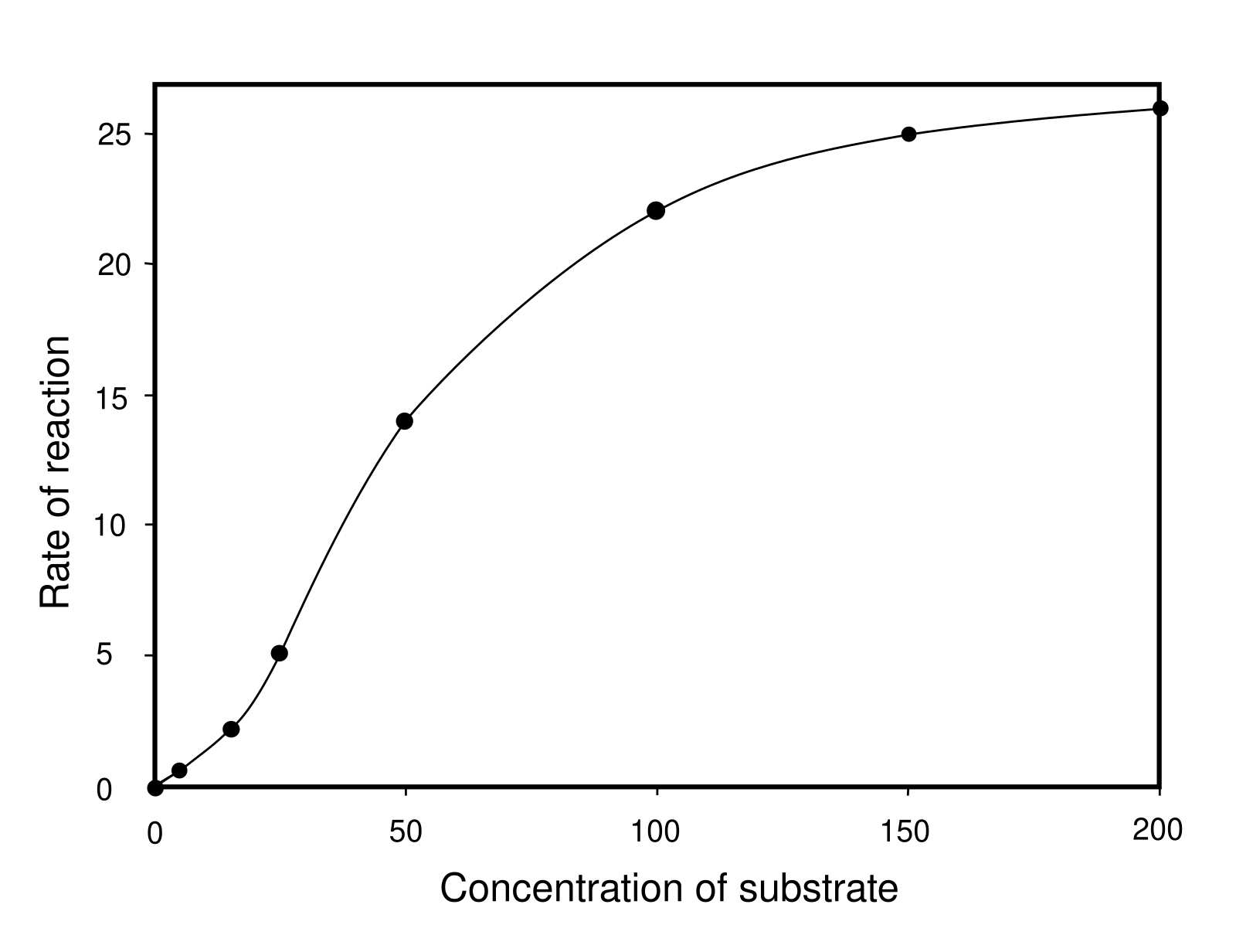
PFK-1 shows sigmoidal (S-shaped) kinetics, the fingerprint of an allosteric enzyme. Low substrate barely activates it; once substrate crosses a threshold, activity climbs steeply. Allosteric inhibitors (ATP, citrate) right-shift this curve - making PFK-1 less responsive. Activators (AMP, F-2,6-BP) left-shift it. Credit: Wikimedia Commons, CC BY-SA
Fructose-2,6-Bisphosphate - The Hormonal Link
F-2,6-BP is not a glycolysis intermediate but a dedicated regulator. It is made by PFK-2 (a different enzyme from PFK-1) from F6P. PFK-2 has two activities: a kinase (makes F-2,6-BP) and a phosphatase (removes it). Insulin and glucagon control which activity dominates:
This reciprocal regulation at the same control point (PFK-1) lets a single hormone shift metabolism between storage (fed) and mobilization (fasted).
Hexokinase and Pyruvate Kinase
Hexokinase: inhibited by its product G6P (end-product inhibition). Glucokinase (liver version) has higher Km and is NOT inhibited by G6P, so the liver continues to phosphorylate glucose at high blood-glucose levels.
Pyruvate kinase: activated by feed-forward signal F-1,6-BP (upstream intermediate); inhibited by ATP and alanine. In liver, pyruvate kinase is also phosphorylated (inactivated) by glucagon-triggered PKA, reducing pyruvate production during fasting.
Which molecule is the strongest allosteric activator of PFK-1?
Click to reveal answer
Fructose-2,6-bisphosphate (F-2,6-BP). It is not an intermediate of glycolysis but a dedicated regulator made by PFK-2. Insulin increases F-2,6-BP (activates glycolysis); glucagon decreases it (activates gluconeogenesis). AMP also activates PFK-1 but F-2,6-BP has a much larger effect in most tissues.
How does glucagon reduce glycolysis in the liver?
Click to reveal answer
Glucagon raises cAMP, activating PKA. PKA phosphorylates PFK-2, shifting it from kinase to phosphatase mode - F-2,6-BP levels drop. PFK-1 is less activated, slowing glycolysis. PKA also phosphorylates pyruvate kinase (inactivating it) and glycogen phosphorylase kinase (activating glycogen breakdown). Net: glucose is released into the blood rather than burned in the liver.
Why is hexokinase inhibited by glucose-6-phosphate, but glucokinase is not?
Click to reveal answer
Hexokinase exists in most cells, which need glucose for routine fuel and should not waste glucose when downstream is backed up. Product inhibition (G6P feedback) stops phosphorylation when glycolysis is slow. Glucokinase is liver-specific, designed to take up excess glucose after a meal - it should NOT be inhibited by G6P so the liver can continue sponging up glucose into glycogen and other pathways when blood glucose is high.
Each pyruvate produced by glycolysis has three possible fates, depending on the presence of oxygen and the cell type. This is the branch point where glucose metabolism splits into “aerobic with full oxidation” or “anaerobic with fermentation.”
Fate 1: Acetyl-CoA (Aerobic) - The Main Pipeline
If oxygen is available, pyruvate enters the mitochondrial matrix and is oxidatively decarboxylated by the pyruvate dehydrogenase (PDH) complex:
Pyruvate + CoA + NAD+→Acetyl-CoA + CO2+NADH
Each glucose produces 2 pyruvate → 2 acetyl-CoA + 2 CO2 + 2 NADH (in the mitochondrial matrix). Acetyl-CoA then enters the TCA cycle (also called the citric acid cycle or Krebs cycle) for full oxidation. The TCA cycle strips more electrons as NADH and FADH2, which feed the electron transport chain (ETC). The ETC pumps protons, and ATP synthase uses the proton gradient to make ATP. This is the main energy pipeline of aerobic respiration, covered in Chapter 10.
PDH requires 5 coenzymes: TPP (B1), FAD (B2), NAD+ (B3), CoA (B5), and lipoic acid. Mnemonic: “Tender Loving Care For Nancy” = TPP, Lipoic acid, CoA, FAD, NAD.
Fate 2: Lactate (Anaerobic in Animals)
When O2 is scarce or mitochondria are absent (red blood cells have no mitochondria), pyruvate is reduced to lactate:
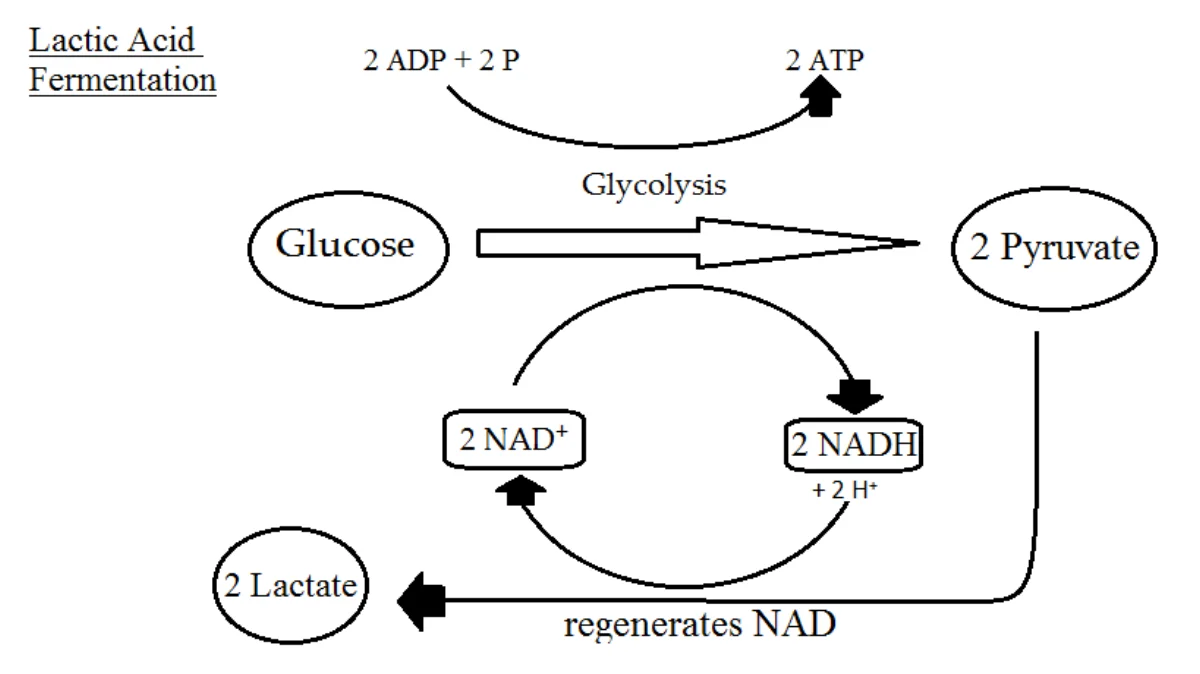
Why bother? Glycolysis requires NAD+ to keep going (step 6, GAPDH). Without the ETC to recycle NADH back to NAD+, the NAD+ pool would run out. Making lactate regenerates NAD+ so glycolysis can continue (~2 ATP per glucose, slow but oxygen-independent).
Lactic acid fermentation. Pyruvate + NADH → lactate + NAD+. The NAD+ is recycled back to glycolysis, allowing continued ATP production when oxygen is unavailable. Credit: Wikimedia Commons, CC BY-SA
Fate 3: Ethanol (Yeast Fermentation)
In yeast, pyruvate is decarboxylated to acetaldehyde, then reduced to ethanol:
Pyruvate→Acetaldehyde + CO2→Ethanol
The second step uses NADH, regenerating NAD+ just like lactic acid fermentation. This is how beer, wine, and bread dough are made - yeast ferment sugar, producing CO2 (bubbles) and ethanol (alcohol).
Why must pyruvate be converted to lactate (or ethanol + CO2) under anaerobic conditions?
Click to reveal answer
Glycolysis requires NAD+ at step 6 (GAPDH). Without oxygen, the electron transport chain cannot regenerate NAD+ from NADH. Converting pyruvate to lactate uses NADH and regenerates NAD+, allowing glycolysis to continue. Without this fermentation step, glycolysis would stop after consuming its small NAD+ pool.
How many coenzymes does the pyruvate dehydrogenase complex require, and what is the mnemonic?
Click to reveal answer
Five. "Tender Loving Care For Nancy" = TPP (B1, thiamine), Lipoic acid, CoA (B5, pantothenate), FAD (B2, riboflavin), NAD+ (B3, niacin). Thiamine deficiency (B1) impairs PDH and other decarboxylase enzymes, causing beriberi and Wernicke-Korsakoff syndrome.
Why do red blood cells produce lactate even at rest?
Click to reveal answer
RBCs lack mitochondria, so they cannot perform the TCA cycle or oxidative phosphorylation. They rely entirely on glycolysis for ATP, producing pyruvate that must be converted to lactate to regenerate NAD+. This produces a continuous basal lactate output into the blood, which is cleared by the liver.
Gluconeogenesis is the synthesis of glucose from non-carbohydrate precursors. It happens mainly in the liver and (to a lesser extent) the kidney. It is essentially glycolysis in reverse, but with four different “bypass” enzymes at the three irreversible steps of glycolysis.
Glycolysis vs gluconeogenesis: the four bypasses
Reciprocal pathways
1
Scroll sideways to see the whole map.
Irreversible in glycolysis Bypass enzyme (gluconeogenesis only) Junction metabolite
CostGluconeogenesis burns 4 ATP + 2 GTP + 2 NADH to make one glucose, while glycolysis returns only 2 ATP + 2 NADH. Running both at once would just heat the cell, which is why the two are reciprocally regulated.
Where it happensLiver mostly, kidney cortex in prolonged fasting. Only these tissues have glucose-6-phosphatase, so only these tissues can release free glucose into the blood.
What can feed itLactate (Cori cycle), glucogenic amino acids (mainly alanine), and glycerol from fat. Acetyl-CoA cannot: PDH is irreversible, so fatty acids can never make net glucose.
Gluconeogenesis is not glycolysis in reverse. Seven of the ten steps are shared, freely reversible enzymes. The other three are one-way, so four separate bypass enzymes are needed to get around them. Learn the four bypasses and you have learned the pathway.
Gluconeogenesis connects to the other metabolic pathways by running them backwards. The glycerol backbone from fat (from triglyceride breakdown) and amino acids (from muscle protein) both feed into gluconeogenesis at different points. Lactate from anaerobic glycolysis in muscle and RBCs is shipped to the liver (Cori cycle) and converted back to glucose here. Essentially, gluconeogenesis is the liver’s reverse-engineering machine that makes glucose when we cannot get it from food.
Why Gluconeogenesis Matters
During fasting, brain and red blood cells still need glucose. Once glycogen stores are depleted (~24 hours), the body must make glucose from precursors. Gluconeogenesis ensures a steady blood-glucose supply even during prolonged fasting.
Glucose-6-phosphatase · liver and kidney only · releases free glucose to blood
hydrolysis
Total cost: 6 ATP equivalents per glucose made (vs. 2 ATP gained by glycolysis). Expensive - run only when glucose is truly needed.
Pyruvate to PEP
This two-step bypass of pyruvate kinase is the most energetically expensive part:
Pyruvate carboxylase (in mitochondrion) adds CO2 to pyruvate, making oxaloacetate. Uses 1 ATP. Requires biotin (vitamin B7).
PEP carboxykinase (PEPCK) decarboxylates oxaloacetate and phosphorylates it to PEP. Uses 1 GTP.
Pyruvate carboxylase is activated by acetyl-CoA - a signal that fat is being burned, so glucose is scarce, and the liver should synthesize more.
Glucose-6-Phosphatase
Only the liver and kidney have glucose-6-phosphatase. This enzyme removes the phosphate from G6P, producing free glucose that can leave the cell via GLUT2. Muscle lacks this enzyme - that is why muscle glycogen cannot directly contribute to blood glucose. Only liver glycogen can be broken down and released as blood glucose.
Cost
Per glucose synthesized from 2 pyruvate: 4 ATP + 2 GTP + 2 NADH consumed. Gluconeogenesis is expensive - the body only runs it when glucose is truly needed (fasting, low blood sugar, post-exercise).
What are the four bypass enzymes of gluconeogenesis, and which glycolysis steps do they bypass?
Click to reveal answer
Pyruvate carboxylase and PEP carboxykinase together bypass pyruvate kinase (step 10). Fructose-1,6-bisphosphatase bypasses PFK-1 (step 3). Glucose-6-phosphatase bypasses hexokinase (step 1). These four enzymes let the pathway run thermodynamically downhill in the opposite direction from glycolysis.
Why can humans not convert fatty acids to glucose?
Click to reveal answer
Beta-oxidation of fatty acids produces acetyl-CoA, not pyruvate. The pyruvate dehydrogenase reaction (pyruvate → acetyl-CoA) is irreversible - there is no enzyme to convert acetyl-CoA back to pyruvate. Without this step, fatty acid carbons cannot enter gluconeogenesis. The glycerol backbone of triglycerides CAN be converted to glucose; only the fatty acid tails cannot.
Why does muscle not contribute directly to blood glucose?
Click to reveal answer
Muscle lacks glucose-6-phosphatase, the enzyme that removes the phosphate from glucose-6-phosphate so glucose can leave the cell. Muscle glycogen breakdown produces G6P, which can be used locally (through glycolysis) but cannot be exported as free glucose. Only the liver and kidney have glucose-6-phosphatase and can directly contribute to blood glucose.
Glycogen is the animal storage form of glucose - highly branched alpha-1,4 + alpha-1,6 polymer found in liver (for blood glucose buffering) and muscle (for local energy). Building it up and breaking it down are separate pathways, reciprocally regulated.
Glycogen: storing and releasing glucose
Pathway map
1
Scroll sideways to see the whole map.
Storing (glycogen synthase) Releasing (glycogen phosphorylase) Junction metabolite Hormone signal
Liver vs muscleOnly the liver has glucose-6-phosphatase, so only liver glycogen can raise blood glucose. Muscle glycogen is a private fuel store: muscle keeps its G6P and burns it locally. This is why liver glycogen runs out in about a day of fasting while muscle glycogen is irrelevant to blood sugar.
One switch, two effectsA single phosphorylation cascade turns synthase off and phosphorylase on. Glucagon and adrenaline raise cAMP, activating protein kinase A; insulin activates protein phosphatase 1, which reverses both. The cell never stores and mobilises at the same time.
Branches matterBranching enzyme creates α-1,6 links roughly every ten residues. Branches multiply the non-reducing ends where phosphorylase can work, so a branched polymer can be dismantled far faster than a straight chain. Debranching enzyme is needed to clear each branch point.
Glycogen is glucose kept in a form that costs nothing osmotically. Free glucose at the same concentration would pull enough water into the cell to burst it. The price is one UTP per residue stored, and a branch point that needs a separate enzyme to take apart.
Glycogen metabolism is a short detour off of glycolysis. When the cell has excess glucose-6-phosphate (the first glycolysis intermediate), it can store it as glycogen rather than running it through glycolysis. When energy is needed later, glycogen is broken down back to glucose-6-phosphate, which can then re-enter glycolysis (in muscle) or be released as blood glucose after G6Pase action (in liver). So glycogen is a short-term reserve that feeds back into the glycolysis → TCA → ETC pipeline when the cell needs it.
Glycogenesis (Building)
Glucose-6-P → glucose-1-P (phosphoglucomutase).
G1P + UTP → UDP-glucose + PPi (UDP-glucose pyrophosphorylase). UDP-glucose is the activated donor.
Glycogen synthase adds UDP-glucose to the non-reducing end of a growing glycogen chain via alpha-1,4 linkage. Rate-limiting enzyme of glycogenesis.
When the chain is ~11 residues long, branching enzyme transfers a short segment to an internal position via an alpha-1,6 bond, creating a branch.
Glycogenolysis (Breaking Down)
Glycogen phosphorylase cleaves glucose-1-P from the non-reducing end of a branch using inorganic phosphate (Pi). Rate-limiting enzyme. Uses pyridoxal phosphate (PLP, vitamin B6) as a cofactor.
When phosphorylase reaches 4 residues from a branch point, it stops.
Debranching enzyme (a dual-function enzyme) moves three residues to the main chain (transferase activity) and then hydrolyzes the remaining alpha-1,6 glucose (glucosidase activity), releasing a free glucose.
G1P → G6P (phosphoglucomutase). In liver, glucose-6-phosphatase converts G6P → free glucose, which leaves the cell. In muscle, G6P enters glycolysis for local ATP.
Hormonal Regulation
Insulin (fed state):
Dephosphorylates glycogen synthase → ACTIVE → storage.
Dephosphorylates glycogen phosphorylase → INACTIVE → no breakdown.
Glucagon (fasted) and epinephrine (stress):
Phosphorylates glycogen synthase → INACTIVE → no storage.
What enzyme is rate-limiting for glycogen synthesis vs. breakdown?
Click to reveal answer
Glycogen synthase is rate-limiting for synthesis (glycogenesis). Glycogen phosphorylase is rate-limiting for breakdown (glycogenolysis). Both are reciprocally controlled by phosphorylation: glucagon/epinephrine activate the PKA cascade, which phosphorylates and ACTIVATES phosphorylase while INACTIVATING synthase. Insulin reverses both.
What are the two activities of debranching enzyme?
Click to reveal answer
Debranching enzyme is bifunctional. It has a transferase activity that moves a short segment (usually three alpha-1,4-linked glucoses) from a branch to the main chain, and a glucosidase (alpha-1,6-glucosidase) activity that hydrolyzes the remaining single alpha-1,6-linked glucose, releasing it as free glucose. Both activities are needed to fully degrade glycogen.
Why does Von Gierke disease produce more severe hypoglycemia than McArdle disease?
Click to reveal answer
Von Gierke is a defect in glucose-6-phosphatase, which is required for BOTH glycogenolysis and gluconeogenesis to produce free blood glucose. Neither pathway can deliver glucose. McArdle is a muscle-specific glycogen phosphorylase defect; the liver (a separate isozyme) is unaffected, so blood glucose can still be maintained. McArdle causes exercise intolerance but not systemic hypoglycemia.
The pentose phosphate pathway (PPP) - also called the hexose monophosphate shunt - runs parallel to glycolysis. It branches off at the first glycolysis intermediate (glucose-6-phosphate) and diverts it away from ATP production toward two different goals: generating NADPH and producing ribose-5-phosphate. The PPP does NOT feed the TCA cycle or ETC directly; it is a side path that produces biosynthetic precursors while glycolysis continues toward pyruvate.
The pentose phosphate pathway
Pathway map
1
Scroll sideways to see the whole map.
G6PD: irreversible and rate-limiting NADPH made Shared with glycolysis Reversible, non-oxidative
Back into glycolysisThe non-oxidative half is fully reversible and its products, fructose-6-P and glyceraldehyde-3-P, are glycolytic intermediates. A cell that needs ribose but not NADPH can therefore run this half backwards from glycolysis and skip the oxidative steps entirely.
What NADPH is forReductive biosynthesis (fatty acids, cholesterol, steroids), keeping glutathione reduced so red cells survive oxidative stress, the respiratory burst in neutrophils, and cytochrome P450 detoxification. NADPH is for building and defending; NADH is for burning.
G6PD deficiencyThe most common enzyme deficiency in humans. Red cells have no nucleus and no other NADPH source, so under oxidative stress (fava beans, sulfa drugs, antimalarials, infection) glutathione cannot be regenerated, hemoglobin precipitates as Heinz bodies, and the cells lyse.
No ATP is made or spent anywhere in this pathway. It exists to produce two things glycolysis cannot: NADPH for building and defending, and ribose-5-phosphate for nucleotides. The cell dials the two halves independently depending on which of them it actually needs.
Why PPP Matters
NADPH: needed for fatty acid synthesis, cholesterol synthesis, nucleotide biosynthesis, and regeneration of glutathione (the main cellular antioxidant).
Ribose-5-phosphate: the sugar backbone for nucleotides (DNA, RNA, ATP, NAD+, FAD, CoA).
Both products are critical. The PPP can produce just NADPH, just ribose-5-P, or both, depending on cellular needs.
6-phosphogluconate → ribulose-5-phosphate + CO2 + NADPH.
Net: 1 G6P → ribulose-5-P + 2 NADPH + CO2.
Non-Oxidative Phase (Reversible)
Ribulose-5-P can be isomerized to ribose-5-P (for nucleotides) or transketolase/transaldolase enzymes can rearrange sugars to produce glycolytic intermediates (F6P, G3P) if ribose is not needed.
G6PD Deficiency
G6PD is X-linked. Deficiency is the most common human enzyme deficiency worldwide. Without G6PD, the oxidative phase of the PPP is impaired, reducing NADPH production. This is a problem for red blood cells because:
RBCs have no nucleus (cannot make new enzymes to replace damaged ones).
RBCs are exposed to oxidative stress in blood.
RBCs depend on NADPH to regenerate reduced glutathione, which detoxifies reactive oxygen species.
Without NADPH, oxidative damage accumulates, hemoglobin precipitates (forming Heinz bodies), and RBCs lyse. This causes hemolytic anemia. Triggers include fava beans, sulfa drugs, antimalarials (primaquine), and infections.
What are the two main products of the pentose phosphate pathway, and what is each used for?
Click to reveal answer
(1) NADPH - used for fatty acid synthesis, cholesterol synthesis, nucleotide biosynthesis, and to reduce glutathione for antioxidant defense. (2) Ribose-5-phosphate - used as the sugar for DNA, RNA, ATP, NAD+, FAD, CoA, and other nucleotide-based molecules. The PPP can produce either or both depending on cellular needs.
Why are red blood cells particularly vulnerable to G6PD deficiency?
Click to reveal answer
RBCs lack a nucleus and cannot synthesize new enzymes to replace damaged ones. Their entire antioxidant defense depends on glutathione, which requires NADPH to be kept in its reduced (active) form. G6PD is the main producer of NADPH in RBCs. Without functional G6PD, RBCs cannot combat oxidative stress; hemoglobin precipitates into Heinz bodies and the cells lyse - hemolytic anemia.
What enzyme catalyzes the rate-limiting step of the pentose phosphate pathway?
Click to reveal answer
Glucose-6-phosphate dehydrogenase (G6PD). It catalyzes the first oxidative step (glucose-6-phosphate + NADP+ → 6-phosphogluconolactone + NADPH). The enzyme is regulated primarily by the NADP+/NADPH ratio - more NADP+ (less NADPH) means more substrate and increased activity.
Whole-body metabolism is coordinated across organs. Two classic inter-organ cycles - the Cori cycle and the glucose-alanine cycle - redistribute carbon and nitrogen between working muscle and the liver.
The Cori and glucose-alanine cycles
Between organs
1
Scroll sideways to see the whole map.
Pyruvate: the shared junction ATP gained by muscle ATP spent by liver Nitrogen carried by alanine
Why muscle exports the problemHard-working muscle outruns its oxygen supply, so it must regenerate NAD⁺ by reducing pyruvate to lactate. Lactate is a dead end in muscle: nothing can be done with it locally, so it leaves in the blood.
What the liver gets out of itNothing, energetically. The liver spends 6 ATP to rebuild one glucose from two lactate, and the muscle got only 2 ATP from burning it. The cycle is a deliberate loss that shifts the metabolic burden to the organ that has oxygen to spare.
What alanine addsThe glucose-alanine cycle carries the same carbon but also ferries nitrogen. Muscle transaminates pyruvate to alanine to move amino groups safely, and the liver strips them off and feeds them straight into the urea cycle.
Both cycles are the same trick: send the carbon somewhere that can afford to fix it. The Cori cycle moves lactate, the glucose-alanine cycle moves lactate's nitrogen-carrying twin. In each case the muscle keeps working anaerobically and the liver pays the bill.
Both cycles are clever ways of linking anaerobic glycolysis (fast ATP, produces lactate) in one tissue with gluconeogenesis (makes glucose from non-carb sources) in another. They show how glycolysis and gluconeogenesis - seemingly opposite pathways from sections 9.2-9.6 - work as a team across the whole body during exercise or fasting.
The Cori Cycle
Muscle at high workload (sprinting) produces lactate via anaerobic glycolysis. Lactate travels through blood to the liver. The liver converts lactate → pyruvate (by lactate dehydrogenase) and runs gluconeogenesis to make glucose, which is released into blood and can return to muscle.
Net energy transfer: muscle gains 2 ATP per glucose (glycolysis). Liver spends 6 ATP per glucose (gluconeogenesis). The muscle “borrows” energy from the liver during intense work - the liver pays the bill.
The Glucose-Alanine Cycle
A parallel cycle that transports nitrogen from muscle to liver. During exercise and fasting, muscle protein is broken down for energy. Amino acid carbons are burned locally, but nitrogen (as NH3) is toxic. Muscle transaminates pyruvate with NH3 to form alanine, which travels through blood to the liver.
In the liver, alanine is transaminated back, releasing NH3 (entering the urea cycle) and producing pyruvate (entering gluconeogenesis to make glucose). The glucose returns to muscle. So the cycle transports BOTH nitrogen (safely to the urea cycle) AND carbon (to make glucose).
What does the Cori cycle transport, and between which organs?
Click to reveal answer
The Cori cycle transports lactate from muscle (where anaerobic glycolysis produces it) to the liver (where gluconeogenesis converts it back to glucose). The glucose is then released into blood and can return to muscle. The cycle lets muscle keep running anaerobically while the liver pays the ATP cost of regenerating glucose.
What does the glucose-alanine cycle accomplish?
Click to reveal answer
It simultaneously transports nitrogen (as -NH2 on alanine) and carbon (pyruvate backbone) from muscle to liver. In the liver, alanine is transaminated to pyruvate (feeding gluconeogenesis) and the nitrogen enters the urea cycle for safe disposal. The glucose produced in the liver returns to muscle. It couples amino acid catabolism with glucose production.
Why is the Cori cycle energetically unfavorable for the body as a whole?
Click to reveal answer
Muscle gains 2 ATP per glucose via glycolysis, but the liver must spend 6 ATP to reconvert lactate back to glucose via gluconeogenesis. Net: -4 ATP per glucose across the whole body. The cycle is run anyway because it is essential - it lets muscle continue working under anaerobic conditions and keeps blood lactate from dangerously accumulating.