Beta-oxidation breaks fatty acids down two carbons at a time, producing acetyl-CoA, NADH, and FADH2. It happens in the mitochondrial matrix. Each round chops off a 2-carbon acetyl-CoA from the carboxyl end of the fatty acid.
β-oxidation: burning a fatty acid
Pathway map
1
Scroll sideways to see the whole map.
FADH₂ per turn NADH per turn Acetyl-CoA out CPT-I: the regulated step
CPT-I is the switchThe carnitine shuttle, not any matrix enzyme, is the rate-limiting step. Malonyl-CoA, the first committed intermediate of fatty acid synthesis, inhibits CPT-I. So the moment the cell starts building fat it stops burning it: one molecule enforces the whole reciprocal relationship.
Where the products goAcetyl-CoA goes to the TCA cycle if oxaloacetate is available, and to ketone bodies if it is not. FADH₂ and NADH go straight to the electron transport chain, which is why β-oxidation is useless without oxygen.
Odd and unsaturated chainsAn odd-numbered chain ends in propionyl-CoA, which becomes succinyl-CoA and enters the TCA cycle. This is the one part of a fatty acid that can contribute to glucose. Double bonds need an extra isomerase and reductase, costing a little yield.
Four steps, over and over, two carbons at a time. Oxidize, hydrate, oxidize, cut. Palmitate (16 carbons) goes round seven times to make 8 acetyl-CoA, 7 NADH, and 7 FADH₂, which is roughly 106 ATP after subtracting the 2 ATP equivalents spent activating it.
The key insight is that beta-oxidation feeds into the same machinery as glucose oxidation. Acetyl-CoA from beta-oxidation enters the TCA cycle (citric acid cycle / Krebs cycle) exactly like acetyl-CoA from PDH. The NADH and FADH2 from both the beta-oxidation cycle and the subsequent TCA cycle donate electrons to the electron transport chain, driving ATP synthesis. So fat and glucose merge at acetyl-CoA and share the same downstream pipeline to ATP.
Getting Fatty Acids Into the Matrix
Short and medium fatty acids diffuse freely into mitochondria. Long-chain fatty acids (>12 carbons) need the carnitine shuttle:
Fatty acid is activated in the cytoplasm to fatty acyl-CoA (uses 2 ATP equivalents).
Carnitine palmitoyltransferase I (CPT-I) on the outer membrane swaps CoA for carnitine.
Fatty acyl-carnitine crosses the inner membrane via a translocase.
CPT-II inside the matrix swaps carnitine back for a matrix CoA.
The Four-Step Cycle
Each round of beta-oxidation chops off one acetyl-CoA.
1
Acyl-CoA → trans-enoyl-CoA
Acyl-CoA dehydrogenase · oxidation (double bond formation)
+1 FADH₂
2
Enoyl-CoA → 3-hydroxyacyl-CoA
Enoyl-CoA hydratase · hydration (water added across double bond)
–
3
3-Hydroxyacyl-CoA → 3-ketoacyl-CoA
3-Hydroxyacyl-CoA dehydrogenase · oxidation (hydroxyl to ketone)
+1 NADH
4
3-Ketoacyl-CoA + CoA → acetyl-CoA + (n−2)-acyl-CoA
Thiolase · cleavage (releases acetyl-CoA, shortens chain by 2C)
+acetyl-CoA
Per round: 1 FADH₂ + 1 NADH + 1 acetyl-CoA, plus a fatty acyl-CoA shortened by 2 carbons. Palmitate (C16) = 7 rounds = ~106 ATP total.
ATP Yield from Palmitate (C16)
Palmitate → 8 acetyl-CoA, 7 FADH2, 7 NADH, minus 2 ATP for initial activation.
8 acetyl-CoA × 10 ATP per TCA turn = 80 ATP
7 FADH2 × 1.5 = 10.5 ATP
7 NADH × 2.5 = 17.5 ATP
Minus 2 ATP (initial activation)
Total: ~106 ATP per palmitate
Compare with glucose (~32 ATP). Palmitate has 2.5x the carbons, but 3.3x the ATP - more efficient per carbon because fatty acid carbons are more reduced.
Odd-Chain Fatty Acids
Most dietary fatty acids are even-chain, so beta-oxidation yields only acetyl-CoA (and therefore cannot contribute to gluconeogenesis). But about 1-5 percent of dietary fatty acids have an odd number of carbons. Beta-oxidation proceeds normally for the first several rounds, but the final round produces one acetyl-CoA plus a 3-carbon propionyl-CoA.
Propionyl-CoA cannot enter the TCA cycle directly. It is converted to succinyl-CoA in three steps:
Propionyl-CoA carboxylase (needs biotin, B7) adds CO2 to make D-methylmalonyl-CoA.
Methylmalonyl-CoA epimerase converts it to L-methylmalonyl-CoA.
Methylmalonyl-CoA mutase (needs cobalamin, B12) rearranges it to succinyl-CoA.
Succinyl-CoA is a TCA intermediate, so its carbons CAN feed gluconeogenesis. That means odd-chain fatty acids, unlike even-chain ones, can produce net glucose. The same propionyl-CoA → succinyl-CoA route is also used by the carbon skeletons of Val, Met, Ile, and Thr.
What are the four steps of a single round of beta-oxidation?
Click to reveal answer
Oxidation (FAD → FADH2), Hydration, Oxidation (NAD+ → NADH), Thiolysis (releases acetyl-CoA and leaves a fatty acyl-CoA shortened by 2 carbons). Mnemonic: O-H-O-T. Each round produces 1 FADH2 + 1 NADH + 1 acetyl-CoA.
Why does fat yield more ATP per gram than carbohydrate?
Click to reveal answer
Fatty acid chains are highly reduced (long stretches of C-H bonds). Beta-oxidation generates many NADH and FADH2 per molecule because every carbon must be oxidized. Carbohydrates are partially oxidized already (C-O bonds), so they carry fewer electrons per gram. Per gram: fat ~9 kcal vs. carb ~4 kcal; per C16 palmitate ~106 ATP vs. per glucose ~32 ATP.
How does malonyl-CoA prevent futile fatty acid cycling?
Click to reveal answer
Malonyl-CoA is the first committed intermediate of fatty acid synthesis. It allosterically inhibits carnitine palmitoyltransferase I (CPT-I), the rate-limiting step of fatty acid import into the mitochondrion for beta-oxidation. When synthesis is active (malonyl-CoA high), CPT-I is inhibited, preventing simultaneous synthesis and breakdown. A classic example of reciprocal regulation at a branch point.
Fatty acid synthesis is the opposite of beta-oxidation. It builds fatty acids two carbons at a time, using acetyl-CoA as the 2-carbon donor. It happens in the cytoplasm, which is a different compartment from beta-oxidation (the matrix) - deliberate separation to allow independent regulation.
Fatty acid synthesis and the citrate shuttle
Pathway map
1
Scroll sideways to see the whole map.
Building direction NADPH consumed Acetyl-CoA carboxylase: the regulated step Mitochondrion
Getting the carbons outAcetyl-CoA cannot cross the inner membrane, so it condenses with oxaloacetate to make citrate, rides the citrate shuttle to the cytosol, and is cut back apart by ATP-citrate lyase. High citrate therefore means two things at once: plenty of fuel, and the raw material for fat.
Where NADPH comes fromMostly the pentose phosphate pathway, plus malic enzyme on the way back from the citrate shuttle. Each two-carbon addition costs 2 NADPH, so 14 NADPH go into one palmitate.
The reciprocal switchMalonyl-CoA is the first committed intermediate here and a direct inhibitor of CPT-I over in β-oxidation. Insulin activates acetyl-CoA carboxylase, glucagon and adrenaline switch it off, and citrate activates it allosterically while palmitate feeds back to shut it down.
Synthesis is not β-oxidation run backwards. It happens in the cytosol rather than the matrix, uses NADPH rather than making NADH, carries the growing chain on acyl carrier protein rather than CoA, and adds carbons via three-carbon malonyl-CoA rather than removing them two at a time. Only the chemistry of the four steps rhymes.
Getting Acetyl-CoA Out of the Matrix
Acetyl-CoA for synthesis is made in the matrix but synthesis happens in the cytoplasm. The citrate shuttle solves this: acetyl-CoA + OAA → citrate (via citrate synthase). Citrate exits the mitochondrion, and in the cytoplasm, citrate lyase regenerates acetyl-CoA + OAA. The cytoplasmic acetyl-CoA is then available for fatty acid synthesis.
Acetyl-CoA Carboxylase (ACC) - The Committed Step
Acetyl-CoA + CO2+ATP→Malonyl-CoA+ADP+Pi
ACC uses biotin (vitamin B7) as a cofactor to carboxylate acetyl-CoA, producing malonyl-CoA. This is the rate-limiting step of fatty acid synthesis. Regulation:
A single multifunctional enzyme (with 7 catalytic activities and an ACP carrier domain) that repeatedly:
Binds acetyl-CoA (or growing acyl chain) and malonyl-CoA.
Condenses them (releasing CO2), making a 4-carbon β-ketoacyl.
Reduces with NADPH.
Dehydrates.
Reduces again with NADPH.
Each cycle adds 2 carbons. Palmitate (C16) requires 7 cycles. After synthesis, palmitate can be elongated and desaturated by other enzymes.
NADPH Source
Fatty acid synthesis requires lots of NADPH (2 per cycle, 14 for palmitate). Main sources:
Pentose phosphate pathway (Chapter 9).
Malic enzyme (in the cytoplasm after citrate shuttle).
Where does fatty acid synthesis occur and where does beta-oxidation occur?
Click to reveal answer
Fatty acid synthesis: cytoplasm. Beta-oxidation: mitochondrial matrix. Compartmentalization prevents simultaneous synthesis and breakdown (futile cycling). Different cofactors also separate them: synthesis uses NADPH; beta-oxidation uses NAD+ and FAD.
What is the rate-limiting enzyme of fatty acid synthesis, and what cofactor does it need?
Click to reveal answer
Acetyl-CoA carboxylase (ACC) catalyzes the committed step: acetyl-CoA + CO2 + ATP → malonyl-CoA. It requires biotin (vitamin B7) as a cofactor. Regulation: citrate activates; palmitoyl-CoA inhibits; insulin dephosphorylates (activates); glucagon/epinephrine phosphorylate (inactivate).
Why does fatty acid synthesis use NADPH while beta-oxidation uses NAD+ and FAD?
Click to reveal answer
The two processes go in opposite directions. Beta-oxidation REMOVES electrons from fatty acids (oxidation), so it needs electron acceptors NAD+ and FAD (reduced to NADH and FADH2). Fatty acid synthesis ADDS electrons to build up the reduced fatty acid chain (reduction), requiring the reducing agent NADPH. Maintaining separate NADH and NADPH pools lets the cell run both without interference.
Fatty acid synthesis and oxidation are tightly coordinated so the cell never wastes ATP by doing both at once. Insulin and glucagon are the main hormonal switches; malonyl-CoA is the key small-molecule regulator.
Insulin (Fed State)
Dephosphorylates ACC → ACTIVE → malonyl-CoA levels rise → synthesis active, CPT-I blocked → no beta-oxidation.
Dephosphorylates and INACTIVATES hormone-sensitive lipase → fat stays stored in adipose.
Activates glucose uptake, glycolysis, and PDH → acetyl-CoA pool for fatty acid synthesis.
Net: storage mode. Carbs and fats stored; nothing mobilized.
Phosphorylates and ACTIVATES hormone-sensitive lipase in adipose → fatty acids released into blood.
Activates gluconeogenesis.
Net: mobilization mode. Fat released from stores; fatty acids burned by muscle and converted to ketone bodies by liver.
Malonyl-CoA: The Switchboard
Malonyl-CoA does two jobs simultaneously:
It is the 2-carbon donor for fatty acid synthesis.
It allosterically inhibits CPT-I, blocking beta-oxidation.
High insulin → high malonyl-CoA → synthesis runs, oxidation stops. Low insulin / high glucagon → low malonyl-CoA → synthesis halts, oxidation starts. One molecule, two opposite effects - textbook reciprocal regulation.
What is the effect of glucagon on fatty acid metabolism in adipose tissue?
Click to reveal answer
Glucagon activates PKA via cAMP, which phosphorylates and activates hormone-sensitive lipase. HSL hydrolyzes stored triglycerides, releasing free fatty acids and glycerol into the blood. Glycerol goes to the liver for gluconeogenesis; fatty acids travel bound to albumin to tissues (muscle, heart, liver) for beta-oxidation.
How does malonyl-CoA reciprocally regulate fatty acid synthesis and oxidation?
Click to reveal answer
Malonyl-CoA (the product of acetyl-CoA carboxylase) serves dual roles: it is the 2-carbon donor for fatty acid synthesis AND it inhibits carnitine palmitoyltransferase I (CPT-I), the rate-limiting step of beta-oxidation. High malonyl-CoA means synthesis on, oxidation off. Low malonyl-CoA means the reverse. A single molecule controls both directions at their respective first committed steps.
What does AMPK do when the cell is energy-starved, and why does this matter for fat burning?
Click to reveal answer
AMPK (AMP-activated protein kinase) senses rising AMP (low energy). It phosphorylates ACC, inactivating it and lowering malonyl-CoA. Reduced malonyl-CoA relieves CPT-I inhibition, so fatty acids are imported into mitochondria for beta-oxidation. This is how exercise activates fat burning. Metformin, a diabetes drug, is partly an AMPK activator.
Ketone bodies are water-soluble fuel molecules made by the liver from acetyl-CoA during prolonged fasting or uncontrolled diabetes. They can cross the blood-brain barrier and fuel the brain when glucose is scarce.
Ketone bodies: made in the liver, burned everywhere else
Pathway map
1
Scroll sideways to see the whole map.
Acetyl-CoA HMG-CoA synthase: the regulated step Mitochondrial matrix Acetone: exhaled, not used
Why they get madeFasting drains oxaloacetate into gluconeogenesis. Without OAA, acetyl-CoA from β-oxidation cannot enter the TCA cycle, so it backs up and is condensed into ketone bodies instead. Ketosis is a traffic problem, not a separate decision.
Why the brain needs themFatty acids cannot cross the blood-brain barrier, so the brain normally runs on glucose alone. Ketone bodies are water-soluble and do cross, and after a few days of fasting they supply most of the brain's fuel. This is what spares muscle protein during a long fast.
Why the liver cannot use themLiver mitochondria lack thiophorase (succinyl-CoA acetoacetate CoA transferase), the enzyme that reactivates acetoacetate. The liver is a factory that cannot consume its own product, which is what makes export the only option.
Ketone bodies are a soluble form of acetyl-CoA. Two acetyl-CoA condense into acetoacetate, which is either reduced to β-hydroxybutyrate or spontaneously decarboxylated to acetone. Only the first two are fuels. Acetone is exhaled, and it is what gives untreated diabetic ketoacidosis its fruity breath.
Ketogenesis is essentially a branch off of beta-oxidation. Beta-oxidation produces acetyl-CoA. Normally, acetyl-CoA enters the TCA cycle. But during prolonged fasting, TCA flux is limited (oxaloacetate is diverted for gluconeogenesis) and acetyl-CoA piles up in the liver. The liver then packages the surplus as ketone bodies and releases them to the blood. Non-liver tissues (especially brain and muscle) take up the ketone bodies, convert them back to acetyl-CoA, and feed them into their own TCA cycles and ETCs. Same final pipeline, just a detour through a water-soluble carrier.
When Ketogenesis Runs
Prolonged fasting (>2-3 days), high-fat/low-carb diets, untreated type 1 diabetes, and alcoholism all promote ketogenesis. The trigger is the same: fatty acid oxidation is active, but TCA cycle flux is limited (low oxaloacetate because OAA is being diverted for gluconeogenesis). Acetyl-CoA cannot enter the TCA cycle efficiently and accumulates. The liver shunts the excess into ketone body synthesis.
The Pathway (in Liver Mitochondria)
2 Acetyl-CoA → acetoacetyl-CoA (thiolase).
Acetoacetyl-CoA + another acetyl-CoA → HMG-CoA (HMG-CoA synthase).
Acetoacetate can be reduced to beta-hydroxybutyrate (BHB, with NADH), or spontaneously decarboxylated to acetone (lost in breath).
The Three Ketone Bodies
Acetoacetate: the primary ketone body.
Beta-hydroxybutyrate: the most abundant form in blood at ketosis; used as fuel by peripheral tissues (not strictly a “keto” body because its ketone has been reduced to an alcohol).
Acetone: a spontaneous byproduct, excreted in breath (fruity smell in DKA).
Clinical Context
Diabetic ketoacidosis (DKA): in uncontrolled type 1 diabetes, insulin is absent. Fat mobilization is unopposed; fatty acid oxidation races; ketone body production soars; blood pH drops (acidosis). The patient presents with fruity breath (acetone), deep rapid breathing (Kussmaul respiration to blow off CO2), and dangerously low blood pH.
Alcoholic ketoacidosis: chronic alcohol + fasting. Ethanol metabolism raises NADH/NAD+, blocking TCA cycle flow; acetyl-CoA piles up and is converted to ketones.
Ketogenic diet: very low carbohydrate intake forces the body into mild ketosis. Therapeutic for refractory epilepsy in children; popular as a weight loss regimen.
Which three molecules are called the ketone bodies?
Click to reveal answer
Acetoacetate, beta-hydroxybutyrate, and acetone. Mnemonic: “ABC.” Beta-hydroxybutyrate is the most abundant in blood during ketosis and serves as the major fuel form. Acetone is a spontaneous byproduct, exhaled in breath (fruity smell in DKA).
Why does the liver make ketone bodies during prolonged fasting?
Click to reveal answer
Fatty acid oxidation produces large amounts of acetyl-CoA. But during fasting, oxaloacetate is diverted for gluconeogenesis, so TCA flux is limited. Acetyl-CoA cannot all enter the TCA cycle and accumulates. The liver converts excess acetyl-CoA into ketone bodies (acetoacetate, beta-hydroxybutyrate, acetone), which are released to the blood and fuel the brain and other tissues when glucose is scarce.
Why does the liver itself not use ketone bodies as fuel?
Click to reveal answer
The liver lacks beta-ketoacyl-CoA transferase (succinyl-CoA:acetoacetate CoA transferase), the enzyme that activates acetoacetate for entry into the TCA cycle. Without this enzyme, ketones cannot be oxidized in the liver. The specialization is by design: it keeps the liver from consuming its own product and leaves the ketones available to fuel the brain, muscle, and heart.
Extrahepatic tissues convert ketone bodies back into acetyl-CoA for oxidation by the TCA cycle. The reverse of ketogenesis, except in a different tissue.
Acetoacetate + succinyl-CoA → acetoacetyl-CoA + succinate (thiophorase, also called succinyl-CoA:acetoacetate CoA transferase). This enzyme is ABSENT in liver.
Acetoacetyl-CoA + CoA → 2 acetyl-CoA (thiolase).
Acetyl-CoA enters the TCA cycle.
Who Uses Ketones
Brain: after 3-4 days of fasting, ketones supply up to 60-75 percent of brain energy.
Heart: always uses ketones when available; the heart is surprisingly flexible in fuel choice.
Skeletal muscle: uses ketones during fasting.
Kidney cortex: uses ketones.
Liver: makes ketones but cannot use them (no thiophorase).
RBCs: do not use ketones (no mitochondria).
How is beta-hydroxybutyrate converted back into acetyl-CoA for use?
Click to reveal answer
BHB + NAD+ → acetoacetate + NADH. Then thiophorase (succinyl-CoA:acetoacetate CoA transferase) transfers CoA from succinyl-CoA to acetoacetate, producing acetoacetyl-CoA. Thiolase then cleaves acetoacetyl-CoA to 2 acetyl-CoA, which enter the TCA cycle. Thiophorase is missing in the liver, which is why the liver cannot use ketones.
What tissues can use ketone bodies as fuel, and what tissue cannot?
Click to reveal answer
Brain, heart, skeletal muscle, kidney cortex, and many other tissues with mitochondria and thiophorase use ketones. Red blood cells cannot (no mitochondria). The liver itself cannot (no thiophorase - this is the reason the liver can make but not use ketones). The brain increasingly shifts to ketones during prolonged fasting, supplying up to 75% of its energy.
Why is ketone body utilization important for survival in prolonged starvation?
Click to reveal answer
The brain depends primarily on glucose and cannot use fatty acids directly. If ketones were unavailable, the body would have to break down muscle protein at a high rate to make glucose via gluconeogenesis - rapidly depleting protein reserves. Ketones let the brain use an alternative fuel derived from fat stores, sparing muscle protein and dramatically extending survival. After 3-4 days of fasting, ketones supply ~60-75% of brain energy.
Cholesterol is synthesized from acetyl-CoA in the cytoplasm of every cell (especially in the liver). The pathway consumes 18 acetyl-CoA, 18 NADPH, and 36 ATP to build one cholesterol molecule - an enormous investment.
Cholesterol: one intermediate, two destinations
Pathway map
1
Scroll sideways to see the whole map.
HMG-CoA reductase: the regulated step Mitochondrion: ketone bodies Cytosol and ER: cholesterol Acetyl-CoA
The same intermediate, two destinationsHMG-CoA is made in both compartments. In the mitochondrion, HMG-CoA lyase cleaves it to acetoacetate and you get ketone bodies. In the cytosol, HMG-CoA reductase reduces it to mevalonate and you get cholesterol. Same molecule, different address, opposite outcome.
Why statins work where they doHMG-CoA reductase is the rate-limiting step and sits in the ER membrane. Statins are competitive inhibitors of it, so the liver makes less cholesterol, compensates by putting more LDL receptors on its surface, and pulls LDL out of the blood. The receptor response, not the synthesis block, is what lowers blood LDL.
Good and bad cholesterolThe cholesterol is identical; only the direction differs. LDL carries it out to tissues and deposits it in artery walls when it is oxidized. HDL runs reverse transport, collecting cholesterol from tissues and returning it to the liver for disposal as bile acids.
Cholesterol is not just a villain: it is the backbone of every steroid hormone, bile acid, and vitamin D, and it is what keeps a membrane from freezing or melting. The body makes far more of it than most diets supply, which is why blocking synthesis lowers blood levels more effectively than eating less of it.
Notice the raw material: acetyl-CoA - the same molecule that feeds the TCA cycle. Cholesterol synthesis is one of several anabolic pathways that COMPETE with the TCA cycle for acetyl-CoA. When the cell has excess acetyl-CoA and plenty of ATP (fed state), acetyl-CoA gets diverted into biosynthesis: cholesterol, fatty acids, and ketones. When ATP is low (fasting), acetyl-CoA is pushed through the TCA cycle → ETC → ATP. The ATP/ADP and NADH/NAD+ ratios dictate which direction acetyl-CoA flows.
Statin drugs (atorvastatin, simvastatin, etc.) are competitive inhibitors.
HMG-CoA Is a Branch Point
HMG-CoA is produced in the cytoplasm for cholesterol synthesis and in liver mitochondria for ketogenesis. Two separate HMG-CoA pools, two separate fates:
What is the rate-limiting enzyme of cholesterol biosynthesis, and what drug class inhibits it?
Click to reveal answer
HMG-CoA reductase, which catalyzes HMG-CoA → mevalonate (using 2 NADPH). Statins (atorvastatin, simvastatin, lovastatin, etc.) are competitive inhibitors. By blocking this enzyme in the liver, statins reduce cholesterol synthesis, upregulate LDL receptors, and lower blood LDL.
HMG-CoA can go to either cholesterol or ketone bodies. What determines the fate?
Click to reveal answer
Compartment. Cytoplasmic HMG-CoA (from cytosolic acetyl-CoA) goes to cholesterol via HMG-CoA reductase. Mitochondrial HMG-CoA (from acetyl-CoA generated by beta-oxidation) goes to acetoacetate (ketone body) via HMG-CoA lyase - only in the liver. The separation of pools prevents crosstalk.
Why does statin therapy lower blood LDL cholesterol?
Click to reveal answer
Statins inhibit hepatic HMG-CoA reductase, reducing cholesterol synthesis in the liver. To maintain its cholesterol supply, the liver upregulates LDL receptors on its surface, importing more LDL from blood. The result: lower circulating LDL and reduced cardiovascular risk. The effect is indirect (via LDL receptor upregulation) rather than direct inhibition of LDL production.
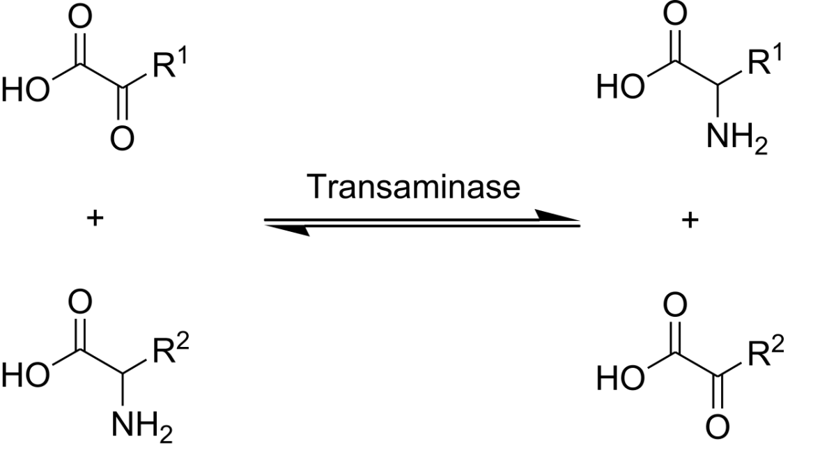
To oxidize an amino acid, the cell must first dispose of its nitrogen. The -NH2 is removed via transamination (shuttled to glutamate) or oxidative deamination (released as ammonia). The remaining carbon skeleton (alpha-keto acid) can enter central metabolism.
The “central metabolism” it enters is always the familiar pipeline: pyruvate or a TCA cycle intermediate or acetyl-CoA. From there, the amino acid’s carbons are oxidized in the TCA cycle (citric acid cycle / Krebs cycle), the resulting NADH and FADH2 feed the electron transport chain, and ATP is made. Exactly the same downstream flow as glucose or fatty acid oxidation - amino acids just enter at different points. Nitrogen is handled separately by the urea cycle (next section) because free ammonia is toxic.
Transamination. An amino acid transfers its -NH2 to alpha-ketoglutarate, producing glutamate and a new alpha-keto acid. PLP (vitamin B6) is the cofactor. Credit: Wikimedia Commons, CC BY-SA
Transamination
The amino group is moved from an amino acid to alpha-ketoglutarate, forming glutamate. The original amino acid becomes an alpha-keto acid. The enzyme family is aminotransferases (or transaminases), and all require pyridoxal phosphate (PLP), the active form of vitamin B6.
Amino acid + α-KG⇌α-keto acid + Glutamate
Two clinically famous aminotransferases:
ALT (alanine aminotransferase): alanine ↔ pyruvate. High in liver. Released in liver damage.
AST (aspartate aminotransferase): aspartate ↔ oxaloacetate. High in liver, heart, muscle. Less liver-specific than ALT.
Oxidative Deamination
Once nitrogen is collected onto glutamate, it can be released as free ammonia by glutamate dehydrogenase in liver mitochondria:
Glutamate + NAD(P)+→α-KG + NH4++NAD(P)H
The ammonia enters the urea cycle (next section) for conversion to urea, which the kidney excretes.
Why Glutamate Is the Nitrogen Hub
Most amino acids transaminate onto alpha-KG (producing glutamate). Glutamate is the single molecule that collects all the nitrogen, so oxidative deamination at just one enzyme (glutamate dehydrogenase) releases free NH3 - and feeds the urea cycle. Simple, elegant architecture.
What is the common acceptor of the amino group in transamination reactions, and what cofactor do all aminotransferases require?
Click to reveal answer
Alpha-ketoglutarate is the universal amino group acceptor, producing glutamate. All aminotransferases require pyridoxal phosphate (PLP), the active form of vitamin B6. PLP forms a Schiff base intermediate with the amino acid to facilitate amino group transfer. B6 deficiency impairs all transamination.
What enzyme performs oxidative deamination of glutamate, and what does it produce?
Click to reveal answer
Glutamate dehydrogenase (GDH). It converts glutamate + NAD(P)+ → alpha-ketoglutarate + NH4+ + NAD(P)H. The ammonia enters the urea cycle in hepatocytes. GDH uniquely uses either NAD+ or NADP+ and is allosterically regulated by ATP (inhibits) and ADP (activates).
Why is glutamate the nitrogen hub of amino acid catabolism?
Click to reveal answer
Most amino acids use alpha-ketoglutarate as their transamination partner, converting it to glutamate. The nitrogen from many different amino acids thus collects on a single carrier, glutamate. One enzyme (glutamate dehydrogenase) can then oxidatively deaminate glutamate to release ammonia for the urea cycle. Glutamate is the funnel that simplifies nitrogen disposal.
Ammonia is toxic, especially to the brain. The liver converts it to urea - non-toxic, water-soluble - for excretion by the kidneys. The urea cycle is the metabolic pathway that does this, spanning the mitochondrial matrix and cytoplasm of hepatocytes.
The urea cycle and the Krebs bicycle
Pathway map
1
Scroll sideways to see the whole map.
Mitochondrial matrix Cytosol CPS-I: the regulated step ATP spent
Aspartate inThe second nitrogen atom of urea does not arrive as free ammonia. It comes in on aspartate, made from oxaloacetate in the TCA cycle. So one nitrogen comes from the mitochondrial ammonia pool and one comes from the amino acid pool.
Fumarate outArgininosuccinate lyase releases fumarate, a TCA intermediate. It becomes malate, then oxaloacetate, which can either restart the cycle as aspartate or leave for gluconeogenesis. This shared traffic is why the two cycles are called the Krebs bicycle.
When it failsAny enzyme deficiency backs ammonia up into the blood. Hyperammonaemia is neurotoxic: ammonia pulls α-ketoglutarate out of the TCA cycle to make glutamate, starving neurons of ATP. OTC deficiency is the most common and is X-linked.
Two nitrogens in, one urea out, at a cost of four ATP equivalents. Ammonia is toxic and cannot be stored or exhaled, so the liver spends real energy to package it as a neutral, water-soluble molecule the kidney can excrete. Everything else on this map exists to serve that one conversion.
The urea cycle connects directly to amino acid catabolism (previous section) and to the TCA cycle. Transamination collects amino-nitrogen onto glutamate. Glutamate dehydrogenase releases that nitrogen as free ammonia, which immediately enters the urea cycle for safe disposal. The urea cycle also generates fumarate as a byproduct - which flows directly into the TCA cycle. So the two cycles share a metabolite and are functionally linked. This link is often called the “Krebs bicycle” because two cycles (urea + TCA) share a common intermediate.
The Net Reaction
NH3+CO2+Aspartate+3 ATP→Urea+Fumarate+2 ADP+AMP+PPi+4 Pi
Two nitrogens end up in urea: one from free NH3, one from aspartate. The fumarate connects the urea cycle to the TCA cycle.
Ornithine transcarbamylase (mitochondrial) · citrulline exits to cytoplasm
–
3
Citrulline + aspartate → argininosuccinate
Argininosuccinate synthetase · second N arrives via aspartate
-1 ATP (→AMP)
4
Argininosuccinate → arginine + fumarate
Argininosuccinate lyase · fumarate feeds the TCA cycle
+fumarate
5
Arginine + H₂O → urea + ornithine
Arginase · ornithine recycles into step 2
+urea
Net cost: 4 high-energy phosphate bonds (2 ATP → 2 ADP; 1 ATP → AMP + PPi). Urea carries 2 nitrogens (one from NH₃, one from aspartate) to the kidney for excretion.
Mnemonic
Urea Cycle Defects
Any urea cycle enzyme deficiency causes hyperammonemia. Ammonia accumulates and damages the brain, causing altered mental status, seizures, vomiting, and potentially coma. Serum levels of specific intermediates help diagnose which enzyme is missing.
Ornithine transcarbamylase (OTC) deficiency: most common. X-linked. High ammonia, high orotate, low citrulline (because the block is before citrulline). Elevated orotate is pathognomonic.
CPS-I deficiency: also high ammonia; low citrulline and low orotate.
Treatment includes dietary protein restriction and medications that divert nitrogen via alternative pathways (phenylbutyrate, benzoate).
What is the function of the urea cycle?
Click to reveal answer
To convert toxic ammonia (from amino acid catabolism) into non-toxic urea for excretion by the kidneys. Each urea molecule contains 2 nitrogens: one from free NH3 (input to CPS-I), one from aspartate (input to argininosuccinate synthetase). The cycle consumes 4 high-energy phosphate bonds per urea.
Which urea cycle enzyme is the most commonly deficient, and what is its diagnostic feature?
Click to reveal answer
Ornithine transcarbamylase (OTC) deficiency. It is X-linked. The enzyme normally converts carbamoyl phosphate + ornithine → citrulline. Without it, carbamoyl phosphate accumulates in mitochondria and diffuses to cytoplasm, where it is diverted into pyrimidine synthesis, producing excess orotate. High serum ammonia + high orotate + low citrulline is the classic triad.
What happens if the urea cycle is impaired?
Click to reveal answer
Hyperammonemia - ammonia accumulates in the blood because it cannot be converted to urea. Ammonia is especially toxic to the brain, causing altered mental status, seizures, vomiting, and coma. Treatments include dietary protein restriction and nitrogen-scavenging drugs (phenylbutyrate, benzoate) that use alternative pathways to excrete nitrogen.
Where amino acid carbon enters, and what it can become
Protein into everything
1
Scroll sideways to see the whole map.
Glucogenic: can become glucose Ketogenic: can only become fat or ketones Both, depending on the fragment Junction metabolite The one-way door
The only rule you needEverything that enters at or above pyruvate, or anywhere on the TCA cycle, can reach oxaloacetate and leave as glucose. Everything that enters as acetyl-CoA or acetoacetate cannot, because PDH and the two decarboxylations of the cycle are irreversible.
The two to memorizeLeucine and lysine are purely ketogenic — the only two of the twenty. Five more (isoleucine, phenylalanine, threonine, tryptophan, tyrosine) are both, because different parts of the same skeleton land in different places. The remaining thirteen are purely glucogenic.
Where the nitrogen goesThis map is only about carbon. The amino group is removed first, usually by transamination onto α-ketoglutarate to make glutamate, which then hands the nitrogen to the urea cycle. Carbon and nitrogen part company at the very first step.
An amino acid's fate is decided entirely by where its carbon skeleton lands. That is why no one memorizes twenty separate answers: you memorize six entry points, note that two amino acids are purely ketogenic, and derive the rest.
After losing its nitrogen, an amino acid’s carbon skeleton enters one of two fates:
Glucogenic: converted to pyruvate or a TCA cycle intermediate. Can be used to make glucose via gluconeogenesis.
Ketogenic: converted to acetyl-CoA or acetoacetate. CANNOT be used to make glucose (because acetyl-CoA cannot be converted back to pyruvate).
Most amino acids are purely glucogenic. A few are both. Only two are purely ketogenic.
Purely Ketogenic
Leucine (L)
Lysine (K)
Both Glucogenic and Ketogenic
Isoleucine (I)
Phenylalanine (F)
Threonine (T)
Tryptophan (W)
Tyrosine (Y)
Purely Glucogenic
All others (13 amino acids).
Entry Points into Central Metabolism
| Entry point | Amino acids |
|-------------|-------------|
| Pyruvate | Ala, Cys, Gly, Ser, Thr, Trp |
| Acetyl-CoA | Ile, Leu, Lys, Phe, Thr, Trp, Tyr |
| Alpha-KG | Arg, Gln, Glu, His, Pro |
| Succinyl-CoA | Ile, Met, Thr, Val |
| Fumarate | Asp, Phe, Tyr |
| Oxaloacetate | Asn, Asp |
You do not need to memorize these entry points for the MCAT, but know the glucogenic/ketogenic classification.
Which two amino acids are purely ketogenic?
Click to reveal answer
Leucine (L) and Lysine (K) - the two L’s. Both are catabolized only to acetyl-CoA and/or acetoacetate, which cannot be converted back to pyruvate and therefore cannot be used for gluconeogenesis. All other amino acids are either purely glucogenic or both.
Why cannot purely ketogenic amino acids contribute to net glucose production?
Click to reveal answer
Their carbon skeletons are broken down only to acetyl-CoA or acetoacetate. Acetyl-CoA cannot be converted back to pyruvate (the pyruvate dehydrogenase reaction is irreversible). Without entry to pyruvate or a TCA intermediate that connects to OAA, no net gluconeogenesis is possible. The only ultimate fate of the carbons is oxidation in the TCA cycle (if paired with OAA) or conversion to ketone bodies/cholesterol.
What are the five amino acids that are both glucogenic and ketogenic?
Click to reveal answer
Isoleucine, Phenylalanine, Threonine, Tryptophan, and Tyrosine. Their catabolism produces both pyruvate or TCA intermediates (glucogenic portion) and acetyl-CoA or acetoacetate (ketogenic portion). Memorize Leu and Lys as the only purely ketogenic; the rest is easier to reason about.
Humans can synthesize 11 of the 20 standard amino acids from scratch. The other 9 are essential - they must come from the diet because we lack the enzymes to synthesize them.
The Nine Essential Amino Acids
Amino acid
One-letter
Phenylalanine
F
Valine
V
Threonine
T
Tryptophan
W
Isoleucine
I
Methionine
M
Histidine
H
Leucine
L
Lysine
K
Conditionally Essential
Some amino acids are nonessential in adults but essential during periods of rapid growth, illness, or specific conditions: arginine, glutamine, and tyrosine (needed from diet when phenylalanine is restricted, as in PKU).
Metabolic Disorders of Amino Acids
Several classic inborn errors of metabolism affect amino acid breakdown. The MCAT occasionally tests recognition.
Result: phenylalanine accumulates, converts to phenylpyruvate and phenyllactate, which damage the developing brain. Intellectual disability if untreated.
Treatment: diet low in phenylalanine (must avoid aspartame), supplement tyrosine.
Newborn screening detects PKU at birth, enabling early dietary management.
Alkaptonuria
Homogentisate oxidase deficiency → homogentisic acid accumulates → dark urine on standing, black connective tissue (ochronosis), arthritis later in life. Famously, one of the first “inborn errors” described by Archibald Garrod.
Maple Syrup Urine Disease (MSUD)
Branched-chain alpha-ketoacid dehydrogenase deficiency → leucine, isoleucine, valine and their alpha-keto acids accumulate. Urine smells like maple syrup. Severe neurological symptoms if untreated.
Phenylalanine, Valine, Threonine, Tryptophan, Isoleucine, Methionine, Histidine, Leucine, Lysine (mnemonic: PVT TIM HALL, where "A" stands for Arginine - conditionally essential). These nine must come from the diet because humans lack the enzymes to synthesize them.
What enzyme is deficient in classic phenylketonuria (PKU), and what is the treatment?
Click to reveal answer
Phenylalanine hydroxylase (PAH), which normally converts phenylalanine to tyrosine. Without PAH, Phe accumulates and damages the developing brain. Treatment: low-phenylalanine diet (avoid aspartame and most protein-rich foods) combined with tyrosine supplementation. Early diagnosis through newborn screening is essential to prevent intellectual disability.
Why is tyrosine considered conditionally essential in patients with PKU?
Click to reveal answer
Tyrosine is normally synthesized from phenylalanine by phenylalanine hydroxylase. In PKU, PAH is deficient, so the patient cannot make tyrosine. Tyrosine must be provided in the diet. This is the general principle of "conditionally essential" - a nonessential amino acid becomes essential when its precursor or synthesis pathway is impaired.
Glucose-6-phosphateThe first fork. G6P can be stored as glycogen, burned through glycolysis, or diverted to the pentose phosphate pathway for NADPH and ribose-5-P. It cannot leave the cell: only liver and kidney have glucose-6-phosphatase to turn it back into free glucose.
PyruvateThe last cytosolic fork. With oxygen it crosses into the mitochondrion for PDH; without oxygen it becomes lactate; in muscle it is transaminated to alanine; and in the fasting liver it is carboxylated to oxaloacetate to start gluconeogenesis.
Acetyl-CoAThe great convergence: carbohydrate, fat, and ketogenic amino acids all arrive here. It burns in the TCA cycle, leaves as citrate for fatty acid and cholesterol synthesis, or condenses into ketone bodies. It can never become glucose, which is why fat is not a gluconeogenic fuel.
OxaloacetateGatekeeper of the TCA cycle and the doorway out of it. Acetyl-CoA cannot enter the cycle without OAA, so when OAA is drained for gluconeogenesis during fasting, acetyl-CoA backs up and is shunted into ketone bodies. OAA also trades with the urea cycle as aspartate and fumarate.
Read it top to bottom for catabolism, bottom to top for anabolism. Fuels enter at the top, carbon funnels through four junction metabolites, and the electron carriers cash out at the electron transport chain. The four navy pills are the only molecules you need to reason from: know what enters and leaves each one and you can rebuild the rest of the map from memory.
All three major fuels converge at acetyl-CoA, which feeds the TCA cycle:
Amino acids → various entry points (pyruvate, TCA intermediates, acetyl-CoA).
Tissue Fuel Preferences
Different tissues prefer different fuels:
| Tissue | Preferred fuels | Notes |
|--------|-----------------|-------|
| Brain | Glucose (always), ketones (starvation) | Cannot use fatty acids directly |
| Heart | Fatty acids (first choice), then ketones, then glucose | Huge aerobic capacity |
| Red blood cells | Glucose (only) | No mitochondria, no alternatives |
| Skeletal muscle | Fatty acids (rest), glycogen (exercise) | Also uses ketones in starvation |
| Liver | Amino acids, fatty acids | Makes glucose and ketones for other tissues |
| Adipose | Fatty acids | Stores and releases them |
Fed vs. Fasted vs. Starvation
Fed state (insulin high): glucose uptake, glycogen and fat storage, protein synthesis, fatty acid synthesis. Liver buffers blood glucose by storing excess.
Fasted state (glucagon high): glycogenolysis, gluconeogenesis, lipolysis. Brain still uses glucose mostly.
Starvation (days without food): glycogen depleted; fat mobilized; ketone bodies produced; brain adapts to use ketones; muscle protein breakdown minimized by this ketone adaptation.
The Pecking Order During Starvation
Hours: blood glucose maintained by liver glycogen.
Day 1-2: liver glycogen depleted; gluconeogenesis from amino acids (muscle protein) and glycerol.
Day 2-3: fat mobilization ramps up; ketone production begins.
Day 3+: ketones rise; brain adapts to use them; protein breakdown slows to spare muscle.
Weeks: fat stores determine survival. Once fat is gone, the body cannibalizes essential proteins, leading to death.
Where do carbohydrate, fat, and protein catabolism converge?
Click to reveal answer
All three converge at acetyl-CoA, which feeds the TCA cycle. Carbohydrates → glycolysis → pyruvate → PDH → acetyl-CoA. Fatty acids → beta-oxidation → acetyl-CoA. Amino acids → various entry points, some directly to acetyl-CoA, some to TCA intermediates. Acetyl-CoA is the central metabolic hub.
How does the body adapt to prolonged starvation after ~3-4 days?
Click to reveal answer
The brain begins using ketone bodies for up to 75% of its energy needs. This reduces the demand for glucose, which had been met by gluconeogenesis from amino acids (muscle protein). The ketone adaptation spares muscle protein and dramatically extends survival. Fat stores then become the main determinant of how long a person can survive without food.
Why is the liver unique in metabolism?
Click to reveal answer
The liver does jobs no other tissue does: gluconeogenesis (releases glucose to blood), ketogenesis (releases ketones), the urea cycle (detoxifies ammonia), bile acid synthesis, lipoprotein synthesis, and drug metabolism. It also uniquely expresses glucose-6-phosphatase (free glucose release) and HMG-CoA lyase (ketone production) while lacking thiophorase (so it cannot use ketones). The liver acts as a metabolic factory for the whole body.