Thermodynamics tells you whether a reaction can happen spontaneously. The key quantity is Gibbs free energy (G). A reaction is spontaneous if its ΔG is negative.
Free energy, coupling, and why ATP works
Bioenergetics
1
Scroll sideways to see the whole map.
Negative ΔG: spontaneous, releases energy Positive ΔG: needs to be paid for ATP, the currency The coupled result
Spontaneous does not mean fastΔG says whether a reaction can happen, not whether it will happen in your lifetime. Glucose burning in air is enormously spontaneous and completely stable on a table. Activation energy is what decides the rate, and enzymes only ever change that, never ΔG.
Why ATP is not a special bondThere is no unusual chemistry in the phosphoanhydride bond. Hydrolysis releases energy because the products are more stable: less charge repulsion between the phosphates, better resonance in free phosphate, and better solvation. ATP is unstable, not magic.
ΔG versus ΔG°′ΔG°′ is the standard value at 1 M, pH 7. ΔG is the actual value under the concentrations in the cell, and it is the one that decides direction. A reaction with positive ΔG°′ runs forward all day if the cell keeps the product concentration low.
The cell never breaks thermodynamics; it just does its accounting in pairs. An unfavorable reaction is run by physically joining it to a favorable one, so what the universe sees is a single reaction with a negative ΔG.
ΔG > 0 (endergonic): non-spontaneous as written (but can be driven by coupling).
ΔG = 0: equilibrium.
Magnitude of ΔG does NOT tell you how fast the reaction goes - only thermodynamics, not kinetics. Enzymes change the rate (by lowering activation energy) without changing ΔG.
ΔG vs. ΔG°’
ΔG°’: standard free energy change at physiologic conditions (pH 7, 1 M substrates). A property of the reaction.
ΔG: actual free energy change under cellular conditions. Depends on actual concentrations. What matters biologically.
A reaction with ΔG°’ > 0 can still have ΔG < 0 in a cell if substrate/product ratios favor forward flow.
ΔG=ΔG∘′+RTlnQ
where Q is the mass action ratio (products/substrates at the moment). Cells maintain non-equilibrium concentrations to keep metabolism flowing.
What does a negative ΔG indicate about a reaction?
Click to reveal answer
The reaction is exergonic - thermodynamically spontaneous as written. Energy is released, and the reaction proceeds toward products until equilibrium. Magnitude of ΔG does not say how fast the reaction runs; a catalyst (enzyme) is needed to achieve biologically useful rates.
How can a reaction with positive ΔG°’ still be favorable in the cell?
Click to reveal answer
Because ΔG depends on actual concentrations, not just ΔG°’. If the cell keeps product concentration low (by consuming it in the next step) or substrate concentration high, the mass action term (RT ln Q) can be strongly negative, making the actual ΔG negative. This is how unfavorable standard ΔG°’ reactions proceed in cells - through concentration control.
How do enzymes affect ΔG of a reaction?
Click to reveal answer
They do not. Enzymes lower the activation energy (Ea), speeding up the rate of both forward and reverse reactions. ΔG is determined only by the energy difference between reactants and products, which enzymes do not change. A reaction that is thermodynamically impossible stays impossible with or without an enzyme.
12.2
ATP Currency
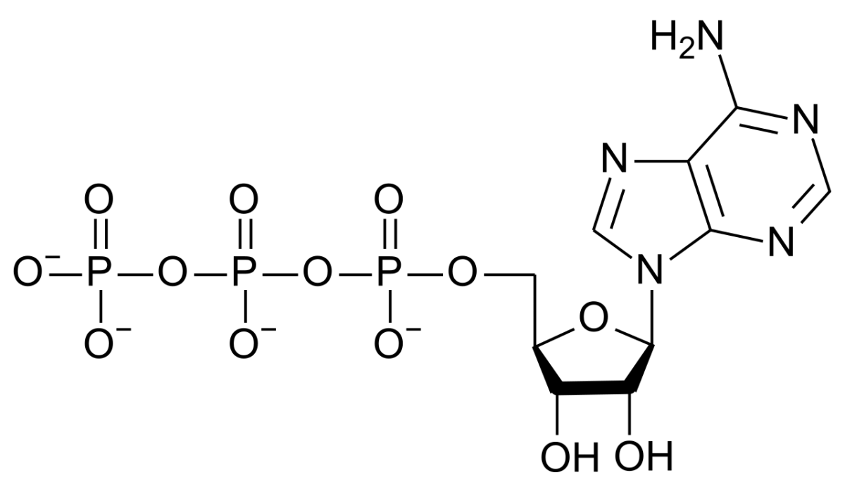
↗ATP structure. Adenine + ribose + three phosphates. The two phosphoanhydride bonds (between phosphates) are the high-energy bonds; breaking either releases ~7.3 kcal/mol. Credit: Wikimedia Commons, CC BY-SA
ATP (adenosine triphosphate) has three phosphates linked in series. The bonds between the second and third phosphates, and between the first and second, are phosphoanhydride bonds. They are “high-energy” because:
Hydrolysis products (ADP + Pi) have lower energy than ATP (resonance stabilization of Pi).
The negatively charged phosphates in ATP repel each other; breaking them reduces strain.
Hydrolysis produces protons and is favored at cellular pH.
Standard ΔG°’ for ATP → ADP + Pi is about -7.3 kcal/mol. Under cellular conditions, the actual ΔG is closer to -11 or -12 kcal/mol because cells keep ATP high relative to ADP + Pi.
The Energy Continuum
ATP → ADP + Pi: ΔG°’ ≈ -7.3 kcal/mol.
ATP → AMP + PPi: ΔG°’ ≈ -10 kcal/mol (extra push from PPi hydrolysis downstream).
Phosphoenolpyruvate (PEP) → pyruvate: ΔG°’ ≈ -14.8 kcal/mol. Higher energy than ATP - can transfer phosphate TO ADP.
Creatine phosphate → creatine: ΔG°’ ≈ -10 kcal/mol. Stores energy for quick ATP regeneration in muscle.
Glucose-6-phosphate → glucose + Pi: ΔG°’ ≈ -3.3 kcal/mol. Lower energy than ATP.
Why Cells Keep Making ATP
Cells never store ATP in huge amounts. ATP half-life in a working cell is a few seconds. Instead, cells maintain a high ATP/ADP ratio (about 10:1 or more) by constantly regenerating ATP at the rate it is consumed. Any drop in ATP immediately activates catabolic pathways to restore the ratio (respiratory control).
Why is the bond between the last two phosphates of ATP called "high-energy"?
Click to reveal answer
Several reasons. Hydrolysis products (ADP + Pi) are resonance-stabilized. The negatively charged phosphates in ATP electrostatically repel each other, so breaking the bond relieves strain. Hydrolysis also releases a proton, which is favored at physiologic pH. The net ΔG°' is about -7.3 kcal/mol - enough to drive most coupled unfavorable reactions.
Why is PEP able to phosphorylate ADP to make ATP?
Click to reveal answer
PEP has a higher phosphate transfer potential (more negative ΔG°' of hydrolysis, ~-14.8 kcal/mol) than ATP (~-7.3 kcal/mol). In pyruvate kinase (step 10 of glycolysis), PEP transfers its phosphate to ADP. The reaction is strongly exergonic because the drop from PEP's energy to ATP's energy is favorable. This is substrate-level phosphorylation.
How do muscles rapidly regenerate ATP during intense exercise?
Click to reveal answer
Creatine phosphate. Creatine kinase transfers the phosphate from creatine-P directly to ADP, regenerating ATP without going through glycolysis or oxidative phosphorylation. This provides a burst of ATP lasting 5-15 seconds, perfect for the start of a sprint. When creatine-P is depleted, glycolysis and eventually oxidative phosphorylation take over.
A reaction with positive ΔG cannot run spontaneously. But if it is coupled to an exergonic reaction (often ATP hydrolysis) and the combined ΔG is negative, the overall process will run. This is how cells drive every biosynthesis and every uphill transport.
The Principle
ΔG values are additive. If Reaction 1 has ΔG = +5 kcal/mol and Reaction 2 has ΔG = -10 kcal/mol, and they share a common intermediate, the coupled reaction has ΔG = -5 kcal/mol - spontaneous.
Example: glutamate + NH3 → glutamine (unfavorable on its own, ΔG°’ = +3.4 kcal/mol). Coupled to ATP → ADP + Pi (ΔG°’ = -7.3 kcal/mol):
Step 1: Glu + ATP → Glu-phosphate + ADP (ΔG°’ ≈ 0, not spontaneous by itself, but drives step 2).
Step 2: Glu-phosphate + NH3 → Glutamine + Pi (strongly exergonic once the phosphate is in place).
Direct ATP hydrolysis: most common. Kinases transfer a phosphate from ATP to substrate, “activating” it for the next step (e.g., hexokinase, pyruvate carboxylase).
ATP → AMP + PPi: the pyrophosphate is then hydrolyzed, producing extra driving force. Used when an especially strong push is needed (tRNA charging, fatty acid activation, DNA synthesis).
NADH / NADPH as reducing equivalents: electrons from glycolysis or TCA drive biosynthesis.
Ion gradients: the proton motive force powers ATP synthase; the sodium gradient powers secondary active transport.
Why ATP Is Perfect for Coupling
ATP sits in the middle of the phosphate transfer hierarchy. It receives phosphate from higher-energy compounds (PEP, creatine-P) in exergonic reactions, and donates phosphate to lower-energy compounds (glucose, amino acids) in reactions that are still favorable. This “intermediate” position makes ATP a universal intermediary.
How do cells drive energetically unfavorable reactions?
Click to reveal answer
By coupling them to a strongly exergonic reaction (most often ATP hydrolysis) through a shared intermediate. ΔG values add. If the unfavorable reaction has ΔG of +4 and ATP hydrolysis has ΔG of -7.3, the coupled reaction has net ΔG of -3.3 - spontaneous. The enzyme catalyzing the pair binds both reactants and channels the energy flow.
Why does ATP hydrolysis to AMP + PPi release more energy than to ADP + Pi?
Click to reveal answer
ATP → AMP + PPi releases one phosphoanhydride bond directly. The pyrophosphate (PPi) is then hydrolyzed by ubiquitous pyrophosphatase to 2 Pi, which is also strongly exergonic and effectively irreversible. Together, the two steps release more energy than a simple ATP → ADP + Pi. Cells use this double hit when they need especially high driving force (tRNA charging, fatty acid activation).
Besides ATP hydrolysis, what are two other ways cells couple energy to drive reactions?
Click to reveal answer
(1) NADH or NADPH as reducing power - electrons transferred from NADPH to a biosynthetic intermediate drive reductive biosynthesis. (2) Ion gradients - the proton gradient across the inner mitochondrial membrane drives ATP synthase; the Na+ gradient drives secondary active transport of glucose, amino acids, and other solutes. All these are forms of energy coupling.
12.4
Biological Redox
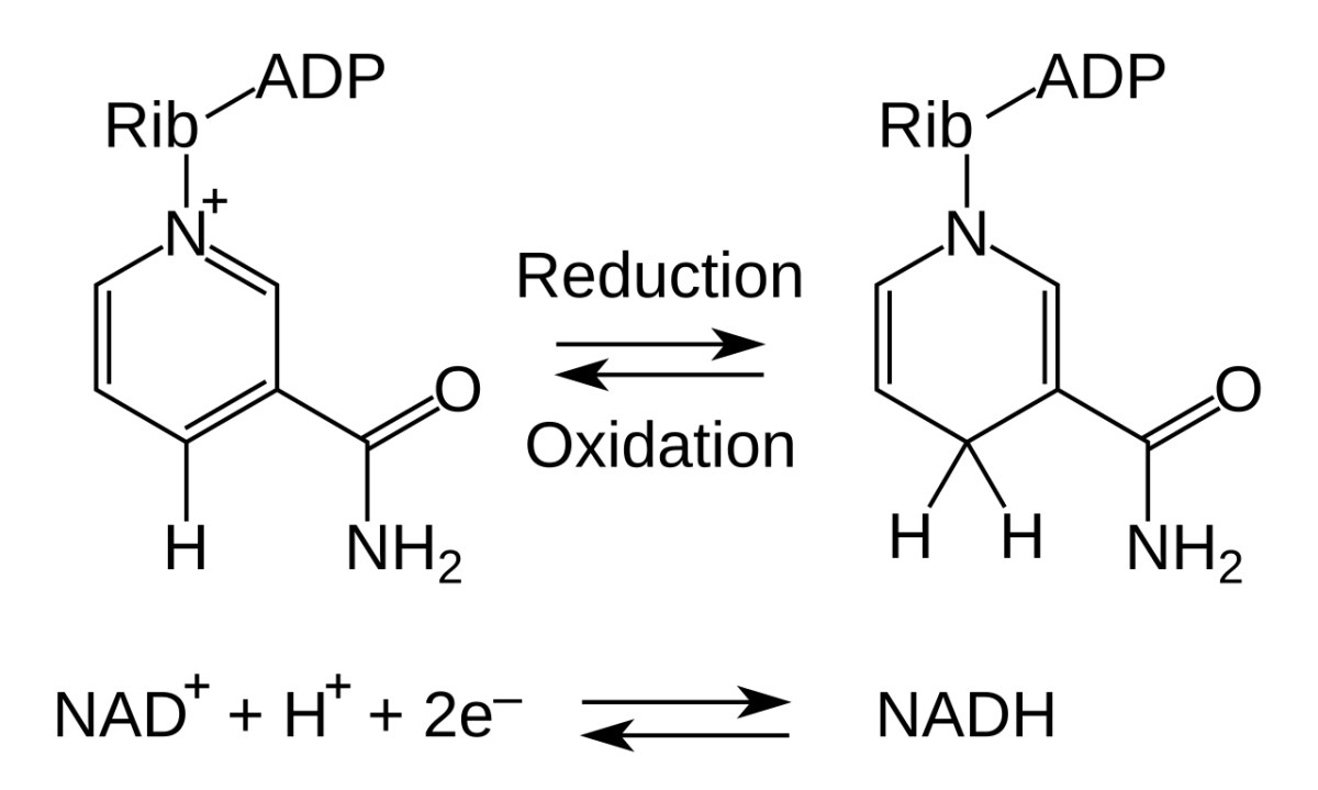
↗NAD+/NADH redox. NAD+ accepts a hydride (2 electrons + 1 proton) to become NADH. The process is reversible and handles most of the cell's oxidative electron transfers. Credit: Wikimedia Commons, CC BY-SA
Oxidation is loss of electrons; reduction is gain. Metabolism is a giant redox system that gradually strips electrons from fuel and hands them to oxygen. The intermediate carriers are NAD+, FAD, and NADP+.
NAD+ and FAD
NAD+ (nicotinamide adenine dinucleotide, from niacin/B3): accepts 2 electrons and 1 proton as a hydride, becoming NADH. Central to catabolism. Each NADH yields ~2.5 ATP at the ETC.
FAD (flavin adenine dinucleotide, from riboflavin/B2): accepts 2 electrons and 2 protons, becoming FADH2. Used by enzymes that need to handle oxidations of less chemically reactive bonds (e.g., alkenes in beta-oxidation, succinate → fumarate in TCA). Each FADH2 yields ~1.5 ATP.
NADPH
NADPH is NAD+ with an extra phosphate on the ribose. Same redox chemistry, but kept in a separate pool for biosynthesis:
NADH/NAD+: high-NADH state means “catabolism is running, feed the ETC.”
NADPH/NADP+: high-NADPH state means “reductive biosynthesis can run” and “antioxidant defense is ready.”
The separation lets the cell run catabolism (which raises NADH) and anabolism (which needs NADPH) in parallel without interfering with each other.
Sources of NADPH
Pentose phosphate pathway (PPP): the main source. Important in liver (fatty acid synthesis) and red blood cells (antioxidant defense).
Malic enzyme: part of the citrate shuttle during fatty acid synthesis.
Isocitrate dehydrogenase: some cytoplasmic IDH isoforms generate NADPH.
Reduction Potential
Each redox pair has a standard reduction potential (E°’). More negative E°’ means the pair donates electrons more strongly. NADH (E°’ = -0.32 V) donates to O2 (E°’ = +0.82 V), falling a total of ~1.14 V. That voltage drop through the ETC is converted to the proton motive force.
Why do cells keep NADH and NADPH as separate pools?
Click to reveal answer
NADH and NADPH have the same redox chemistry but different metabolic roles. NADH feeds the ETC for ATP production (catabolism). NADPH provides reducing power for biosynthesis (fatty acid synthesis, cholesterol synthesis) and antioxidant defense (regeneration of reduced glutathione). Separating the pools lets the cell manage catabolic energy production and anabolic/reductive needs independently.
What vitamin is the precursor for NAD+, and what condition results from severe deficiency?
Click to reveal answer
Niacin (vitamin B3) is the precursor for both NAD+ and NADP+. Severe deficiency causes pellagra, characterized by the 3 D's: dermatitis (sun-exposed skin), diarrhea, dementia. Pellagra is rare in developed countries but still occurs in settings of chronic alcoholism or malabsorption.
Why does NADH yield more ATP than FADH2 via the ETC?
Click to reveal answer
NADH has a more negative reduction potential than FADH2 and enters the ETC at Complex I (which pumps protons). FADH2 enters at Complex II (which does NOT pump protons). More protons pumped per NADH means more ATP. Approximate yields: 2.5 ATP per NADH, 1.5 ATP per FADH2.
Most coenzymes are derived from vitamins. Memorizing the vitamin-coenzyme-enzyme-deficiency connections lets you reason about a lot of MCAT biochemistry at once.
Water-Soluble Vitamins (B Complex and C)
Vitamin
Coenzyme / Role
Key enzymes
Deficiency
B1 (Thiamine)
TPP
PDH, α-KG dehydrogenase, branched-chain KG dehydrogenase, transketolase
Beriberi, Wernicke-Korsakoff
B2 (Riboflavin)
FAD, FMN
Succinate dehydrogenase, acyl-CoA dehydrogenase, many oxidases
Gamma-carboxylation of clotting factors II, VII, IX, X
Bleeding; newborns given K at birth
B12 and Folate
Both are needed for DNA synthesis, and deficiency of either causes megaloblastic anemia. But B12 deficiency additionally causes neurological problems (peripheral neuropathy, subacute combined degeneration) because it is needed for methylmalonyl-CoA mutase in nerve myelin synthesis. Folate replacement alone can mask B12 deficiency and allow the neurological damage to progress - hence the caution about folate supplementation without B12 checking.
Which vitamin is the coenzyme for transaminases and glycogen phosphorylase?
Click to reveal answer
Vitamin B6 (pyridoxine), as pyridoxal phosphate (PLP). PLP is required by all aminotransferases (ALT, AST, etc.) and by glycogen phosphorylase. B6 deficiency impairs both amino acid metabolism and glycogen breakdown, with clinical features including sideroblastic anemia and peripheral neuropathy.
Why should thiamine (B1) be given before glucose in a chronic alcoholic presenting with altered mental status?
Click to reveal answer
Thiamine is the cofactor for PDH and α-KG dehydrogenase. Alcoholics are often thiamine-deficient. Glucose loads drive glycolysis and produce more pyruvate that requires PDH to proceed. Without thiamine, pyruvate cannot be oxidized, lactic acid accumulates, and Wernicke encephalopathy is precipitated (confusion, ataxia, ophthalmoplegia). Giving thiamine first prevents this catastrophe.
Why does B12 deficiency cause both megaloblastic anemia and neurological symptoms, while folate deficiency causes only anemia?
Click to reveal answer
Both B12 and folate are needed for DNA synthesis (thymidine synthesis in particular), and deficiency of either causes megaloblastic anemia. But B12 is additionally required for methylmalonyl-CoA mutase (important for myelin integrity). B12 deficiency causes demyelination (peripheral neuropathy, subacute combined degeneration of the spinal cord). Folate alone does not have this neurological role.
The body cycles between three main metabolic states, each with characteristic hormonal signals and pathway activities.
Fed to starving: the same pathways, re-weighted
Over time
1
Scroll sideways to see the whole map.
Glycogen breakdown Gluconeogenesis Ketone body production What the brain is running on
The one constraintThe brain cannot burn fatty acids, and red blood cells have no mitochondria at all. Between them they demand a floor of glucose no matter how long you fast, and every other decision on this chart follows from that single requirement.
Why muscle gets spent, then sparedOnce liver glycogen runs out, the only remaining source of new glucose is amino acids from muscle. Burning your own muscle is survivable for days, not weeks, so the body switches the brain onto ketone bodies. Ketosis is not a failure state; it is the adaptation that stops you consuming yourself.
What the hormones actually doInsulin means store: it activates glycogen synthase, acetyl-CoA carboxylase, and PDH phosphatase. Glucagon means mobilise: it raises cAMP, which activates glycogen phosphorylase and hormone-sensitive lipase and shuts down glycolysis via PFK-2. Every arrow in this book obeys those two.
Nothing new happens as you fast; the same pathways are just re-weighted. Glycogen covers the first day, gluconeogenesis covers the second, and ketone bodies take over the brain from there so that muscle protein survives. Read the three rails as a relay, not as three separate topics.
Fed State (after a meal)
Hormonal signal: high insulin, low glucagon.
Glucose uptake into cells (via GLUT4 in muscle and adipose).
Fat storage in adipose (via LPL) - insulin activates lipoprotein lipase at capillaries, so chylomicron and VLDL triglycerides are hydrolyzed and fatty acids absorbed.
Muscle protein breakdown → amino acids → gluconeogenesis (Cori cycle and glucose-alanine cycle).
Ketogenesis ramps up; ketone bodies rise in blood.
After 3-4 days, brain adapts to use ketones for ~60-75% of energy.
Muscle protein breakdown slows as ketones replace brain’s glucose need.
Fat stores determine how long the body survives.
Which hormone drives the metabolic shift from fed to fasted state?
Click to reveal answer
Glucagon. Rising glucagon (and falling insulin) activates glycogenolysis, gluconeogenesis, lipolysis, and fatty acid oxidation. The main targets include glycogen phosphorylase (on), glycogen synthase (off), hormone-sensitive lipase (on), PFK-2/F-2,6-BP (lowered), and ACC (off). Glucagon is the “fasting” signal.
What is the main adaptation during prolonged starvation that lets survival extend for weeks?
Click to reveal answer
The brain’s shift to using ketone bodies for the majority of its energy. Without this adaptation, the body would need to continuously break down muscle protein to make glucose via gluconeogenesis, rapidly depleting protein stores. Ketones spare protein, and fat stores become the main determinant of survival duration. Most adults have enough fat to fuel weeks of fasting; glycogen lasts only a day.
Why does the body preferentially break down muscle protein (rather than, say, collagen) during fasting?
Click to reveal answer
Muscle has the largest pool of readily catabolized protein. It is metabolically active and can release amino acids (especially alanine) relatively quickly. Structural proteins like collagen, elastin, and most enzymes are not easily mobilized. The body will also spare certain critical proteins (hemoglobin, albumin, enzymes) unless starvation is very severe. The glucose-alanine cycle lets muscle provide carbon and nitrogen to the liver for gluconeogenesis and urea cycle.
Four hormones dominate metabolic regulation: insulin, glucagon, epinephrine, and cortisol. Knowing what each does in each major tissue lets you reason through almost every MCAT metabolism passage.
Insulin
Secreted by pancreatic beta cells in response to high blood glucose (and incretins, amino acids). Signal: “store energy.”
Muscle / adipose: translocates GLUT4 to the cell surface, letting glucose enter; activates glycogen synthesis; activates lipoprotein lipase for fat storage.
Secreted by adrenal medulla in response to stress, exercise, low blood glucose. Signal: “fight or flight - mobilize everything fast.”
Similar to glucagon but faster, broader tissue distribution (including muscle).
Acts through beta-adrenergic receptors (Gs → cAMP → PKA) and some alpha receptors.
Activates glycogenolysis in liver AND muscle; activates lipolysis in adipose; increases heart rate and contractility.
Cortisol
Secreted by the adrenal cortex in response to chronic stress. Signal: “sustained mobilization + shift toward glucose conservation.”
Activates gluconeogenesis (liver) and proteolysis (muscle) - provides amino acids for glucose production.
Promotes lipolysis.
Immunosuppressive.
Slow-acting (steroid hormone, transcriptional effects over hours to days).
Who gives the orders: the fuel hormones
Regulation map
1
Scroll sideways to see the whole map.
Hormone increases this process Hormone decreases it Little direct effect The enzyme each one acts through
Phosphorylated or notGlucagon and epinephrine both work through cAMP and PKA, so both end up phosphorylating the same enzymes. Insulin activates phosphatases, so it strips those phosphates off. This is why one rule covers most of metabolism: phosphorylation switches glycogen synthase off and glycogen phosphorylase on, and dephosphorylation does the reverse. Learn the rule once and you no longer have to memorize each enzyme.
Why cortisol looks slowCortisol is a steroid, so it crosses the membrane, binds a nuclear receptor, and changes transcription. Its effects take hours and work by changing how much enzyme exists rather than how active it is. That is why cortisol dominates prolonged fasting and chronic stress while epinephrine dominates the first minute of a sprint.
Epinephrine is tissue-specificIn the liver it drives glycogenolysis and the glucose leaves the cell, because the liver has glucose-6-phosphatase. In muscle the same signal drives glycogenolysis but the glucose-6-phosphate is trapped, so it can only be burned locally. Muscle glycogen never raises anyone else's blood sugar.
Four hormones, one grid. Insulin is the only one that says store; the other three all say release, for different reasons and on different timescales. Read a column to learn one hormone, read a row to answer the usual exam question, which is what happens to a single process when the hormonal state changes.
Muscle and adipose tissue lack glucagon receptors; only the liver expresses them. This is physiologically appropriate because only the liver has glucose-6-phosphatase and can release glucose into the blood. Muscle’s parallel mobilization of glycogen during stress is triggered by epinephrine, which does reach muscle via beta-adrenergic receptors.
How does insulin increase glucose uptake into muscle and adipose?
Click to reveal answer
Insulin binding to the receptor tyrosine kinase triggers a PI3K/AKT cascade that translocates GLUT4 transporters from intracellular vesicles to the plasma membrane. More GLUT4 at the surface means more glucose flowing into the cell down its concentration gradient. This mechanism is specific to muscle and adipose; the brain and liver use non-insulin-dependent GLUTs (GLUT1, GLUT2, GLUT3).
What is cortisol’s main metabolic effect during prolonged stress?
Click to reveal answer
Cortisol promotes proteolysis in muscle (to supply amino acids for gluconeogenesis) and gluconeogenesis in liver. It also stimulates lipolysis in adipose. The net effect is sustained mobilization of glucose and amino acids, with immune suppression as a side effect. Cortisol’s effects are slower than insulin and glucagon because it is a steroid hormone acting at the level of gene transcription (hours to days).
Tissues have specialized metabolic roles and fuel preferences. Knowing the characteristic fuel and function of each tissue helps you navigate MCAT passages.
Liver
The metabolic factory for the whole body.
Performs glycogenolysis, gluconeogenesis, ketogenesis, urea cycle, cholesterol and bile acid synthesis, lipoprotein assembly (VLDL), drug metabolism.
Fed state (insulin): lipoprotein lipase (LPL) at capillaries hydrolyzes circulating triglycerides; fatty acids enter adipocytes and re-esterified into triglyceride.
Fasting state (glucagon/epinephrine): hormone-sensitive lipase (HSL) breaks down stored triglycerides; fatty acids released to blood, bound to albumin, delivered to other tissues.
Glycerol released from adipose goes to liver for gluconeogenesis.
Heart
Cardiac muscle is highly aerobic (many mitochondria).
Preferred fuel: fatty acids (most of the time).
Also uses ketones well (always), lactate, and glucose.
Very flexible - can switch fuels based on availability.
Red Blood Cells
Lack mitochondria and nuclei.
Use glucose ONLY, via glycolysis.
Produce lactate continuously (sent to liver for Cori cycle).
Cannot use fatty acids, ketones, or amino acids for fuel.
Why do red blood cells rely entirely on glucose via glycolysis?
Click to reveal answer
RBCs lack mitochondria. They cannot use the TCA cycle, oxidative phosphorylation, or beta-oxidation. They also lack a nucleus and cannot synthesize new proteins. Their only ATP source is cytoplasmic glycolysis, converting glucose to lactate to regenerate NAD+. This produces ongoing lactate output that the liver recycles via the Cori cycle.
Why cannot muscle glycogen contribute directly to blood glucose?
Click to reveal answer
Muscle lacks glucose-6-phosphatase, the enzyme that removes phosphate from glucose-6-phosphate so free glucose can leave the cell. Muscle glycogen breakdown produces G6P that can only feed glycolysis locally. Only liver (and kidney cortex) have glucose-6-phosphatase and can release glucose to blood.
What fuel does the heart prefer under most conditions?
Click to reveal answer
Fatty acids. Cardiac muscle has abundant mitochondria and heavily depends on beta-oxidation. The heart also readily uses ketone bodies (always) and lactate (especially during rest). It is a metabolic generalist - can switch fuel sources based on availability. In ischemia, it switches to anaerobic glycolysis, building up lactate.
A classic MCAT topic: given a patient with specific symptoms, identify the deficient enzyme or metabolic defect. Below are the most-tested disorders.
Diabetes Mellitus
Type 1 diabetes: autoimmune destruction of pancreatic beta cells. Absolute insulin deficiency. Early onset, thin patients, ketoacidosis-prone. Treatment: insulin replacement.
Type 2 diabetes: insulin resistance + relative insulin deficiency. Later onset, typically associated with obesity. Hyperglycemia without severe ketoacidosis (some insulin still present suppresses lipolysis). Treatment: lifestyle, metformin, eventually insulin.
Diabetic ketoacidosis (DKA): insulin absent → unopposed lipolysis and ketogenesis → acidosis, dehydration, hyperglycemia. Kussmaul respiration (deep, rapid breathing to exhale CO2), fruity breath (acetone), and altered mental status. Life-threatening if untreated.
What is the biochemical defect in phenylketonuria (PKU), and what accumulates?
Click to reveal answer
Phenylalanine hydroxylase (PAH) deficiency. PAH normally converts phenylalanine to tyrosine. Without it, phenylalanine accumulates. Excess Phe is converted to phenylpyruvate and phenyllactate, which damage the developing brain. Treatment: low-Phe diet and tyrosine supplementation. Newborn screening prevents the severe intellectual disability seen in untreated PKU.
Why does DKA occur almost exclusively in type 1 diabetes rather than type 2?
Click to reveal answer
Type 1 diabetes has absolute insulin deficiency. Without any insulin, adipose lipolysis is unopposed, fatty acids flood the liver, and ketogenesis runs full speed - producing the severe ketoacidosis. Type 2 patients usually retain some residual insulin, which is enough to suppress lipolysis and prevent full-blown DKA. In severe type 2 decompensation (HHS), hyperglycemia is severe but ketoacidosis is absent or mild.
Which glycogen storage disease causes severe fasting hypoglycemia and hepatomegaly?
Click to reveal answer
Von Gierke disease (Type I, glucose-6-phosphatase deficiency). Without G6Pase, neither glycogenolysis nor gluconeogenesis can produce free glucose for the blood. Glycogen and G6P accumulate in the liver (hepatomegaly). Fasting causes severe hypoglycemia because no pathway can deliver glucose to the blood. Also produces hyperuricemia and hyperlipidemia from intermediary metabolism being redirected.
12.10
Exercise Metabolism
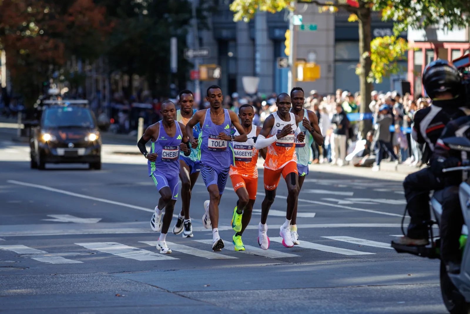
↗Elite marathon runners mid-race. Sustained exercise like marathon running demands aerobic respiration - glycogen and fatty acids feeding the TCA cycle for hours. Three overlapping energy systems (creatine phosphate, anaerobic glycolysis, aerobic respiration) layer by duration and intensity. Credit: Wikimedia Commons, CC BY-SA
Muscle uses three energy systems that layer on top of each other as exercise continues. Their relative contributions depend on duration and intensity.
System 1: Immediate (0-15 seconds)
Source: stored ATP + creatine phosphate.
Duration: seconds.
Creatine kinase transfers phosphate from creatine-P to ADP, regenerating ATP instantly. No oxygen needed.
Used in: sprints, power lifts, very brief efforts.
System 2: Anaerobic Glycolysis (10 seconds to 2 minutes)
Produces 2 ATP per glucose; pyruvate → lactate (regenerates NAD+, no O2 needed).
Lactate accumulation eventually limits this system - the “lactate threshold.”
Used in: 400-800 meter runs, 100m swim, hard cycling intervals.
System 3: Aerobic Respiration (2 minutes to hours)
Source: glucose, fatty acids, eventually ketones.
Duration: sustainable as long as fuel and O2 are present.
Full oxidation: glucose gives ~30-32 ATP, palmitate ~106 ATP.
Used in: 5K, marathon, long bike rides.
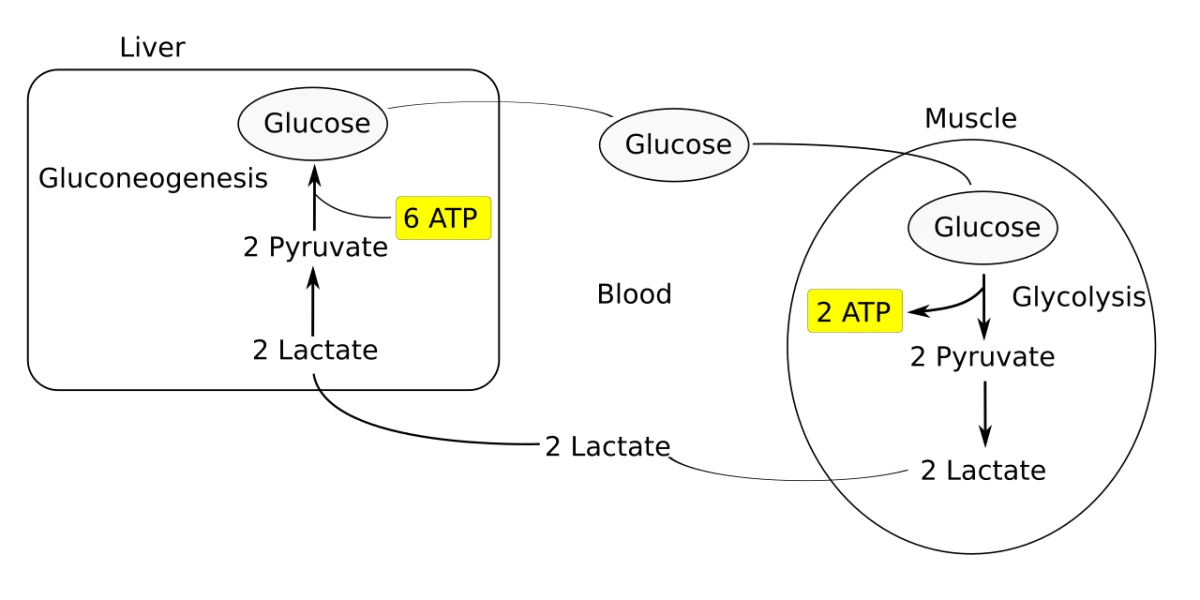
The Cori cycle during exercise. Muscle produces lactate anaerobically; liver converts it back to glucose via gluconeogenesis. Muscle keeps running while the liver pays the ATP cost. Credit: Wikimedia Commons, CC BY-SA
The Marathon Wall
Marathoners “hit the wall” around mile 18-22. Why? Muscle glycogen is depleted. The runner must transition to fat oxidation, which is slower and cannot sustain the same pace. Brain glucose also drops, causing hypoglycemia-like symptoms (dizziness, tunnel vision). Training (more mitochondria, better fat oxidation) and carbohydrate loading push this wall farther.
Lactate Fate
Lactate from working muscle travels to liver (Cori cycle) and to oxidative muscle fibers (that can reconvert it to pyruvate and oxidize it). Lactate is not purely waste; it is a mobile fuel. Blood lactate rises during exercise then falls after, as liver and muscle consume it.
What fuel does creatine phosphate provide, and for how long?
Click to reveal answer
Creatine phosphate donates its phosphate to ADP (catalyzed by creatine kinase), regenerating ATP instantly. This is the "immediate" energy system, used in the first 5-15 seconds of intense effort (sprints, lifts). It needs no oxygen and no glycolysis. When creatine-P is depleted, the muscle shifts to anaerobic glycolysis.
Why do marathoners "hit the wall" around mile 20?
Click to reveal answer
Muscle glycogen runs out. The runner must transition to fat oxidation, which is slower (fatty acids produce ATP more slowly than glucose and require more oxygen per ATP). Brain glucose also drops, adding hypoglycemia-like symptoms. Training and pre-race carbohydrate loading increase glycogen stores, pushing the wall farther. The wall is a real biochemical transition from fast to slow metabolism.
What is the lactate threshold?
Click to reveal answer
The exercise intensity at which lactate begins to accumulate rapidly in blood because muscle cells are running anaerobic glycolysis faster than oxidative capacity can consume the pyruvate. Above this threshold, fatigue accelerates. Below it, the aerobic system can handle the pace indefinitely. Training raises the lactate threshold - a primary aim of endurance conditioning.