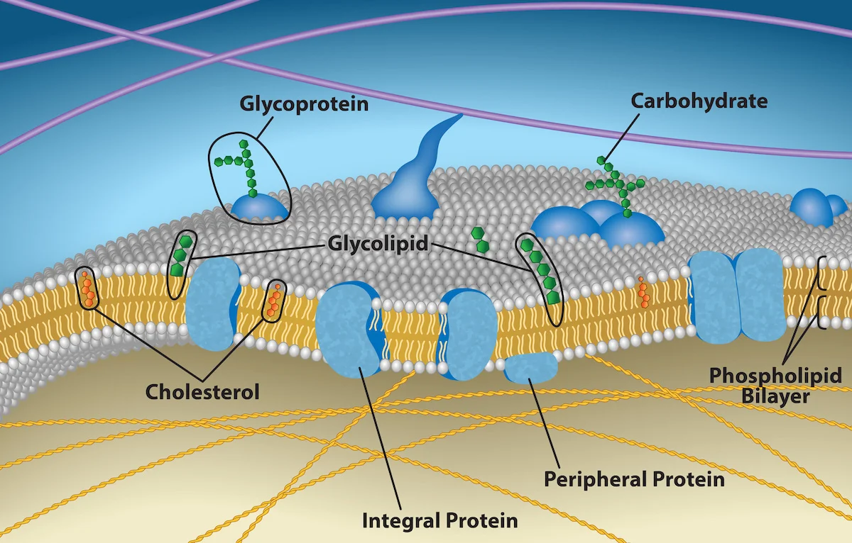
The fluid mosaic model (Singer and Nicolson, 1972) is the standard picture of a cell membrane. Two leaflets of phospholipids with embedded proteins. “Fluid” because lipids and proteins can move laterally; “mosaic” because proteins are scattered throughout the lipid sea.
The fluid mosaic model. Phospholipid bilayer with embedded proteins, cholesterol interleaved between fatty acid tails, and surface glycans facing the extracellular space. Credit: OpenStax Biology 2e, CC BY 4.0
Components
Component
Location
Role
Phospholipids
Both leaflets
Main structural matrix
Cholesterol (animals)
Throughout
Modulates fluidity
Sphingolipids
Mostly outer leaflet
Lipid rafts, signaling
Membrane proteins
Integral, peripheral
Transport, signaling, enzymes, anchors
Glycans on lipids and proteins
Outer leaflet only
Recognition, signaling
Asymmetry
The two leaflets are not identical. Phosphatidylcholine and sphingomyelin are in the outer leaflet. Phosphatidylethanolamine, phosphatidylserine, and phosphatidylinositol are in the inner leaflet. Glycans are always on the outer leaflet. Flippases and floppases (ATP-driven) maintain this asymmetry. Apoptosis flips phosphatidylserine to the outer leaflet as an “eat me” signal.
Membrane Fluidity
Fluidity depends on:
Temperature: higher T → more fluid.
Saturation: more cis double bonds (kinks) → more fluid.
Chain length: shorter tails → more fluid.
Cholesterol: bidirectional buffer - reduces fluidity at high T, prevents gel formation at low T.
Lipid Rafts
Special microdomains enriched in cholesterol, sphingolipids, and certain proteins. Rafts are more ordered than the surrounding membrane. Many signaling receptors cluster in rafts, concentrating their downstream signaling partners together.
Why does increasing the proportion of unsaturated fatty acids increase membrane fluidity?
Click to reveal answer
Cis double bonds create kinks in fatty acid tails. Kinked tails cannot pack as tightly together as straight saturated tails, leaving more space and permitting more lateral movement. The result is a more fluid, less ordered membrane.
How does cholesterol buffer membrane fluidity at both high and low temperatures?
Click to reveal answer
At high temperature, cholesterol's rigid ring restricts the motion of adjacent fatty acid tails, making the membrane less fluid. At low temperature, cholesterol disrupts the tight packing of fatty acid tails, preventing them from freezing into a gel. The net effect is stabilization in both directions.
What determines the asymmetry of the plasma membrane's two leaflets?
Click to reveal answer
ATP-dependent enzymes called flippases and floppases actively move specific lipids between leaflets, maintaining asymmetry. Phosphatidylserine is kept on the inner leaflet normally; scramblases flip it outward during apoptosis as an "eat me" signal for phagocytes. Glycolipids and glycoproteins are always on the outer leaflet with sugars facing the extracellular space.
Half of every plasma membrane by mass is protein. Proteins do most of the work: transport, signaling, catalysis, structural anchoring. They sit in the membrane in specific ways depending on their structure.
Integral vs. Peripheral
Integral membrane proteins: embedded in the bilayer, held there by hydrophobic amino acid interactions with the lipid tails. They can only be removed by disrupting the membrane (detergents). Most span the entire membrane (transmembrane proteins).
Peripheral membrane proteins: stuck to the surface via interactions with integral proteins or with lipid head groups. They can be removed by mild treatments (salt washes, pH changes) without disrupting the membrane.
Transmembrane Structure
A transmembrane protein crosses the bilayer through one or more membrane-spanning segments. Two common structural motifs:
Alpha helices: the most common transmembrane motif. A 20-residue hydrophobic alpha helix is long enough to span the 30 Å hydrophobic core of the bilayer. Examples: most receptors, channels, transporters.
Beta barrels: stacks of antiparallel beta strands form a cylindrical pore. Rare in the plasma membrane but common in the outer membranes of bacteria, mitochondria, and chloroplasts. Example: porins.
Lipid-Anchored Proteins
Some proteins that are not transmembrane nonetheless attach to the membrane via a covalent lipid modification. Examples include:
GPI anchor (glycosylphosphatidylinositol): links the protein’s C-terminus to the outer leaflet via a GPI moiety. Prion protein (PrP) is GPI-anchored.
Myristoylation: 14-carbon myristate attached to the N-terminal glycine. Signaling kinases like Src.
Palmitoylation: 16-carbon palmitate attached to cysteine. Reversible and regulatory.
Prenylation: attachment of a farnesyl (15C) or geranylgeranyl (20C) isoprenoid. Small GTPases like Ras.
Four Functional Classes
Transporters and channels: move molecules across the membrane. Covered in the next sections.
Receptors: bind extracellular signals and transmit them inside (GPCRs, RTKs, ion channel receptors).
Enzymes: catalyze reactions at the membrane (adenylyl cyclase, phospholipase C, acetylcholinesterase).
Anchors and structural proteins: connect the membrane to the cytoskeleton or extracellular matrix (integrins, cadherins, spectrin-ankyrin network).
What distinguishes an integral from a peripheral membrane protein?
Click to reveal answer
Integral membrane proteins are embedded in the lipid bilayer via hydrophobic interactions with the fatty acid tails and can only be removed by disrupting the membrane (e.g., with detergents). Peripheral proteins are associated with the membrane surface via non-covalent interactions with integral proteins or lipid head groups, and can be removed by salt washes or pH changes without disrupting the membrane.
What structural motif is typical of transmembrane proteins in the plasma membrane?
Click to reveal answer
Alpha helices of about 20 hydrophobic amino acids that span the ~30 Å hydrophobic core of the bilayer. Proteins can have one (single-pass) or many (multi-pass, like 7-TM GPCRs) such helices. Beta-barrel transmembrane proteins exist in outer membranes of bacteria and mitochondria but are rare in the plasma membrane.
How are lipid-anchored proteins attached to the membrane?
Click to reveal answer
A lipid group is covalently attached to the protein and inserts into one leaflet of the bilayer. Examples: GPI anchors (glycosylphosphatidylinositol), myristoylation (14C myristate on N-terminal glycine), palmitoylation (16C palmitate on cysteine, reversible), and prenylation (farnesyl or geranylgeranyl on cysteine near the C-terminus). The protein itself is not transmembrane.
Passive transport moves substances down their concentration or electrochemical gradient without consuming energy. Two varieties: simple diffusion (through the lipid bilayer) and facilitated diffusion (through a protein channel or carrier).
Simple Diffusion
Small, nonpolar molecules cross the bilayer by dissolving through it. O2, CO2, N2, steroid hormones, small uncharged molecules like ethanol - all cross freely. Rate depends on concentration gradient, temperature, and the molecule’s lipid solubility.
Charged molecules and large polar molecules (ions, glucose, amino acids) cannot cross the hydrophobic core effectively. They need a protein to help.
Facilitated Diffusion
Still passive (down the gradient, no ATP), but requires a protein. Two types of proteins:
Channels: hollow tunnels. Ions or small polar molecules flow through. Rate is fast (107-108 per second). Channels can be gated (open/close in response to voltage, ligand, or mechanical stimulus). Examples: voltage-gated Na+ channels, aquaporins for water.
Carriers: bind the substrate, change conformation, release it on the other side. Slower than channels (102-104 per second) but more selective. Examples: GLUT transporters for glucose, GABA transporters.
Transport types across the plasma membrane. Simple diffusion (no protein), facilitated diffusion (channels, carriers - still passive), primary active transport (ATP-driven pumps), and secondary active transport (symporters, antiporters using ion gradients). Credit: Wikimedia Commons, CC BY-SA
Factors Affecting Rate
Gradient: steeper gradient, faster transport.
Temperature: higher T, faster (Arrhenius).
Surface area: more membrane, more transporters, faster.
Molecule properties: for simple diffusion, smaller and more lipophilic = faster. For facilitated diffusion, specificity matters.
Aquaporins
Water can slowly diffuse through the bilayer but much faster through aquaporins - protein channels dedicated to water. Kidneys and some brain regions have high aquaporin expression and can move water rapidly across membranes. Antidiuretic hormone (ADH) inserts aquaporin-2 into collecting duct cells, concentrating urine.
What is the fundamental difference between passive and active transport?
Click to reveal answer
Passive transport moves a substance DOWN its concentration or electrochemical gradient and does not consume energy. Active transport moves a substance AGAINST its gradient and requires energy (typically from ATP hydrolysis or from a coupled ion gradient).
Why do facilitated diffusion carriers show saturation kinetics like enzymes?
Click to reveal answer
A carrier binds its substrate, changes shape, releases it on the other side, and resets. The number of carriers is finite. When substrate is high enough to keep every carrier busy, the rate plateaus at a Vmax. This produces the same Michaelis-Menten-style hyperbolic curve seen with enzymes. Channels, which allow many ions to flow through simultaneously, generally do not show this saturation under physiologic conditions.
Which molecules can cross the lipid bilayer by simple diffusion without protein help?
Click to reveal answer
Small nonpolar molecules (O2, CO2, N2), small uncharged polar molecules at slow rates (water, urea), and lipid-soluble molecules like steroid hormones and ethanol. Ions, large polar molecules (glucose, amino acids), and charged species generally cannot cross without a protein transporter or channel.
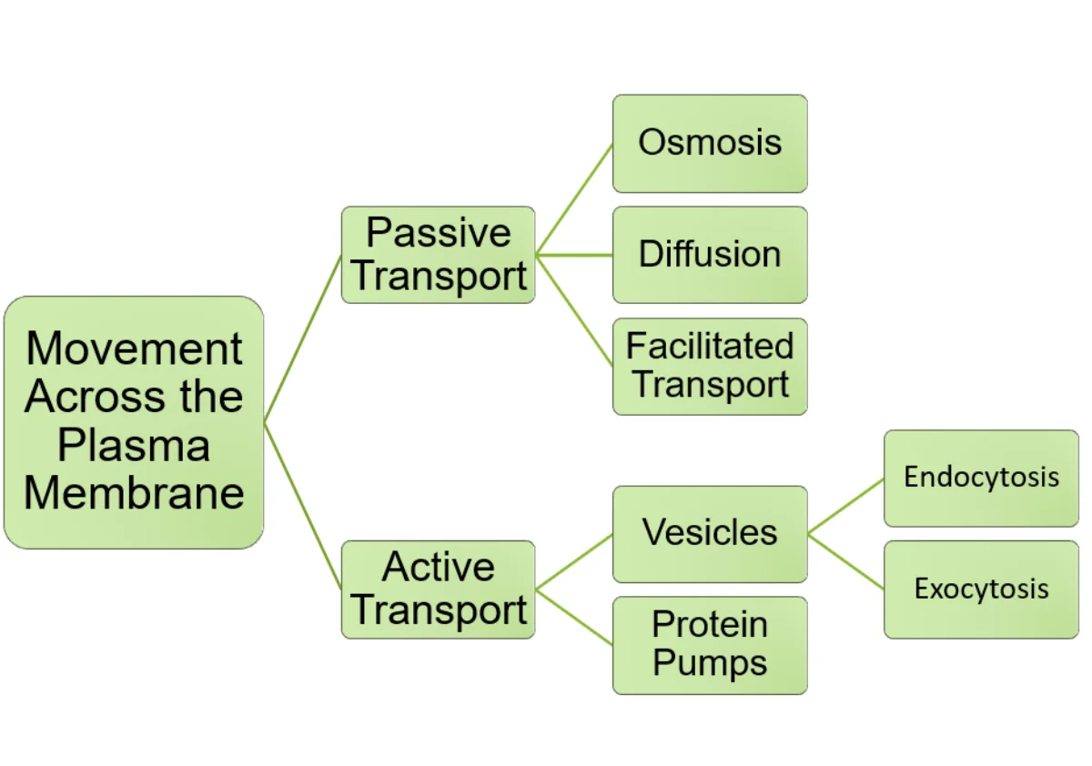
Active transport moves molecules against their gradient. This requires energy, either directly from ATP (primary) or from another ion gradient (secondary).
Five ways across a membrane
Transport
1
Scroll sideways to see the whole map.
Passive: down the gradient, no ATP Active: against the gradient, costs energy Bulk transport by vesicle The membrane
The one question that sorts themAsk which way relative to the gradient. Down the gradient is passive and free, whether or not a protein helps. Against the gradient always costs energy, and the only argument is whether the cell pays with ATP directly or spends a gradient it already built.
Why secondary active is still activeThe cotransporter itself uses no ATP. But the sodium gradient it rides was built by the Na⁺/K⁺-ATPase, which does. Stop that pump and secondary active transport stops within minutes, which is exactly how ouabain and digoxin work.
Saturation is the tellAnything that uses a protein has a finite number of them, so its rate plateaus like an enzyme, complete with a Km-equivalent. Simple diffusion has nothing to saturate, so its rate rises linearly with the gradient forever. A graph that plateaus means a protein is involved.
Two questions answer every transport problem: is a protein involved, and is it moving with or against the gradient. Protein plus with-the-gradient is facilitated diffusion; protein plus against is active, and then you only have to say who pays.
Primary Active Transport
The pump uses ATP directly. Classic example: Na+/K+ ATPase.
Each cycle:
Pump binds 3 Na+ from inside + 1 ATP.
ATP hydrolysis phosphorylates the pump; it changes shape and expels 3 Na+ to outside.
Pump binds 2 K+ from outside.
Phosphate is released; the pump changes shape again and releases 2 K+ inside.
Net result per ATP: 3 Na+ out, 2 K+ in. Because 3 positive charges leave while only 2 enter, the pump is electrogenic and contributes directly to the resting membrane potential (more negative inside).
Other major ATP-driven pumps:
Ca2+ ATPase (SERCA): pumps Ca2+ from cytoplasm into the ER. Keeps cytoplasmic Ca2+ extremely low (~100 nM) so small releases produce dramatic signaling.
H+/K+ ATPase: pumps H+ into stomach lumen, producing gastric acid. Target of proton pump inhibitors.
V-type ATPase: pumps H+ into lysosomes and other acidic compartments.
Secondary Active Transport
The transporter does NOT use ATP directly. Instead, it uses the energy stored in an ion gradient (usually Na+, built by the Na+/K+ ATPase). The Na+ moves down its gradient through the transporter, and the energy drags another molecule UP its gradient as a passenger.
Two classes:
Symporter (cotransporter): Na+ and the co-transported molecule move in the SAME direction. Example: SGLT1 in intestinal cells absorbs glucose against its gradient by coupling to Na+ flowing down its gradient.
Antiporter (exchanger): Na+ and the co-transported molecule move in OPPOSITE directions. Example: Na+/Ca2+ exchanger in heart cells pumps Ca2+ out using the Na+ gradient.
How many Na+ and K+ does the Na+/K+ ATPase pump per ATP, and in which directions?
Click to reveal answer
3 Na+ are pumped OUT of the cell and 2 K+ are pumped INTO the cell per ATP hydrolyzed. Net loss of one positive charge from inside the cell per cycle makes the pump electrogenic, contributing to the negative resting membrane potential.
What is the difference between a symporter and an antiporter?
Click to reveal answer
A symporter moves two different molecules in the SAME direction (e.g., SGLT1 moves Na+ and glucose both into the cell). An antiporter moves two different molecules in OPPOSITE directions (e.g., Na+/Ca2+ exchanger moves Na+ in and Ca2+ out). Both are secondary active transporters harnessing an ion gradient.
How can blocking the Na+/K+ ATPase indirectly increase intracellular calcium?
Click to reveal answer
The Na+/K+ ATPase normally keeps intracellular Na+ low. The Na+/Ca2+ exchanger (antiporter) pumps Ca2+ out of the cell using the Na+ gradient - flowing Na+ in, Ca2+ out. If the pump is blocked (e.g., by digoxin), intracellular Na+ rises. The Na+ gradient weakens, so the Na+/Ca2+ exchanger cannot pump Ca2+ out as effectively. Cytoplasmic Ca2+ rises, increasing cardiac contractility.
Osmosis is the movement of water across a semipermeable membrane from low solute concentration to high solute concentration. It is passive - driven by the concentration gradient of water itself. Tonicity describes what happens to a cell placed in a solution relative to the cell’s own interior.
Osmosis: three solutions, three outcomes
Tonicity
1
Scroll sideways to see the whole map.
Water moving in Water moving out The cell The surrounding solution
Osmolarity is not tonicityOsmolarity counts every dissolved particle. Tonicity counts only the ones that cannot cross the membrane. A urea solution can have the same osmolarity as the cell and still be hypotonic, because urea crosses freely and water follows it in. Only impermeant solutes pull water.
Water follows soluteWater moves toward the side with more solute, which is the side with lower water concentration. Saying water moves down its own concentration gradient and saying it moves toward solute are the same sentence.
Why it matters clinicallyGive pure water intravenously and red cells lyse. Give concentrated saline and they shrivel. Isotonic saline at about 0.9% exists precisely so that neither happens, and it is why a drip bag says what it says.
Name the solution, then move the water toward the solute. Hypotonic outside means water goes in and the cell swells; hypertonic outside means water goes out and it shrinks. The prefix always describes the solution, never the cell.
Osmosis
A semipermeable membrane allows water through but blocks solute. Water moves from the side with less solute (high water concentration) to the side with more solute (low water concentration) until the solute concentrations equalize or until a physical pressure stops the flow.
Water can cross the bilayer directly at a slow rate, but most physiologic water movement goes through aquaporins - specialized channel proteins that let water pour through at much higher rates while excluding ions and other solutes. Kidney collecting-duct cells express aquaporin-2 under vasopressin (ADH) control, which is how urine is concentrated on demand.
Osmotic Pressure
The pressure needed to prevent osmosis is called osmotic pressure. For dilute solutions:
Π=iMRT
Π = osmotic pressure.
i = van’t Hoff factor (number of particles a solute dissociates into; NaCl → Na+ and Cl-, i = 2).
M = molar concentration of solute.
R = gas constant.
T = absolute temperature.
A solution with more dissolved particles exerts more osmotic pressure. This is why IV solutions are designed to match blood osmolarity (~300 mOsm/L).
The Three Tonicities
Always stated from the perspective of the SOLUTION compared to the CELL’s cytoplasm.
| Tonicity | Solution vs. cell | Water flows | RBC outcome |
|----------|-------------------|-------------|-------------|
| Hypertonic | Higher solute | Water OUT of cell | Cell shrinks (crenation) |
| Hypotonic | Lower solute | Water INTO cell | Cell swells (lysis, hemolysis) |
| Isotonic | Equal solute | Balanced | No net change |
Plant Cells and Turgor Pressure
Plant cells have a rigid cell wall outside the membrane. When placed in a hypotonic solution, they absorb water and expand against the wall until the wall resists further expansion. That pressure inside the cell is turgor pressure - it is what keeps non-woody plants upright. In a hypertonic solution, plant cells lose water and the membrane pulls away from the wall (plasmolysis) - wilting.
What happens to a red blood cell placed in a hypotonic solution, and why?
Click to reveal answer
Water flows INTO the cell because the interior has a higher solute concentration than the hypotonic surrounding solution. The cell swells. RBCs lack a cell wall, so if the volume increases enough, the membrane ruptures - hemolysis. This is why pure water cannot be given intravenously; it would hemolyze RBCs.
What is the van’t Hoff factor for NaCl, and why does it matter for osmotic pressure?
Click to reveal answer
i = 2 because NaCl dissociates into two ions (Na+ and Cl-). Osmotic pressure depends on the total concentration of dissolved PARTICLES. A 0.5 M NaCl solution has the same osmotic pressure as a 1 M glucose solution (glucose does not dissociate, i = 1). Always multiply by i when comparing osmotic effects.
Why do plant cells placed in a hypotonic solution not burst?
Click to reveal answer
Plant cells have a rigid cell wall outside the plasma membrane. As water flows into the cell, the cell expands until the wall resists further expansion. The pressure inside the cell (turgor pressure) rises to balance the osmotic driving force. This is what keeps non-woody plants rigid and upright; wilted plants have lost turgor.
Some cargoes are too big to cross the membrane. The cell wraps the membrane around them (endocytosis) or wraps the membrane around something inside to release it (exocytosis). Both use vesicles.
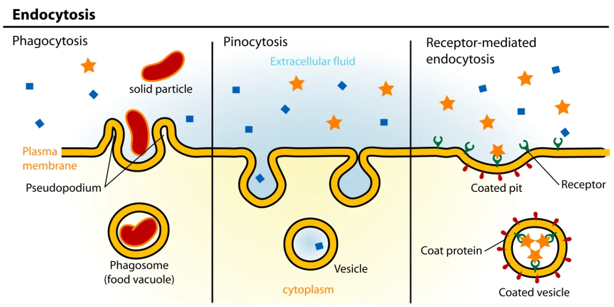
Three endocytosis types. Phagocytosis: engulfing large particles. Pinocytosis: drinking fluid. Receptor-mediated endocytosis: selective uptake via clathrin-coated pits. Credit: Wikimedia Commons, CC BY-SA
Endocytosis Types
Phagocytosis (“cell eating”)
Engulfing solid particles (bacteria, dead cells, debris). Used mostly by specialized immune cells (macrophages, neutrophils). The cell extends pseudopodia around the target, forming a large vesicle (phagosome). The phagosome fuses with a lysosome for degradation.
Pinocytosis (“cell drinking”)
Non-specific uptake of fluid and small dissolved molecules. The cell surface pinches in continuously, engulfing whatever fluid is nearby. No specific receptors involved. Happens constantly in most cells.
Receptor-Mediated Endocytosis
Highly specific. A receptor on the cell surface binds a specific ligand; the receptor-ligand complex clusters in a clathrin-coated pit; the pit pinches off into a vesicle coated with clathrin. The vesicle uncoats and enters the endosomal pathway. This is how cells efficiently import specific cargo (LDL, transferrin, many hormones) even when their blood concentrations are low.
Exocytosis
The reverse of endocytosis. An intracellular vesicle (usually carrying secretory protein cargo, like insulin, a neurotransmitter, or a hormone) fuses with the plasma membrane and releases its contents to the outside. SNARE proteins mediate the fusion. Exocytosis is how cells secrete proteins, discard waste, or deliver new membrane components to the cell surface.
Regulated: vesicles wait near the membrane until a stimulus triggers fusion. Examples: neurotransmitter release at synapses (triggered by Ca2+), insulin release from beta cells (triggered by glucose), hormone secretion.
What distinguishes receptor-mediated endocytosis from pinocytosis?
Click to reveal answer
Receptor-mediated endocytosis requires specific cell-surface receptors that bind specific ligands. Clathrin-coated pits form around clusters of bound receptors and internalize them as vesicles. The result is selective, high-efficiency uptake of specific molecules (e.g., LDL, iron-transferrin). Pinocytosis is non-specific - the cell drinks in whatever fluid and solutes happen to be nearby, without targeting any particular molecule.
How does exocytosis deliver secreted proteins to the outside of the cell?
Click to reveal answer
Proteins destined for secretion are packaged into vesicles (by the Golgi). The vesicle travels to the plasma membrane, where SNARE proteins mediate fusion between the vesicle and the plasma membrane. The fusion event opens the vesicle lumen to the extracellular space, releasing its cargo. The vesicle's membrane is incorporated into the plasma membrane.
Why is familial hypercholesterolemia caused by a defect in endocytosis rather than in LDL production?
Click to reveal answer
LDL is cleared from the blood primarily by receptor-mediated endocytosis. The LDL receptor on hepatocyte surfaces binds LDL particles and internalizes them via clathrin-coated pits. When the LDL receptor is mutated (as in familial hypercholesterolemia), LDL cannot be internalized and accumulates in plasma. The defect is in uptake, not synthesis, of LDL.
Most cells have a negative voltage inside (about -70 mV in neurons at rest). This membrane potential powers signaling in neurons and muscle, drives secondary active transport, and keeps membranes ready to respond to stimuli.
Why the Inside Is Negative
Two main reasons:
The Na+/K+ ATPase pumps 3 positive charges out per 2 positive charges in, directly making the inside more negative (electrogenic).
K+ has high cytoplasmic concentration and is the most permeable ion at rest (because K+ leak channels are open). K+ tends to flow down its gradient out of the cell, leaving behind a net negative charge inside. This sets the resting potential close to the K+ equilibrium potential.
The Nernst Equation
For a single ion at equilibrium, the voltage difference across the membrane that would stop net ion flow is the equilibrium potential (Eion). The Nernst equation calculates it:
Eion=zFRTln[ion]in[ion]out
R = gas constant, T = temperature in K, z = ion charge, F = Faraday constant.
At 37°C, this simplifies to E (mV) = (61.5 / z) × log([out]/[in]) for a single ion.
At typical cellular concentrations:
EK ≈ -90 mV
ENa ≈ +60 mV
ECl ≈ -65 mV
ECa ≈ +120 mV
The Goldman Equation
The Nernst equation handles one ion at a time. The actual resting potential depends on all permeable ions. The Goldman-Hodgkin-Katz equation weighs each ion by its permeability:
At rest, K+ has the highest permeability, so Vm is close to EK but not exactly equal because some Na+ and Cl- leak through too. When Na+ channels open (action potential), permeability shifts toward Na+, and Vm swings toward ENa.
What does the Nernst equation calculate?
Click to reveal answer
The equilibrium potential for a single ion - the membrane voltage at which the concentration gradient's driving force on that ion is exactly balanced by the electrical gradient's driving force, producing zero net flow. It assumes the ion is the only permeable species. At 37°C, Nernst simplifies to E = (61.5 mV / z) log([out]/[in]).
Why is the resting membrane potential closer to the K+ equilibrium potential than to the Na+ equilibrium potential?
Click to reveal answer
At rest, the membrane is much more permeable to K+ (through leak channels) than to Na+. In the Goldman equation, the weights are permeabilities, so the resting potential is pulled close to EK (~-90 mV). A small Na+ permeability pulls it slightly positive, giving a resting value near -70 mV in neurons.
Why is the Na+/K+ ATPase called electrogenic?
Click to reveal answer
Because each cycle pumps 3 positive charges out and only 2 in, producing a net loss of one positive charge from the cell per ATP. Over time this contributes directly to the negative resting potential. Inhibiting the pump (e.g., with ouabain) causes the membrane potential to drift toward zero.
Most extracellular signals do not enter the cell directly. They bind receptors on the outside of the plasma membrane, which transmit the signal to the inside. Three main classes of cell surface receptors dominate the MCAT:
GPCRs (Reviewed from Chapter 3)
Seven transmembrane alpha helices. Coupled to heterotrimeric G proteins inside. Ligand binding triggers GDP-GTP exchange on the Gα subunit, which dissociates from Gβγ and activates downstream effectors (adenylyl cyclase, phospholipase C, etc.). Major second messengers: cAMP (Gs), decreased cAMP (Gi), IP3/DAG/Ca2+ (Gq).
Single transmembrane protein. The cytoplasmic domain is a tyrosine kinase. Ligand binding dimerizes two receptor monomers, triggering cross-phosphorylation on specific tyrosine residues. Phosphotyrosines become docking sites for SH2-domain proteins that activate downstream pathways (Ras/MAPK, PI3K/AKT, PLCγ).
Receptors that double as ion channels. Ligand binding opens or closes the channel directly. Fast electrical response (milliseconds).
Examples:
Nicotinic acetylcholine receptor: nonselective cation channel at the neuromuscular junction. Opening depolarizes the muscle cell.
GABA-A receptor: Cl- channel in the CNS. Opening hyperpolarizes neurons (inhibitory). Target of benzodiazepines and many anesthetics.
NMDA receptor: Ca2+ and Na+ channel. Requires both glutamate and membrane depolarization to open. Critical for learning and memory (long-term potentiation).
What are the three main classes of cell surface receptors, and which is fastest?
Click to reveal answer
(1) Ligand-gated ion channels - fastest, milliseconds (direct electrical effect). (2) GPCRs - seconds (second messenger cascades). (3) Receptor tyrosine kinases - minutes to hours (phosphotyrosine signaling and transcriptional changes). Steroid hormone receptors (intracellular, not surface) are slower still.
How does a receptor tyrosine kinase activate itself upon ligand binding?
Click to reveal answer
Ligand binding dimerizes two receptor monomers. The intracellular kinase domains cross-phosphorylate each other on tyrosine residues. The resulting phosphotyrosines serve as docking sites for SH2-domain-containing proteins (e.g., Grb2), initiating downstream cascades like Ras/MAPK and PI3K/AKT.
Why is the GABA-A receptor considered inhibitory in the CNS?
Click to reveal answer
GABA-A is a ligand-gated Cl- channel. When GABA binds, the channel opens and Cl- flows into the cell (because ECl is near or below the resting potential). The influx of negative charge hyperpolarizes the cell, making it less likely to fire an action potential - inhibitory. Benzodiazepines and barbiturates enhance GABA-A activity, increasing this inhibition.
A second messenger is a small intracellular molecule produced in response to an extracellular signal. It carries the signal from the receptor into the cell and amplifies it. Five main ones dominate the MCAT.
Three ways a signal gets through the wall
Cell signalling
1
Scroll sideways to see the whole map.
G-protein coupled: slow, amplified Receptor tyrosine kinase: growth and metabolism Ion channel: fast, electrical Second messenger
Why amplification mattersOne hormone molecule activates one receptor, which activates many G proteins, each of which drives a cyclase making many cAMP, each activating a kinase that phosphorylates many targets. A handful of molecules outside the cell becomes millions of altered molecules inside it. That is why hormones work at nanomolar concentrations.
The other second messengerscAMP is not the only one. IP₃ releases calcium from the endoplasmic reticulum, DAG activates protein kinase C, calcium itself acts through calmodulin, and cGMP carries nitric oxide's signal. All do the same job: turn one event at the surface into many events inside.
Steroids skip all of itSteroid hormones are lipids, so they cross the membrane and bind receptors inside the cell. The complex then acts as a transcription factor directly. No second messenger, no amplification cascade, and a response measured in hours rather than seconds.
Water-soluble signals cannot get in, so they shout through the wall and something inside repeats the message. The receptor family determines how loud the shout gets and how fast: milliseconds for a channel, seconds for a kinase cascade, hours for a steroid.
The Five
| Second messenger | Made by | Activates | Receptor type |
|------------------|---------|-----------|---------------|
| cAMP | Adenylyl cyclase (from ATP) | Protein kinase A | Gs-coupled GPCR |
| cGMP | Guanylyl cyclase (from GTP) | Protein kinase G | Some hormones, NO |
| IP3 | Phospholipase C (from PIP2) | Opens ER Ca2+ channels | Gq-coupled GPCR |
| DAG | Phospholipase C (from PIP2) | Protein kinase C | Gq-coupled GPCR |
| Ca2+ | Released from ER by IP3 or entering via channels | Calmodulin, PKC, many enzymes | Various |
cAMP
Adenylyl cyclase converts ATP to cAMP (+ pyrophosphate). cAMP activates PKA, which phosphorylates many targets. cAMP is degraded by phosphodiesterase (PDE). Caffeine inhibits PDE, so caffeine raises cAMP indirectly.
Cholera toxin locks Gs in the active form, producing persistent cAMP in gut cells and massive watery diarrhea.
IP3 and DAG
Phospholipase C (PLC) cleaves PIP2 into IP3 (diffusible, cytoplasmic) and DAG (stays in the membrane). IP3 binds receptors on the ER and releases stored Ca2+. DAG, together with Ca2+, activates PKC. PKC phosphorylates a different set of substrates than PKA.
Calcium as Second Messenger
Cytoplasmic free Ca2+ is kept very low (~100 nM) by Ca2+ ATPases and Na+/Ca2+ exchangers. Small Ca2+ releases produce dramatic changes. Ca2+ binds calmodulin, activating many calmodulin-dependent kinases (CaMK). Ca2+ also activates PKC (with DAG), triggers muscle contraction (binds troponin C), and triggers neurotransmitter release at synapses.
Amplification Numbers
For a typical GPCR-cAMP-PKA cascade:
1 hormone → 1 active receptor
1 active receptor activates ~100 G proteins
Each G protein’s Gα produces ~1000 cAMPs (over its active lifetime)
Net: one hormone molecule → ~10,000-1,000,000 phosphorylation events, depending on kinetics.
Which second messengers does a Gq-coupled receptor produce, and how?
Click to reveal answer
Gq activates phospholipase C, which cleaves PIP2 into IP3 and DAG. IP3 binds ER-membrane receptors and releases stored Ca2+ into the cytoplasm. DAG stays in the membrane and activates protein kinase C (which also needs Ca2+). Net second messengers: IP3, DAG, and Ca2+.
Why is cholera toxin so disruptive at the cellular level?
Click to reveal answer
Cholera toxin ADP-ribosylates Gα(s), locking it in its GTP-bound (active) state. Adenylyl cyclase stays continuously active, cAMP accumulates to pathologic levels, and in gut epithelial cells this causes massive fluid secretion by opening chloride channels. The result is severe watery diarrhea. Removing the toxin does not immediately reverse the damage because the G protein is chemically modified.
How does signal amplification occur in a GPCR-cAMP cascade?
Click to reveal answer
Each step amplifies. One active receptor activates many G proteins. One Gα activates adenylyl cyclase to produce many cAMPs. Each PKA activated by cAMPs phosphorylates many targets. One hormone can produce tens of thousands to millions of downstream phosphorylation events. This multi-stage gain is why low hormone concentrations (nanomolar) produce strong cellular responses.