RNA is not just an intermediate between DNA and protein. There are multiple classes of RNA, each with a specific structural and functional role.
The central dogma, with the compartments
Information flow
1
Scroll sideways to see the whole map.
The three information-carrying molecules Cytoplasm Nucleus Where the exam sets its traps
The one arrow that runs backwardsReverse transcriptase makes DNA from RNA, which is how retroviruses such as HIV work and how a cDNA library is built. It is the reason the word dogma is a historical embarrassment rather than a law.
Why processing exists at allA eukaryotic transcript is not ready to use. The 5' cap protects it and helps the ribosome find it, the poly-A tail sets how long it survives, and splicing removes introns. Alternative splicing means one gene can produce several different proteins, which is how twenty thousand genes make far more than twenty thousand products.
Prokaryotes skip all of itNo nucleus means no separation, so a bacterial ribosome can start translating a transcript that is still being made. No cap, no tail, no introns, and polycistronic messages carrying several genes at once. Every difference follows from the missing membrane.
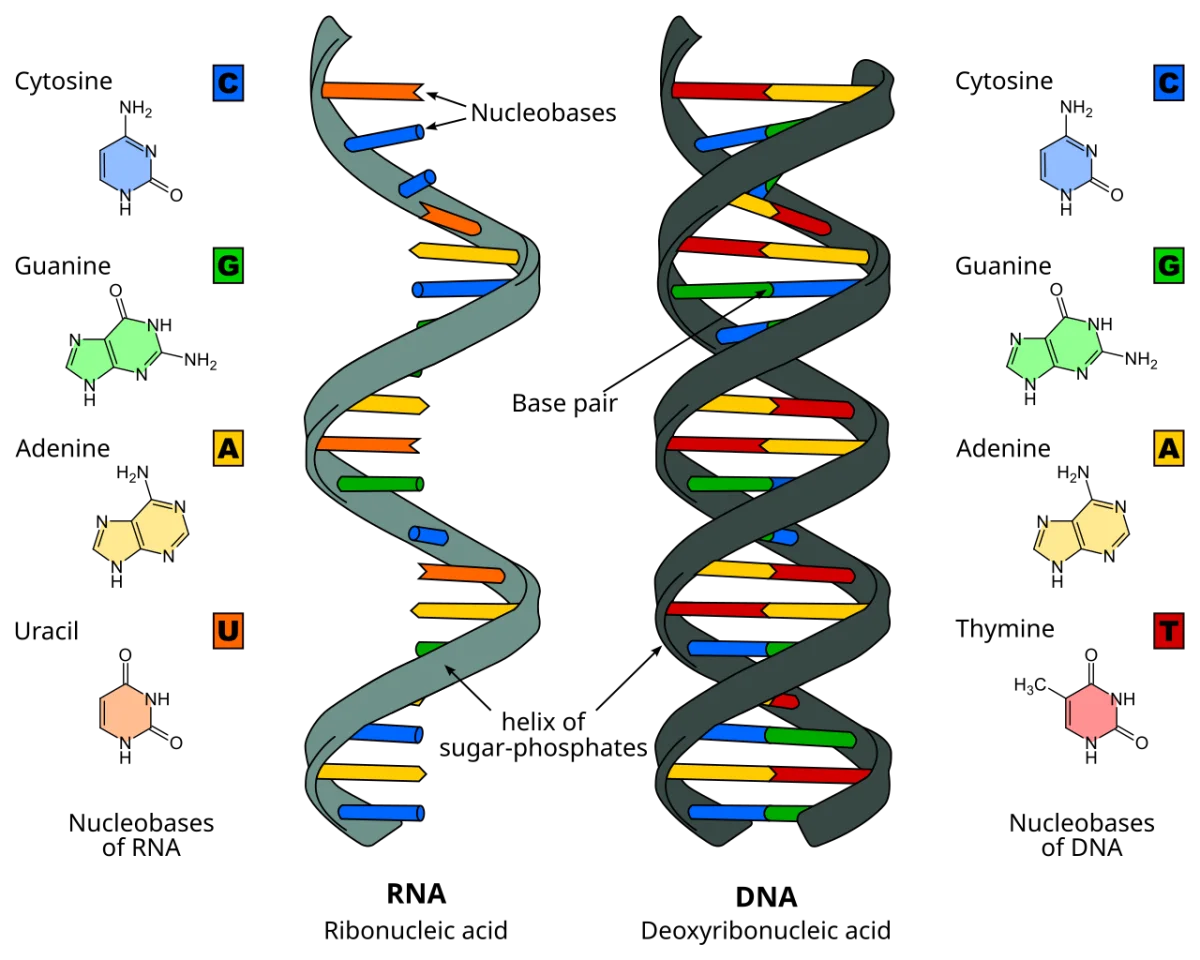
Information flows one way through three molecules, and the compartment tells you which organism you are in. If a question mentions splicing, a cap, or a tail, it is eukaryotic. If it mentions transcription and translation happening together, it is not.Key differences between DNA and RNA: ribose vs. deoxyribose sugar, uracil replaces thymine, and RNA is typically single-stranded. Credit: Wikimedia Commons, CC BY-SA
The Main Classes
| RNA type | Job | Length / notes |
|----------|-----|----------------|
| mRNA (messenger) | Encodes protein sequence | Variable, short-lived |
| tRNA (transfer) | Carries amino acids to the ribosome | ~75-90 nt, cloverleaf shape |
| rRNA (ribosomal) | Structural and catalytic part of the ribosome | Several sizes; most abundant RNA |
| snRNA (small nuclear) | Components of the spliceosome | ~100-300 nt |
| snoRNA (small nucleolar) | Guide chemical modifications of rRNA | ~60-300 nt |
| miRNA (microRNA) | Regulate gene expression by binding mRNA and silencing or degrading it | ~22 nt |
| siRNA (small interfering) | Similar to miRNA but typically exogenous | ~21-23 nt |
DNA vs. RNA at a Glance
Sugar: RNA has ribose (with 2’-OH). DNA has deoxyribose.
Bases: RNA uses uracil (U) instead of thymine (T). A, G, and C are the same.
Strand: RNA is usually single-stranded, although it can fold into complex secondary structures. DNA is almost always double-stranded.
Stability: RNA is less stable due to the 2’-OH. DNA is far more stable and therefore used for long-term storage.
Which three RNAs are the main participants in translation?
Click to reveal answer
mRNA, tRNA, and rRNA. mRNA carries the coding message. tRNA brings amino acids matching each codon. rRNA is a major structural and catalytic component of the ribosome (peptidyl transferase activity is in the 23S/28S rRNA).
What do miRNAs do?
Click to reveal answer
MicroRNAs are ~22 nt RNAs that bind complementary sequences in target mRNAs, typically in the 3’ UTR. The miRNA-RISC complex either inhibits translation or promotes degradation of the target. miRNAs are widespread regulators of gene expression and many are implicated in cancer and development.
Which sugar does RNA contain, and what key structural difference does that create vs. DNA?
Click to reveal answer
RNA contains ribose, which has a 2’-hydroxyl. DNA uses 2’-deoxyribose, which lacks the 2’-OH. The 2’-OH makes RNA more chemically reactive - it can attack the phosphodiester backbone intramolecularly - so RNA is far less stable than DNA.
Transcription is the synthesis of RNA from a DNA template. The product is an RNA copy of one strand of DNA.
Transcription elongation. RNA polymerase unwinds a short stretch of DNA, reads the template strand 3' to 5', and synthesizes RNA 5' to 3' using ribonucleotides. Credit: Wikimedia Commons, CC BY-SA
Template vs. Coding Strand
Only one of the two DNA strands is copied at each locus:
Template strand (antisense): the strand that RNA polymerase reads. The RNA is complementary to this strand (3’ to 5’ read, 5’ to 3’ written).
Coding strand (sense): the other strand. The RNA has the same sequence as the coding strand (except U replaces T).
The Three Stages
Initiation
RNA polymerase recognizes a promoter upstream of the gene. In bacteria, the sigma factor helps the polymerase find the promoter (-10 and -35 consensus boxes). In eukaryotes, general transcription factors (TFIID, TFIIB, etc.) assemble at the promoter (TATA box) and recruit RNA Pol II. The polymerase unwinds about 14 bp of DNA to expose the template.
Elongation
The polymerase reads the template 3’ to 5’ and synthesizes RNA 5’ to 3’. Each new ribonucleotide is added to the 3’-OH of the growing chain. The polymerase moves along the DNA, maintaining a short “transcription bubble” of unwound DNA. About 10-20 nucleotides are added per second in eukaryotes.
Termination
In bacteria: rho-independent termination uses a GC-rich hairpin followed by a stretch of U’s that destabilizes the RNA-DNA hybrid. Rho-dependent termination uses the rho protein to pull the RNA off the polymerase.
In eukaryotes: termination of Pol II is less clean - the polymerase transcribes past the polyadenylation signal (AAUAAA), and a cleavage factor cuts the RNA. The polymerase eventually falls off.
Does RNA polymerase need a primer to begin synthesis, and what direction does it read/write?
Click to reveal answer
No primer needed. RNA polymerase can start RNA synthesis from scratch at a promoter. It reads the template DNA 3' to 5' and synthesizes RNA 5' to 3'. This is unlike DNA polymerase, which requires a primer.
Which RNA polymerase in eukaryotes transcribes mRNA?
Click to reveal answer
RNA polymerase II. Pol I handles rRNA (in the nucleolus), and Pol III handles tRNA and 5S rRNA. Bacteria have just one RNA polymerase that handles all RNA types.
If the coding strand of a gene reads 5'-ATGGCATTC-3', what is the mRNA sequence?
Click to reveal answer
5'-AUGGCAUUC-3'. The mRNA has the same sequence as the coding strand but with U in place of T. The template strand (3'-TACCGTAAG-5') is what RNA polymerase actually reads; the resulting RNA is complementary to the template and matches the coding strand.
A promoter is the DNA sequence that tells RNA polymerase where to start transcribing. Promoter elements are conserved near the transcription start site, and transcription factors recognize them and recruit the polymerase.
Bacterial Promoters
Bacterial (E. coli) promoters have two main elements upstream of the start site:
Pribnow box (-10): TATAAT consensus, about 10 bp upstream.
-35 box: TTGACA consensus, about 35 bp upstream.
The sigma factor of RNA polymerase recognizes these two boxes and positions the polymerase for initiation. Different sigma factors can recognize different promoters, allowing bacteria to activate different gene programs.
Eukaryotic Promoters (Pol II)
Eukaryotic promoters are more varied. The most common element is the TATA box, about 25 bp upstream, recognized by TATA-binding protein (TBP, part of TFIID). Additional upstream elements (CAAT box, GC box) bind other factors that help recruit the polymerase. Farther upstream, enhancers can activate transcription from thousands of bp away through DNA looping.
Three Eukaryotic RNA Polymerases
Eukaryotes divide the work among three polymerases:
Polymerase
Transcribes
Location
Sensitivity to alpha-amanitin
Pol I
rRNA (except 5S)
Nucleolus
Insensitive
Pol II
mRNA, most snRNAs
Nucleus
Very sensitive
Pol III
tRNA, 5S rRNA
Nucleus
Moderately sensitive
Alpha-amanitin is a toxin from death cap mushrooms that strongly inhibits Pol II. This property is used in the lab to distinguish which polymerase made a given RNA.
Which eukaryotic RNA polymerase makes mRNA, and what toxin strongly inhibits it?
Click to reveal answer
RNA polymerase II transcribes mRNA. Alpha-amanitin, a toxin from the death cap mushroom (Amanita phalloides), strongly inhibits Pol II. This is why death cap poisoning causes broad cellular dysfunction - protein synthesis stops throughout the body.
What is the function of a sigma factor in bacterial transcription?
Click to reveal answer
Sigma factors help RNA polymerase recognize promoter sequences (-10 and -35 boxes). Different sigma factors recognize different classes of promoters, allowing the cell to switch between gene expression programs (e.g., under heat shock, sporulation). After initiation, sigma dissociates and the core polymerase continues elongation on its own.
Why can bacteria couple transcription and translation while eukaryotes cannot?
Click to reveal answer
Bacteria have no nucleus, so ribosomes can attach to nascent mRNA as it is still being transcribed. Eukaryotic transcription happens inside the nucleus, and the mRNA must be processed (cap, poly-A tail, splicing) and exported before it can be translated. The physical compartmentalization and required processing decouple the two steps in eukaryotes.
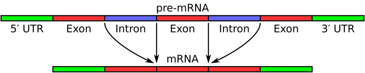
A eukaryotic pre-mRNA has to be processed before it can leave the nucleus and be translated. Three modifications happen, roughly in order: 5’ capping, splicing, and 3’ polyadenylation.
mRNA processing. A pre-mRNA gains a 5' cap, has introns removed by splicing, and gets a poly-A tail before being exported to the cytoplasm. Credit: Wikimedia Commons, CC BY-SA
5’ Cap
As soon as RNA Pol II makes the first ~25 nucleotides, a 7-methylguanosine cap is added to the 5’ end via a 5’-5’ triphosphate linkage. The cap:
Protects the 5’ end from exonucleases.
Serves as a recognition site for the ribosome during translation initiation (via cap-binding proteins).
Signals successful transcription initiation to the processing machinery.
3’ Poly-A Tail
Near the end of transcription, the AAUAAA polyadenylation signal is recognized. A cleavage factor cuts the RNA about 10-35 nucleotides downstream, and poly-A polymerase adds 100-250 A residues. The poly-A tail:
Protects the 3’ end from degradation.
Is required for efficient nuclear export.
Is progressively shortened during the mRNA’s life in the cytoplasm; when it becomes too short, the mRNA is degraded.
Splicing
Eukaryotic genes are split into coding exons and non-coding introns. Splicing removes introns and joins exons together. The machinery is the spliceosome, a large ribonucleoprotein complex made of snRNAs (U1, U2, U4, U5, U6) and associated proteins. Splicing recognizes conserved GU at the 5’ splice site and AG at the 3’ splice site.
Splicing proceeds in two transesterification steps:
A special adenosine within the intron (the branch point) attacks the 5’ splice site, forming a lariat.
The released 3’-OH of the upstream exon attacks the 3’ splice site, joining the two exons and releasing the intron lariat.
What three modifications convert a pre-mRNA into a mature eukaryotic mRNA?
Click to reveal answer
(1) Addition of a 5' cap (7-methylguanosine linked via 5'-5' triphosphate). (2) Splicing out of introns and joining of exons. (3) Cleavage near the AAUAAA signal and addition of a 100-250-nt poly-A tail at the 3' end. Together these modifications stabilize the mRNA, enable nuclear export, and prepare it for translation.
What is the spliceosome made of, and what does it do?
Click to reveal answer
The spliceosome is a large ribonucleoprotein complex made of five snRNAs (U1, U2, U4, U5, U6) and many associated proteins. It recognizes 5' (GU) and 3' (AG) splice sites and catalyzes two transesterification steps to remove each intron as a lariat and join the flanking exons.
Why is the poly-A tail important for mRNA stability?
Click to reveal answer
The poly-A tail is a buffer against 3' exonuclease degradation. Deadenylases progressively shorten the tail in the cytoplasm. When the tail becomes too short, the mRNA is rapidly degraded. Thus the poly-A tail sets the mRNA's half-life: longer initial tails and slower deadenylation produce a longer-lived mRNA.
Most eukaryotic genes are split by introns - non-coding sequences between exons. The spliceosome removes introns. By including or excluding different exons, the cell can produce multiple different proteins from a single gene. This is alternative splicing.
The Numbers
The human genome has about 20,000 protein-coding genes but produces about 100,000+ distinct proteins. Alternative splicing is the main reason. Over 90% of multi-exon human genes undergo alternative splicing in at least some tissue or condition.
Types of Alternative Splicing
Exon skipping: an exon is included in some tissues, excluded in others.
Alternative 5’ or 3’ splice sites: the same exon is used but with a shifted boundary.
Intron retention: an intron is left in the mature mRNA.
Mutually exclusive exons: exon A or exon B is included, never both.
Why Introns Exist
Introns are evolutionarily ancient but vary widely. Their functional value is debated, but they clearly:
Enable alternative splicing to produce proteome diversity.
Allow exon shuffling over evolutionary time (introns let exons recombine without disrupting coding sequence).
Sometimes contain regulatory elements like enhancers or noncoding RNAs.
Bacterial genes lack introns. Eukaryotes tolerate the costs of splicing because alternative splicing is so valuable.
Why can the human genome encode about 20,000 genes but produce ~100,000 proteins?
Click to reveal answer
Alternative splicing. By including or excluding different exons during splicing, a single gene can produce multiple mRNA isoforms, each encoding a different protein. Combined with post-translational modifications, this produces the much larger proteome diversity from a relatively small number of genes.
What is exon skipping?
Click to reveal answer
Exon skipping is an alternative splicing pattern in which a normally included exon is skipped in some transcripts, producing an mRNA missing that exon. The resulting protein lacks the amino acids encoded by the skipped exon. Cells use exon skipping to produce tissue-specific protein variants from a single gene.
Why might a single amino acid mutation in a gene produce no protein at all?
Click to reveal answer
Splice site mutations. If a mutation destroys a splice site (the GU at a 5' site or AG at a 3' site), splicing fails. Introns are retained or wrong junctions are used, producing frameshifts and premature stop codons that trigger nonsense-mediated decay of the mRNA. The gene essentially produces no functional protein. Beta-thalassemia includes famous examples.
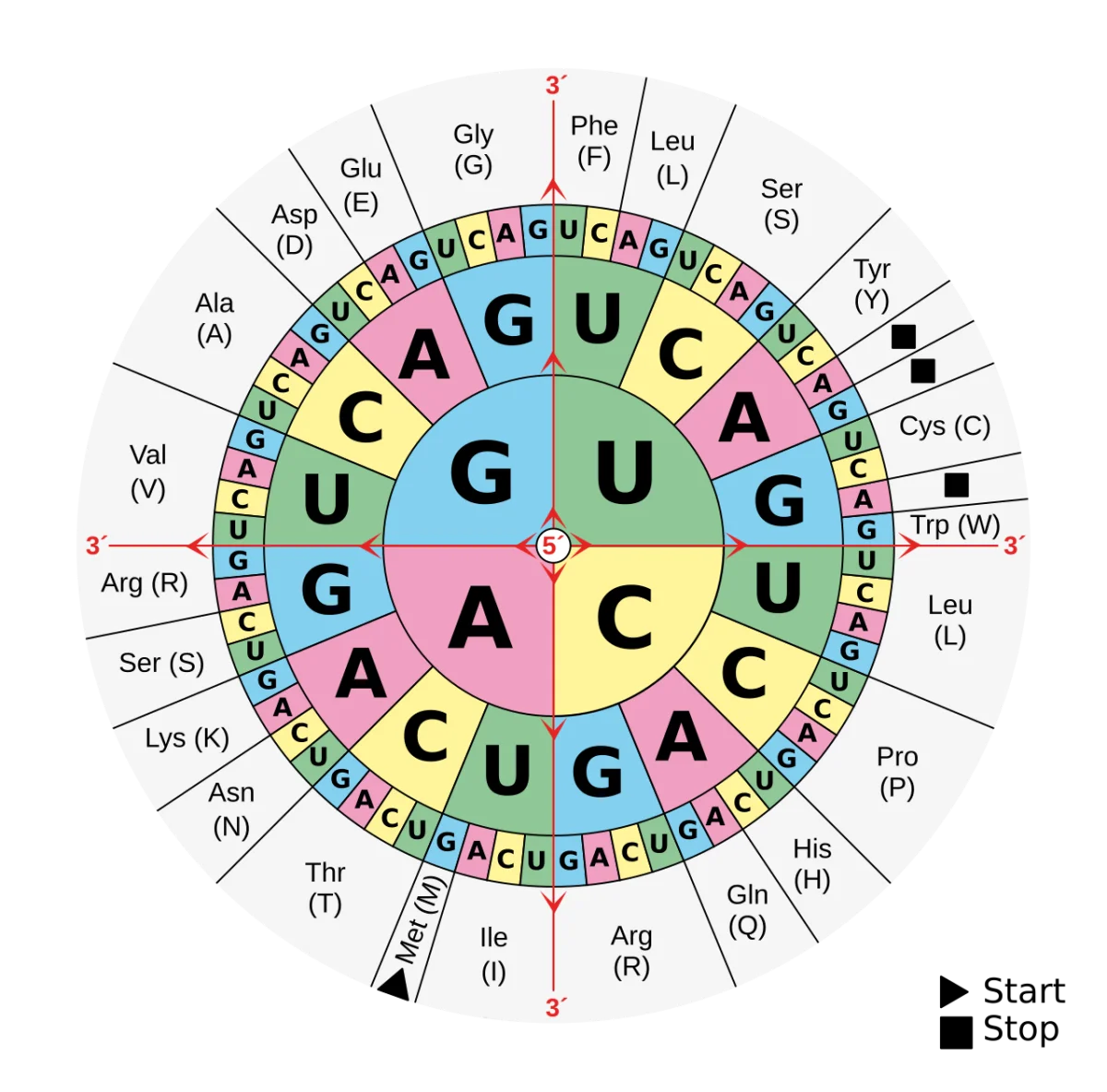
The genetic code maps 3-letter mRNA codons onto amino acids. With 4 bases and 3 positions, there are 43 = 64 possible codons. Since only 20 amino acids are encoded, most amino acids have multiple codons - the code is “degenerate.”
The standard genetic code. 64 codons encode 20 amino acids plus 3 stop codons. Most amino acids have multiple codons (degeneracy). Credit: Wikimedia Commons, CC BY-SA
Key Features
Triplet code: each amino acid is specified by 3 consecutive mRNA nucleotides.
Non-overlapping: codons are read one after another without sharing nucleotides.
Unambiguous: each codon specifies only one amino acid.
Degenerate: most amino acids are encoded by multiple codons. Only methionine (AUG) and tryptophan (UGG) have single codons.
Universal: the code is nearly identical across all organisms. A few exceptions exist in mitochondria and some protists.
Start and Stop Codons
AUG is the start codon. It codes for methionine. Translation begins with a specific initiator methionine (formyl-methionine in bacteria).
UAA, UAG, UGA are the three stop codons. They do not code for any amino acid; instead, release factors recognize them and terminate translation.
Wobble
Francis Crick proposed the wobble hypothesis in 1966. The third position of a codon (and the first position of the corresponding anticodon on tRNA) has relaxed base pairing. Non-standard pairings (G with U, inosine with A/U/C) are allowed.
Wobble lets one tRNA recognize multiple codons, reducing the number of tRNAs needed. It also makes the code more robust - mutations in the third codon position often produce silent mutations (same amino acid).
Mutation Types from the Code
Mutation
Effect
Silent
Same amino acid (often a wobble-position change)
Missense
Different amino acid
Nonsense
Creates a premature stop codon → truncated protein
Frameshift
Insertion or deletion of bases (not multiples of 3); shifts the reading frame
Frameshift mutations typically destroy the protein entirely because every downstream codon is misread and usually a premature stop arises quickly.
Which two amino acids are encoded by a single codon each, and what are those codons?
Click to reveal answer
Methionine (AUG) and tryptophan (UGG). All other amino acids have two or more codons. Because AUG is also the start codon, every protein starts with methionine (though it is often cleaved off after translation).
What are the three stop codons?
Click to reveal answer
UAA, UAG, UGA. They do not code for any amino acid. When a ribosome reads one, release factors bind instead of a tRNA, and the completed polypeptide is released. The mnemonic "U Go Away, U Are Away, U Are Gone" captures them all.
What is the wobble position, and why does it matter for the genetic code?
Click to reveal answer
The third position of a codon (and the first position of the anticodon) has relaxed base pairing - G can pair with U, inosine can pair with A/U/C, etc. Wobble lets a single tRNA recognize multiple codons, reducing the tRNA requirement. It also makes the code robust to point mutations: about 25% of random single-base changes in a coding region are silent because they fall at the wobble position.
Translation turns mRNA into protein. A ribosome, loaded with mRNA and tRNAs carrying amino acids, reads codons in sequence and links amino acids into a polypeptide chain. The process has three stages: initiation, elongation, and termination.
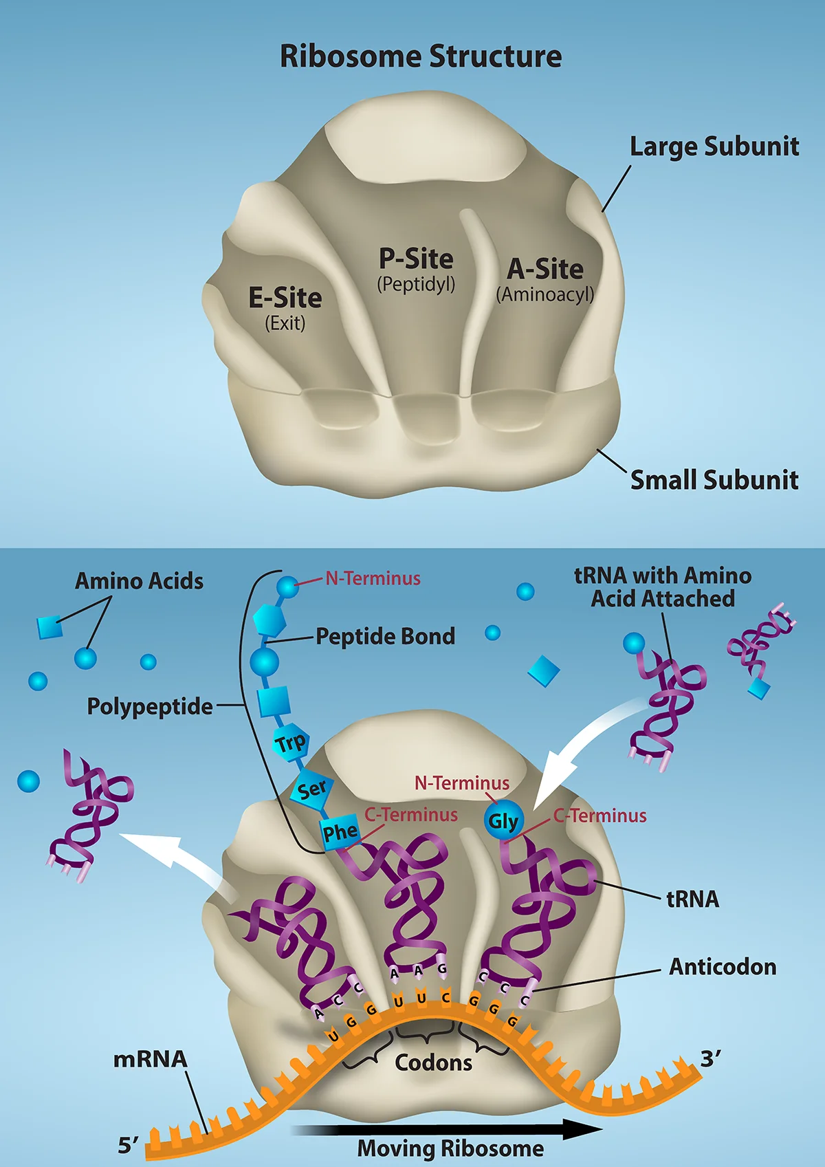
The ribosome: three sites, one movement
Translation
1
Scroll sideways to see the whole map.
The growing polypeptide Charged tRNA arriving Stop codon: no tRNA exists for it GTP spent
The catalyst is RNA, not proteinPeptide bond formation is done by peptidyl transferase, which is part of the large subunit's ribosomal RNA. The ribosome is a ribozyme, and that is a favorite exam fact because it undercuts the assumption that catalysis means protein.
Reading frame is everythingThe code is read three bases at a time with no punctuation, so an insertion or deletion that is not a multiple of three shifts the frame and garbles everything downstream. A substitution changes at most one amino acid; a frameshift changes the whole tail of the protein.
Where the ribosome sitsTranslation starts free in the cytosol. If the emerging chain begins with a signal sequence, the whole ribosome is towed to the rough ER and finishes there, which sends the protein into the secretory pathway. No signal sequence means the protein stays in the cytosol.
One tRNA moves right to left through three sites, and the chain it carries grows by one residue each time. Everything else, the factors, the GTP, the start and stop signals, is arrangement around that single movement.
Initiation
The small ribosomal subunit binds the mRNA near the start codon. In bacteria, the Shine-Dalgarno sequence (AGGAGG, upstream of AUG) base-pairs with 16S rRNA. In eukaryotes, the small subunit binds the 5’ cap and scans along the mRNA until it finds the first AUG in a favorable context (the Kozak sequence, GCCACCAUGG).
The initiator tRNA carrying methionine (or formyl-methionine in bacteria) base-pairs with the start AUG in the P site. The large ribosomal subunit joins, completing the ribosome.
Elongation
Elongation repeats three steps for every codon:
A site entry: an aminoacyl-tRNA with an anticodon matching the current codon binds in the A site (with GTP hydrolysis by EF-Tu / eEF1A).
Peptide bond formation: the peptidyl transferase activity of the large subunit (catalyzed by 23S/28S rRNA - a ribozyme) transfers the growing chain from the P-site tRNA to the amino acid on the A-site tRNA. The growing chain now hangs from the A-site tRNA.
Translocation: the ribosome shifts one codon (with GTP hydrolysis by EF-G / eEF2). The A-site tRNA moves to the P site; the P-site tRNA moves to the E site and leaves. A new codon is exposed in the A site.
The ribosome moves along the mRNA 5’ to 3’, elongating the polypeptide from N-terminus to C-terminus.
Termination
When the A site encounters a stop codon (UAA, UAG, or UGA), a release factor enters the A site instead of a tRNA. Release factors trigger hydrolysis of the bond linking the polypeptide to the P-site tRNA. The finished polypeptide is released. The ribosome dissociates.
Energy Cost
Translation is expensive. Per amino acid added:
1 ATP to charge the tRNA with its amino acid (aminoacyl-tRNA synthetase).
1 GTP during A-site entry (EF-Tu / eEF1A).
1 GTP during translocation (EF-G / eEF2).
Plus initiation and termination costs. Building a 300-residue protein burns about 1200 high-energy phosphate bonds.
What are the three steps of each elongation cycle on the ribosome?
Click to reveal answer
(1) A-site entry: aminoacyl-tRNA matches the codon in the A site. (2) Peptide bond formation: the peptidyl transferase (rRNA catalytic activity) joins the growing chain to the new amino acid. (3) Translocation: the ribosome moves one codon, shifting tRNAs from A → P → E.
Why is the ribosome called a ribozyme?
Click to reveal answer
Because its catalytic activity (peptidyl transferase - forming peptide bonds) is performed by rRNA, not protein. The 23S rRNA in bacteria (28S in eukaryotes) catalyzes peptide bond formation. This makes the ribosome an RNA-based enzyme, supporting the idea that RNA-catalyzed reactions predated protein enzymes.
What happens when the ribosome reaches a stop codon?
Click to reveal answer
A release factor enters the A site (no tRNA anticodon matches a stop codon). The release factor triggers hydrolysis of the bond between the P-site tRNA and the polypeptide, releasing the finished protein. The ribosome then dissociates into large and small subunits, which recycle to initiate new translation.
The ribosome has two subunits. tRNAs carry amino acids. Aminoacyl-tRNA synthetases make sure each tRNA is charged with the correct amino acid. These three components, together with mRNA, do translation.
Ribosome Subunits
Cell type
Small subunit
Large subunit
Assembled
Bacterial
30S
50S
70S
Eukaryotic
40S
60S
80S
“S” is a Svedberg unit - a measure of how fast a particle sediments in a centrifuge. Svedberg units are not additive (hence 30S + 50S = 70S, not 80S - it is shape-dependent).
Each subunit contains rRNA and many ribosomal proteins. Bacterial ribosomes have 16S rRNA (small) and 23S + 5S rRNAs (large). Eukaryotic ribosomes have 18S (small) and 28S + 5.8S + 5S rRNAs (large).
The three tRNA binding sites on the ribosome: A (aminoacyl), P (peptidyl), E (exit). tRNAs move A → P → E as the ribosome translocates. Credit: OpenStax Biology 2e, CC BY 4.0
tRNA Structure
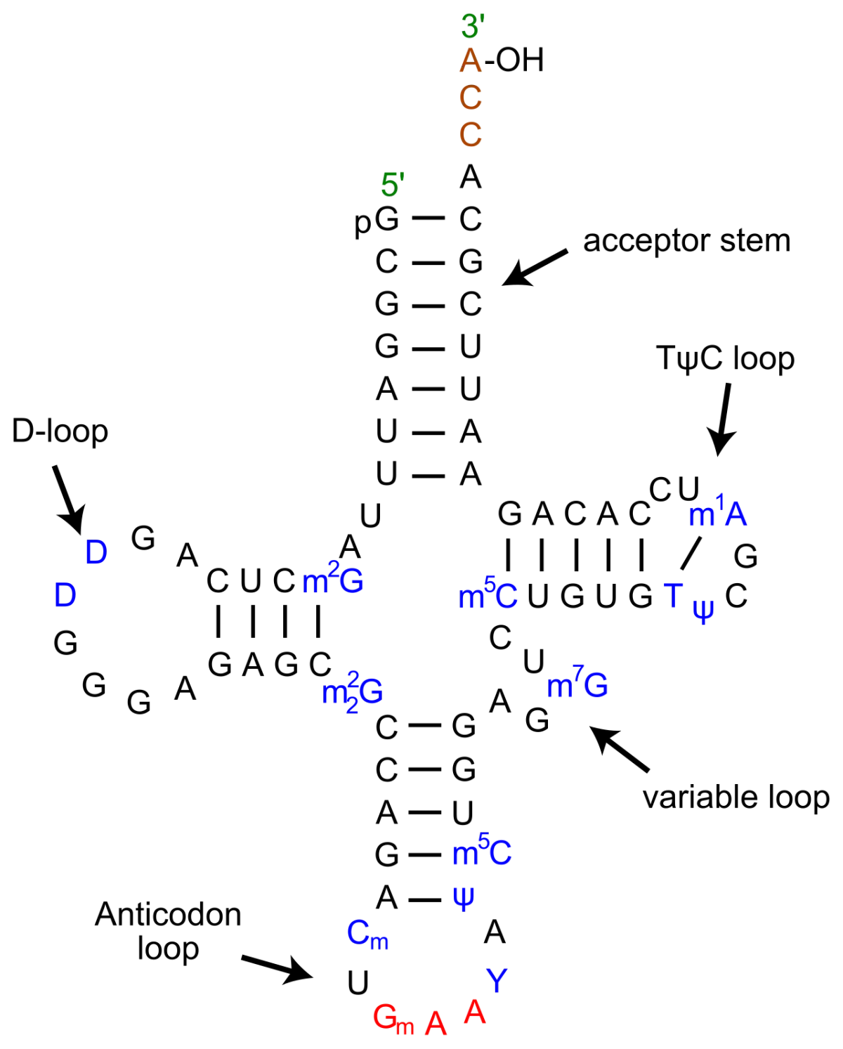
A tRNA is ~75-90 nucleotides long, folded into a cloverleaf in 2D and an L-shape in 3D. Key features:
Acceptor stem: at the 3’ end, always ending in CCA-3’. The amino acid is covalently attached to the 3’-OH of the final A.
Anticodon loop: contains the 3-nucleotide anticodon that base-pairs antiparallel with the mRNA codon.
Other loops: D-loop, T-loop, variable loop - structural and regulatory.
tRNA cloverleaf. The 3' acceptor stem ends in CCA where the amino acid is attached. The anticodon loop reads the mRNA codon. Credit: Wikimedia Commons, CC BY-SA
Aminoacyl-tRNA Synthetase - The “Second Genetic Code”
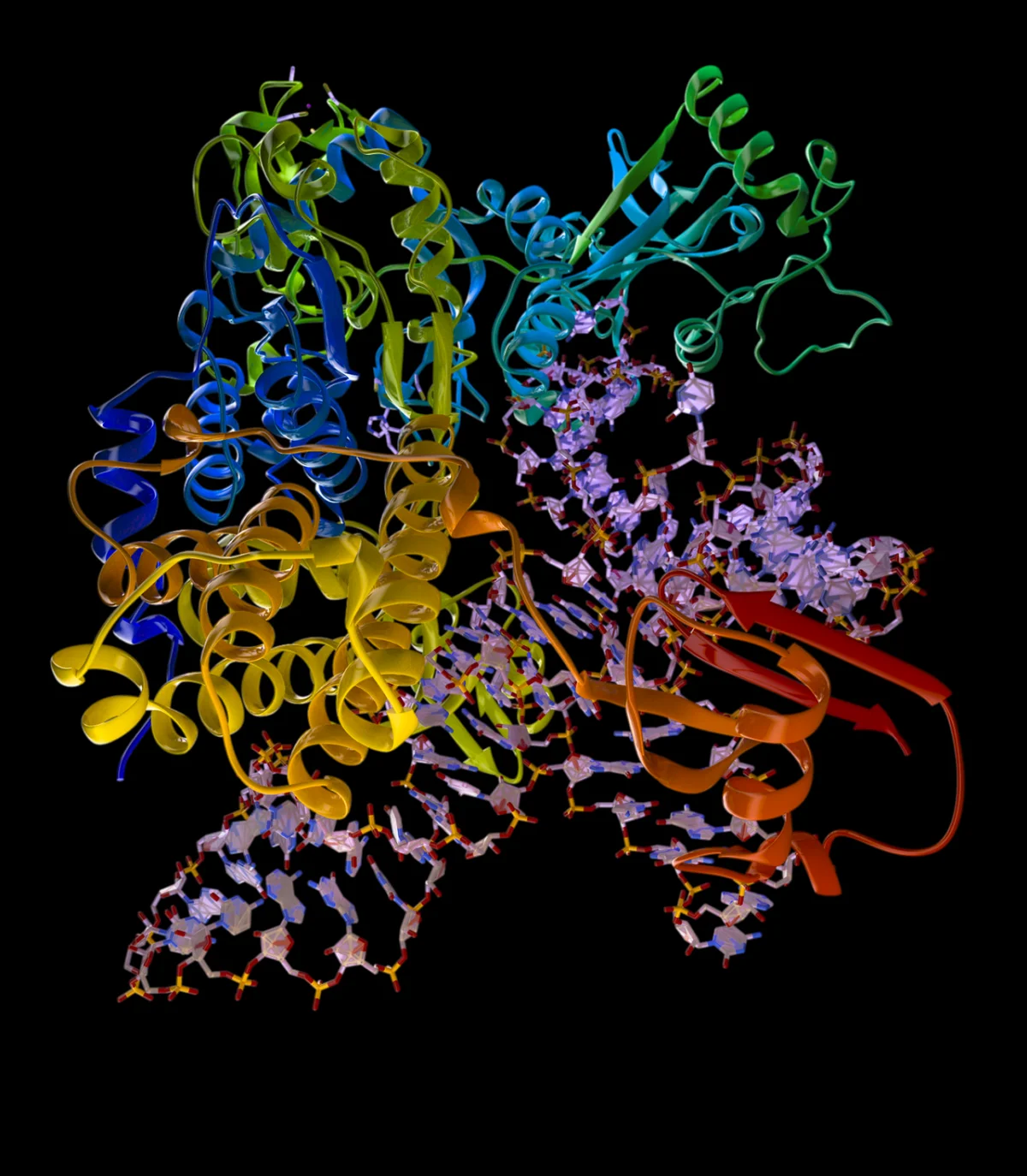
Each amino acid has its own aminoacyl-tRNA synthetase that attaches the correct amino acid to the correct tRNA. There are 20 synthetases, one per amino acid. The synthetase reads the tRNA’s structure and the amino acid’s structure, and uses ATP to catalyze the covalent attachment (aminoacyl-tRNA formation, costing 2 ATP equivalents).
An aminoacyl-tRNA synthetase binding its cognate tRNA. The enzyme discriminates by reading structural features of both the tRNA and the amino acid, then catalyzes the covalent attachment of the amino acid to the 3'-CCA end of the tRNA. Credit: Wikimedia Commons, CC BY-SA
This charging step is the “second genetic code” - if the wrong amino acid is loaded onto a tRNA, the ribosome cannot detect the error and will insert the wrong amino acid. Some synthetases have proofreading activity to catch mischarging.
What is the Svedberg size of bacterial vs. eukaryotic ribosomes?
Click to reveal answer
Bacterial: 30S + 50S = 70S. Eukaryotic: 40S + 60S = 80S. Svedberg units are not additive (they depend on shape and mass). Bacterial mRNA typically uses the Shine-Dalgarno sequence; eukaryotic mRNA uses the Kozak sequence and cap-dependent scanning.
How many aminoacyl-tRNA synthetases does a cell need, and why?
Click to reveal answer
Twenty - one per amino acid. Each synthetase selects its amino acid and the correct tRNA, then covalently attaches the amino acid to the tRNA's 3'-CCA acceptor end. Because the ribosome cannot double-check the amino acid once attached, synthetase accuracy is critical - some have proofreading activity to reject wrong amino acids.
Where does the peptidyl transferase activity of the ribosome reside?
Click to reveal answer
In the large subunit's rRNA - specifically the 23S rRNA in bacteria (or 28S rRNA in eukaryotes). Peptide bond formation is catalyzed by RNA, not by ribosomal protein. This makes the ribosome a ribozyme (RNA-based enzyme) and supports the ancient RNA world hypothesis.
After a protein is made, most proteins are further modified. Post-translational modifications (PTMs) expand the proteome’s functional diversity and regulate protein activity, location, and lifespan.
The Main Modifications
Modification
Chemistry
Purpose
Phosphorylation
Phosphate on Ser/Thr/Tyr
On/off switch for activity; fast, reversible
Glycosylation
Sugar chain on Asn (N-linked) or Ser/Thr (O-linked)
Activates zymogens, cleaves signal peptides, matures hormones
Signal Peptide and Secretion
Proteins destined for membranes, lysosomes, or secretion carry an N-terminal signal peptide. Signal recognition particle (SRP) recognizes the emerging peptide on the ribosome and docks it to the ER. The ribosome finishes translation while the nascent protein threads into the ER lumen. The signal peptide is then cleaved by signal peptidase.
Zymogen Activation (Review)
Chapter 2 introduced zymogens - inactive precursors activated by cleavage. Digestive proteases, clotting factors, and caspases are all activated this way. Irreversible cleavage is a committed “on” signal that cannot easily be undone.
What does polyubiquitination signal for a target protein?
Click to reveal answer
Degradation by the 26S proteasome. Short or single ubiquitin tags can alter protein location or activity, but long polyubiquitin chains (especially K48-linked) are recognized by the proteasome, which unfolds and cleaves the tagged protein into small peptides. Polyubiquitination is the cell's primary signal for regulated protein destruction.
How does a signal peptide direct a protein to the ER?
Click to reveal answer
The signal peptide is an N-terminal sequence of about 15-30 amino acids, often rich in hydrophobic residues. As it emerges from the ribosome, the signal recognition particle (SRP) binds it and pauses translation. The SRP-ribosome complex docks at the ER's SRP receptor; translation resumes as the peptide threads into the ER lumen. Signal peptidase then cleaves the signal peptide.
Why does histone acetylation activate transcription?
Click to reveal answer
Histones are positively charged (rich in lysine and arginine) and bind tightly to negatively charged DNA. Acetylating a lysine neutralizes its positive charge, weakening the histone-DNA interaction. The chromatin relaxes, making DNA more accessible to RNA polymerase and transcription factors. Histone deacetylases (HDACs) reverse this.
Bacteria often organize several related genes as an operon - a cluster under a single promoter, producing a polycistronic mRNA. Operons let bacteria switch multiple genes on or off at once in response to environmental signals. The lac and trp operons are the textbook examples.
Two operons, opposite defaults
Gene regulation
1
Scroll sideways to see the whole map.
Inducible: off by default, substrate switches it on Repressible: on by default, product switches it off Operator: where control happens Positive control (CAP and cAMP)
The rule that generates bothCatabolic pathways are inducible and anabolic pathways are repressible. A pathway that breaks something down should only run when that something is present. A pathway that builds something should run until there is enough. Both operons follow from that one sentence.
Why glucose beats lactoseThe lac operon has a second, positive control. CAP only binds when cAMP is high, and cAMP is only high when glucose is low. So the cell eats glucose first and only turns to lactose when glucose runs out, which is exactly what diauxic growth curves show.
Eukaryotes do it differentlyNo operons, because each gene has its own promoter. Control is spread across chromatin state (histone acetylation opens it, methylation usually closes it), distant enhancers looped in by transcription factors, alternative splicing, and microRNAs acting after the message is made.
Both operons use a repressor and an operator; only the default differs. Ask what the pathway is for. If it breaks something down, the substrate turns it on. If it builds something, the product turns it off.
The Lac Operon
Controls three genes needed to metabolize lactose:
lacZ: beta-galactosidase (splits lactose into glucose + galactose).
lacY: lactose permease (imports lactose into the cell).
lacA: transacetylase (role less clear).
Regulation logic:
Negative regulation: The lac repressor (product of lacI gene) binds the operator and blocks transcription when lactose is absent. When lactose is present, allolactose (a lactose derivative) binds the repressor, causing it to release the operator - transcription turns ON. Lactose is the inducer.
Positive regulation: When glucose is scarce, cAMP levels rise. cAMP binds the catabolite activator protein (CAP / CRP), which binds upstream of the promoter and enhances transcription. When glucose is abundant, cAMP is low, CAP is inactive, and even with lactose, the lac operon expresses only weakly. This is catabolite repression - the cell prefers glucose, and only turns to lactose when glucose is gone.
The Trp Operon
Controls five genes needed to synthesize tryptophan from chorismate. When tryptophan is plentiful, the cell does not need to make more, so the operon should be OFF.
Regulation logic:
Repression: The trp repressor is inactive on its own. When tryptophan is abundant, Trp binds the repressor as a corepressor, activating it. The active repressor binds the operator and shuts off transcription. When Trp is scarce, the repressor is inactive, and the operon expresses.
Attenuation: An additional fine-tuning mechanism using the leader region of the trp mRNA. If Trp is abundant, ribosomes translating a leader peptide coast smoothly over trp codons, and the mRNA forms a structure that terminates transcription early. If Trp is scarce, ribosomes stall at trp codons, a different mRNA structure forms, and transcription continues. The MCAT does not typically require deep attenuation knowledge, but recognizing that the trp operon uses attenuation is a useful concept.
Induction vs. Repression
Inducible operon: normally OFF, turned ON by a substrate (inducer). Lac is the classic example. The substrate is scarce most of the time, and the cell only expresses the enzymes when the substrate appears.
Repressible operon: normally ON, turned OFF by the pathway’s product (corepressor). Trp is the classic example. The cell makes tryptophan constantly unless there is already plenty, in which case it shuts down synthesis.
How does the lac operon behave when both glucose and lactose are present in the medium?
Click to reveal answer
Lactose inactivates the repressor, so the operator is unbound. But glucose keeps cAMP levels low, so CAP does not activate the promoter strongly. The operon expresses only weakly. The cell uses the available glucose first; once glucose is gone, cAMP rises, CAP activates, and lac expression turns fully on. This is catabolite repression - the diauxic growth pattern.
Why is the trp operon called “repressible” rather than “inducible”?
Click to reveal answer
A repressible operon is ON by default and is turned OFF when a signal is present. The trp operon defaults to ON (because the cell usually needs to make Trp). When Trp accumulates, it binds the trp repressor as a corepressor, activating the repressor and turning the operon OFF. “Repressed by its end product” is the signature of a biosynthetic operon.
What would a lacI- mutation cause phenotypically?
Click to reveal answer
Constitutive expression of lacZ, lacY, lacA. lacI encodes the lac repressor. If repressor cannot be made or cannot bind the operator, the operon expresses regardless of whether lactose is present. The cell wastes energy producing enzymes it may not need. The same phenotype arises from an operator mutation that prevents repressor binding.
Eukaryotic gene regulation happens at multiple levels. A gene can be silenced via chromatin state, blocked from transcription by repressors, subjected to alternative splicing, regulated at translation, or tagged for rapid degradation after being made. Cells use all of these simultaneously.
The Five Levels
Chromatin (epigenetic): DNA methylation, histone modifications, chromatin remodeling. Determines whether a gene can be accessed at all.
Post-translational: phosphorylation, ubiquitination, etc.
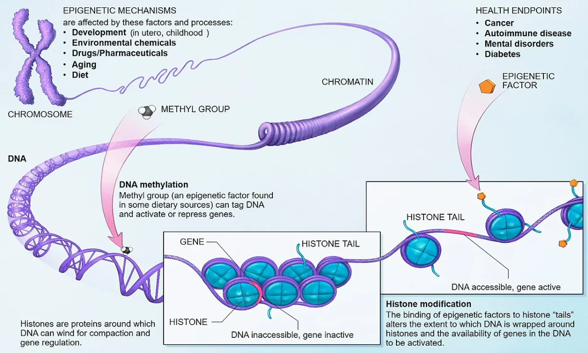
Chromatin and Epigenetics
Epigenetic control: DNA methylation, histone tail modifications (acetylation, methylation, phosphorylation), and nucleosome positioning together determine which genes are accessible. Credit: Wikimedia Commons, public domain
DNA methylation: methyl groups added to cytosine in CpG dinucleotides. Heavy methylation at a gene’s promoter typically silences the gene. CpG island hypermethylation is a common mechanism for silencing tumor suppressor genes in cancer.
Histone modifications: acetylation (generally activates), methylation (can activate or repress depending on site), phosphorylation, and ubiquitination. Histone tails stick out and are the substrates.
Chromatin remodeling: ATP-dependent complexes slide, eject, or restructure nucleosomes, exposing or hiding DNA.
Transcription Factors, Enhancers, and Silencers
Enhancers are DNA elements that can activate transcription from thousands of base pairs away. They loop through space to contact the promoter. Activator transcription factors bind enhancers and recruit coactivators (including chromatin remodelers and general transcription machinery).
Silencers are the opposite - they bind repressor transcription factors and reduce gene expression, sometimes through recruitment of histone deacetylases or DNA methyltransferases.
Other Regulation
Alternative splicing: same gene, different mRNAs, different proteins (Section 7.5).
mRNA stability: miRNAs, AU-rich elements, and deadenylation control how long an mRNA lasts in the cytoplasm.
Translation initiation: eIF2 phosphorylation halts global translation during stress (integrated stress response).
Protein degradation: ubiquitin-proteasome system marks specific proteins for destruction.
X-Inactivation
In female mammals, one of the two X chromosomes is silenced in each cell early in development. The silenced X forms a Barr body and is heavily methylated and histone-modified. This dosage compensation ensures females and males produce similar amounts of X-linked gene products. The choice of which X is inactivated is random and heritable within a cell lineage.
What are the five main levels at which eukaryotic gene expression can be regulated?
How does DNA methylation typically affect gene expression?
Click to reveal answer
Methylation of cytosines in CpG dinucleotides at a gene's promoter region usually silences transcription. Methyl-CpG-binding proteins recruit histone deacetylases and other repressive factors, producing compact chromatin. Aberrant hypermethylation of tumor suppressor promoters is a common mechanism of gene silencing in cancer.
What is a Barr body, and what is its biological significance?
Click to reveal answer
A Barr body is an inactivated X chromosome in the nuclei of female mammal cells. One of the two X chromosomes in each cell is randomly silenced early in development - dosage compensation. The silenced X is heavily methylated and histone-modified, appearing as a dense structure under a microscope. This is a classic example of epigenetic regulation and gives rise to mosaic phenotypes in X-linked conditions.