
Introduction
Spectroscopy measures how matter interacts with electromagnetic radiation. Different wavelengths excite different molecular motions: radio waves flip nuclear spins (NMR), infrared drives vibrations (IR), visible/UV promotes electrons to excited states (UV-Vis). Mass spectrometry is not technically a spectroscopy but is grouped with it as a structure-determination technique.
The Electromagnetic Spectrum and What It Tells Us
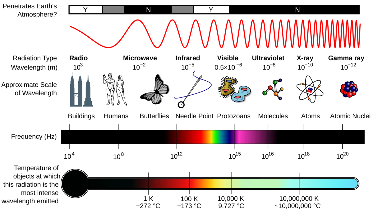
| Radiation | Energy | Excites | Technique |
|---|---|---|---|
| Radio (~100 MHz) | Very low | Nuclear spin flips | NMR |
| Microwave (~10 GHz) | Low | Rotational transitions | Microwave spectroscopy |
| Infrared (~10¹³ Hz) | Medium | Molecular vibrations | IR |
| Visible (~10¹⁴ Hz) | Medium-high | Electronic transitions | Vis absorption |
| UV (~10¹⁵ Hz) | High | Electronic transitions | UV absorption |
| X-ray (~10¹⁸ Hz) | Very high | Inner-shell electrons | X-ray diffraction |
Higher-frequency light has higher photon energy: E = hν = hc/λ. A photon matching the energy gap between two molecular states can be absorbed, exciting the molecule from the lower state to the higher state.
What Each Technique Tells You
IR spectroscopy: which functional groups are present. Each bond type (C=O, O-H, N-H, C≡N, etc.) vibrates at a characteristic frequency. A peak at the right frequency = that bond is there.
UV-Vis spectroscopy: degree of conjugation (for organic molecules); also concentration (via Beer-Lambert). Double bonds, aromatic rings, and conjugated systems absorb UV light. Free transition metal complexes absorb visible light.
NMR: complete hydrogen (and carbon) framework. Chemical shift locates each hydrogen relative to adjacent functional groups. Splitting counts neighboring hydrogens. Integration counts each type of hydrogen.
Mass spectrometry: molecular weight + fragmentation pattern. The molecular ion (M⁺) gives MW. Fragment ions reveal which pieces of the molecule have been lost, giving clues about structure.
The Power of Combining Techniques
No single technique is sufficient for complete structure determination. Each answers a different question:
- MS: “How heavy is it?” (molecular formula)
- IR: “What groups are there?” (functional group inventory)
- NMR: “How is it connected?” (hydrogen framework)
Used together, you can solve most organic structures in minutes. Section 11.11 covers the systematic strategy.
Instrumentation Basics
All spectroscopy instruments share a similar layout:
- Source: produces radiation (tungsten lamp for Vis, deuterium for UV, globar/Nernst for IR, electromagnet for NMR, electron beam for MS).
- Sample chamber: holds the sample in the path of the radiation.
- Detector: measures the radiation that comes through (absorbance) or emerges from the sample (emission).
- Computer/readout: digitizes the signal and produces the spectrum.
You do not need to memorize instrument details for the MCAT; focus on what the data mean.