You are trying to hear a phone vibrate across the room while your roommate runs the vacuum. Sometimes you catch it. Sometimes you swear you heard it and check your phone to find nothing there. Sometimes you miss a real call. This is not a trivial, everyday annoyance. This is the textbook setup for everything in signal detection theory, and the MCAT loves it.
Before we get to detection, we need to define what “sensing something” even means.
Sensation vs. Perception
Sensation is the physical conversion of a stimulus into a neural signal. A rod in your retina flips a molecule, fires a neuron. That is sensation.
Perception is what your brain makes of that signal. Your brain decides the neural blip means “my phone is vibrating.” That is perception.
The physical step of converting stimulus energy into neural energy is called transduction. Rods and cones transduce light. Hair cells transduce sound. Free nerve endings transduce pain. Every sense has its own transducers.
Absolute Threshold of Sensation
Your phone vibrates so gently you catch it only half the time. That crossover point - the intensity at which you detect a stimulus 50% of the time - is your absolute threshold of sensation. Below the threshold, you would correctly detect the stimulus less than half the time (it is below the line you called “detectable”). Above the threshold, you would detect it more than half the time.
Stimuli below absolute threshold are called subliminal. They still reach your sensory organs, but you do not consciously detect them more than half the time.
Absolute threshold is not a hard, fixed number. It slides around based on:
Expectations. If you are waiting for a text, your threshold drops.
Experience. A new parent can hear a baby’s whimper through two closed doors.
Motivation. If you’re waiting to hear from someone important, you’re more sensitive to notifications.
Alertness. A drowsy person misses signals a well-rested person would catch.
Just Noticeable Difference and Weber’s Law
Absolute threshold asks, “Can you hear the phone at all?” The just-noticeable difference (JND) asks, “Can you hear the phone getting louder?” JND is the smallest change in a stimulus that a person can detect 50% of the time.
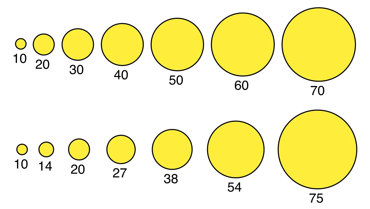
Here is the trick that the MCAT wants you to internalize: the JND is not a fixed amount, it is a fixed proportion. A 2 lb weight next to a 2.2 lb weight feels different - a 10% change. A 50 lb weight next to a 50.2 lb weight feels identical, because 0.2 is a tiny fraction of 50. To feel a change against 50 lb, you would need to add about 5 lb.
That proportionality is Weber’s Law:
IΔI=k
where ΔI is the JND, I is the starting intensity, and k is a constant (the “Weber fraction”) specific to the sense. For weight, k is roughly 0.02 (a 2% change). For brightness, k is closer to 0.08.
Weber's law in pictures. Top row: equal absolute increments produce shrinking perceived steps (the gap between 60 and 70 looks tiny). Bottom row: equal proportional increments feel like equal perceptual steps, because the JND scales with the baseline intensity. Credit: MrPomidor via Wikimedia Commons (CC BY-SA 4.0).
Signal Detection Theory
Now to your vacuum-vs-vibrating-phone problem. Signal detection theory (SDT) models how we decide whether a stimulus is present when conditions are noisy. Your brain is constantly trying to distinguish signal (the phone) from noise (the vacuum, the dog, the wind).
There are four possible outcomes on any trial:
Signal Present
Signal Absent
You say “yes”
Hit
False Alarm
You say “no”
Miss
Correct Rejection
Hit. You say yes, the phone really was buzzing.
Miss. You say no, but the phone actually was buzzing. You missed the call.
False alarm. You say yes, but the phone never buzzed. You check and feel silly.
Correct rejection. You say no, and nothing was there.
d’ (Sensitivity) and c (Bias)
Signal detection splits performance into two independent pieces:
d’ (d-prime) measures sensitivity. How far apart are the “signal” and “noise” distributions in the brain? A bigger d’ means the signal stands out clearly from noise. A small d’ means they overlap and you cannot reliably tell them apart no matter how carefully you try.
c measures response bias (strategy). At what intensity do you decide to say “yes”? This is a personality trait of a detector, not a feature of the signal.
Conservative strategy. “I’ll only say yes when I’m 100% sure.” Many misses, few false alarms.
Liberal strategy. “I’ll say yes if I even suspect it.” Many hits, but also many false alarms.
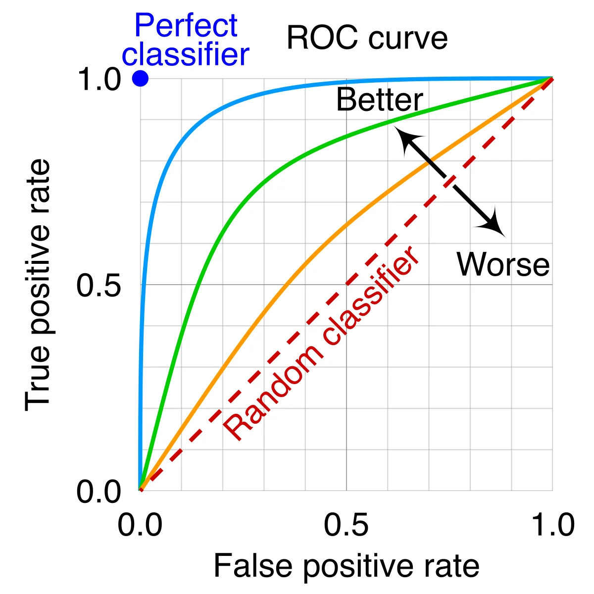
The ROC curve plots hit rate (true positives) against false-alarm rate (false positives) as the response criterion shifts from very conservative (bottom-left) to very liberal (top-right). The further the curve bows toward the upper-left corner, the higher the underlying sensitivity (d'). The diagonal is chance performance. Credit: cmglee & MartinThoma via Wikimedia Commons (CC BY-SA 4.0).
What is the absolute threshold of sensation?
Click to reveal answer
The minimum stimulus intensity needed to detect a stimulus 50% of the time. Below threshold = subliminal.
If you can just barely feel a 1 oz change on a 10 oz weight, how many ounces must you add to a 100 oz weight to feel a change? (Weber's Law)
Click to reveal answer
10 oz. Weber's Law says ΔI/I = constant. 101 = 0.10, so ΔI on a 100 oz weight is 0.10 × 100 = 10 oz.
In signal detection theory, what are the four outcomes?
Click to reveal answer
Hit (signal present, said yes), miss (signal present, said no), false alarm (signal absent, said yes), correct rejection (signal absent, said no).
What does a liberal response strategy in signal detection look like in terms of hits and false alarms?
Click to reveal answer
High hit rate AND high false alarm rate. A liberal subject says "yes" readily, catching real signals but also imagining phantom ones.
Your phone camera has a lens that focuses light, a sensor that converts light into voltage, and a processor that interprets those voltages as an image. Your eye is the same architecture, assembled by biology instead of engineers. Understand the camera and the eye follows.
The Anatomy: Front to Back
Light takes a short journey from the outside world to your retina. Memorize the pathway in this order and you have the structure locked down.
Cornea. The clear, thick dome at the front of the eye. Does about two thirds of all light focusing. Covered by a thin mucus membrane called the conjunctiva.
Aqueous humor. Watery fluid in the anterior chamber (in front of the lens). Nourishes the cornea and lens, which have no blood vessels of their own.
Pupil. The hole in the middle of the iris. Dilates in the dark (let more light in), constricts in the sun (let less light in).
Iris. The colored ring. A muscle that changes the size of the pupil.
Lens. Fine-tunes the focus. The ciliary muscles attached to the lens via suspensory ligaments change the lens’s shape (accommodation) to focus on near vs. far objects.
Vitreous humor. A jelly that fills most of the eyeball and keeps it inflated.
Retina. The back wall of the eye, packed with photoreceptors (rods and cones). This is where transduction happens.
Macula. A central patch of retina specialized for high-detail vision.
Fovea. The very center of the macula. Packed with cones, no rods. This is where you see the word you are reading right now.
Optic nerve. Bundles the output of all retinal ganglion cells and ships it to the brain.
Between the retina and the tough outer sclera (the white of your eye) is the choroid, a layer of blood vessels that feeds the retina and absorbs stray light so it does not bounce around.
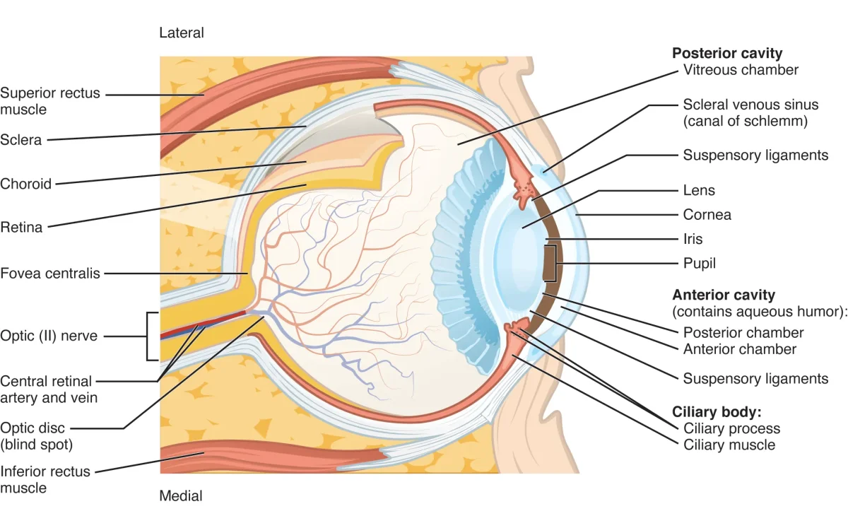
Anatomy of the eye, front to back. Light passes through cornea → aqueous humor → pupil → lens → vitreous humor → retina, where rods and cones transduce the signal. Credit: OpenStax Anatomy & Physiology 2e, CC BY 4.0.
Rods and Cones: Two Sensors in One Retina
Your retina has two kinds of photoreceptors, and they have almost opposite jobs.
Feature
Rods
Cones
How many?
~120 million
~6 million
Location
Periphery
Fovea / macula
Sensitivity
~1000× more sensitive
Less sensitive
Color?
No (black and white only)
Yes (R, G, B)
Detail?
Low resolution
High resolution
Recovery
Slow
Fast
Best for
Night vision, motion
Daylight, color, detail
This is why you cannot see color clearly in the dark: cones need bright light, and at night only your rods are firing. It is also why motion catches your eye in peripheral vision: rods dominate the periphery and are wired for rapid, transient changes. To actually read something, you have to point your fovea at it - one reason reading a wall clock is easy but reading peripheral text is not.
The three cone types are not evenly distributed. Roughly 60% are red-sensitive, 30% green-sensitive, and 10% blue-sensitive. This uneven mix is why we can resolve red and green subtleties better than blue ones, and it becomes load-bearing in the color-vision theories covered in the next section.
Phototransduction: How a Photon Becomes a Nerve Signal
This is the single highest-yield mechanism in the whole sensory chapter. Get it right and you pick up multiple MCAT points.
The big counterintuitive twist: in the dark, photoreceptors are depolarized and constantly releasing neurotransmitter. Light turns them OFF. Your retina reports light by going silent. Every other sense reports a stimulus by firing more; photoreceptors are the exception.
Here is the cascade, in the dark and in the light.
In the Dark (Baseline, Depolarized)
cGMP (cyclic GMP) is abundant inside the rod.
cGMP binds sodium channels and keeps them open.
Na⁺ flows in. The rod stays depolarized.
The rod continuously releases glutamate onto bipolar cells.
When Light Hits a Rod
A photon strikes rhodopsin (the photopigment in rods; cones use photopsin). Rhodopsin is a G-protein-coupled receptor with a small molecule called 11-cis retinal tucked inside.
Light converts 11-cis retinal into all-trans retinal (the shape changes). This activates rhodopsin.
Activated rhodopsin triggers transducin, a G-protein. The alpha subunit of transducin splits off.
The alpha subunit activates phosphodiesterase (PDE).
PDE converts cGMP → GMP. cGMP levels plummet.
With cGMP gone, the Na⁺ channels close. Less Na⁺ enters.
The rod hyperpolarizes and stops releasing glutamate.
Downstream bipolar cells sense the drop in glutamate. ON-center bipolar cells (which are normally inhibited by glutamate) become more active.
Active ON-center bipolar cells excite retinal ganglion cells, which fire action potentials down the optic nerve. The brain now knows a photon arrived.
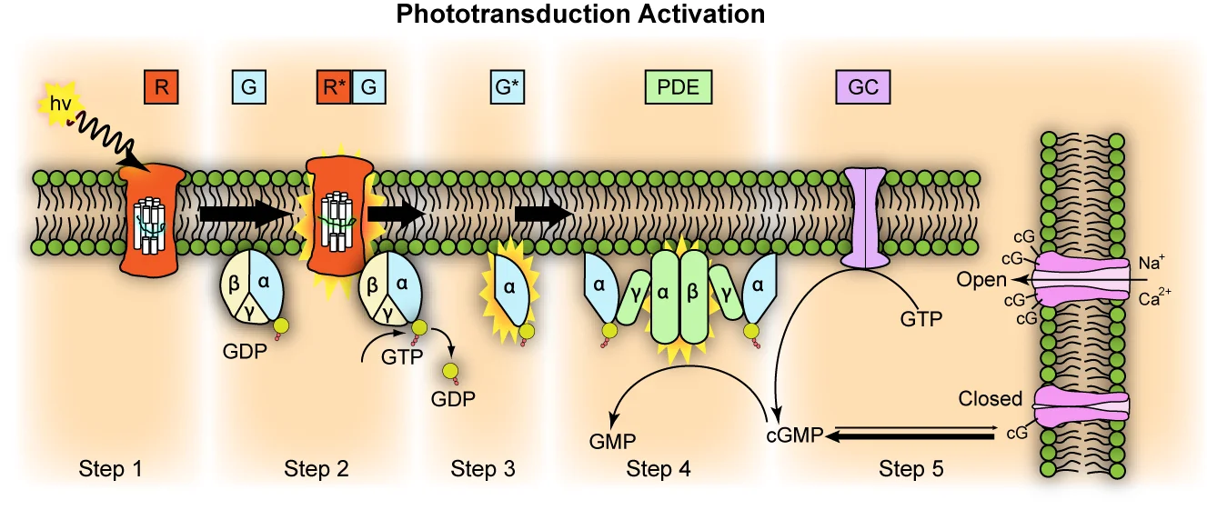
The phototransduction cascade. Light activates rhodopsin → transducin → PDE → cGMP breakdown → Na⁺ channels close → hyperpolarization. One activated rhodopsin amplifies the signal across ~800 transducin molecules. Credit: Jason J. Corneveaux via Wikimedia Commons, CC BY 3.0.
Light Adaptation: Dark Room, Bright Sunlight
When you walk from a sunny sidewalk into a dark theater, it takes minutes to see. That delay is the cascade running in reverse: rhodopsin was mostly bleached (11-cis retinal all used up) in bright light, and your rods need time to regenerate it. Once rhodopsin is restocked, rods become sensitive again.
Going the other way - dark room to bright hallway - is fast because cones adjust quickly (high-activity pathways recover faster), and because pupil constriction happens in seconds. This is part of why rods take longer to adapt than cones: rods have to resynthesize more photopigment before they can fire again.
What structure does most of the eye's light refraction, the cornea or the lens?
Click to reveal answer
The cornea does roughly two-thirds of the bending. The lens fine-tunes focus via accommodation (changing shape through ciliary muscle action) so we can see near and far.
What is the fovea, and what photoreceptors live there?
Click to reveal answer
The fovea is the central pit of the macula. It contains ONLY cones (no rods). It is where high-acuity, color vision is sharpest - you point it at whatever you want to see clearly.
Why are rods better than cones for night vision?
Click to reveal answer
Rods are about 1000× more light-sensitive than cones. Cones need bright light to fire; rods can respond to a handful of photons. Rods are also far more numerous (120M vs. 6M).
In phototransduction, what happens to cGMP when light hits a rod?
Click to reveal answer
cGMP levels DROP. Rhodopsin activates transducin, which activates phosphodiesterase (PDE), which converts cGMP to GMP. With less cGMP, Na⁺ channels close and the rod hyperpolarizes.
Which cell in the visual pathway fires the first action potential?
Click to reveal answer
The retinal ganglion cell. Rods, cones, and bipolar cells use graded potentials. Action potentials begin at the ganglion cells, whose axons form the optic nerve.
The raw photon-to-neuron step happens in the retina. From there, the signal takes a highly organized trip to your visual cortex, and different features of the image get routed down different wires. This section is about what happens after the retinal ganglion cells fire.
The Visual Field Crosses Over
Your visual field splits into a left half and a right half. Here is the rule to memorize: the right half of the visual field is processed by the left brain, and the left half by the right brain. It is the same crossover you see with motor and somatosensory cortex.
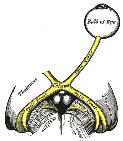
How does the crossover actually happen? By the optic chiasm.
Each retina has a nasal side (closer to the nose) and a temporal side (closer to the temple).
Light from the right visual field lands on the nasal side of the right eye and the temporal side of the left eye.
At the optic chiasm, the axons from the nasal retinas cross to the opposite hemisphere; axons from the temporal retinas stay on the same side.
The net effect: all signals carrying the right visual field end up in the left occipital lobe, and vice versa.
The optic chiasm. Fibers from the nasal half of each retina cross to the opposite hemisphere; temporal fibers stay on the same side. A tumor pressing on the chiasm from below knocks out only the crossing fibers and produces bitemporal hemianopia. Credit: Henry Vandyke Carter, Gray's Anatomy (Public Domain).
Feature Detection: Color, Form, Motion
After the optic chiasm, signals travel through the lateral geniculate nucleus (LGN) and up to the primary visual cortex in the occipital lobe. Along the way, different features of what you are seeing ride on different “wires”:
The parvocellular pathway handles color and fine spatial detail (form). It has good spatial resolution but poor temporal resolution - it cannot track fast movement, only stationary detail. Driven mostly by cones.
The magnocellular pathway handles motion and temporal detail. It is color-blind and low-resolution but excellent at tracking things that move. Driven mostly by rods and the larger ganglion cells.
Parallel processing means your brain handles color, form, and motion simultaneously on different tracks, then stitches them back together upstream. You do not see color first, then motion. You see them at once.
The Two Theories of Color Vision
The MCAT will absolutely ask you which theory explains which phenomenon. They are not competing - modern neuroscience says both are right at different stages.
Trichromatic Theory (Young-Helmholtz)
Three cone types exist: red-sensitive, green-sensitive, and blue-sensitive. Any perceived color is a mixture of the activations of these three. Purple? Red + blue cones firing. Orange? Red + green. White? All three.
This explains how a TV screen with only red, green, and blue subpixels can produce any color you see on it. Trichromatic theory lives at the level of the retina (the cones themselves).
Red-green color blindness - the most common form - usually comes from a missing or defective red or green cone. It is X-linked, which is why it hits men more often. Those individuals still distinguish blue from red or green; they just cannot tell red from green.
Opponent Process Theory (Hering)
Three opposing pairs of colors are processed together: red vs. green, blue vs. yellow, and black vs. white. In any pair, one color inhibits the other. You can have reddish-blue (magenta/purple) or yellowish-red (orange), but you can never have reddish-green or bluish-yellow. The pairs are mutually exclusive.
This is why after staring at a green square for 30 seconds and then looking at a white wall, you see a red afterimage. The green channel became fatigued; its opposing channel (red) rebounds. Opponent process theory explains afterimages, simultaneous color contrast, and why certain color combinations look “impossible.” It lives in the retinal ganglion cells and beyond - downstream of the cones.
Bonus: Color Constancy
Take a white sheet of paper outside at noon (blue-sky lighting) and then inside under a warm incandescent bulb (orange-ish lighting). The light hitting your retina is wildly different, but you still perceive the paper as white. That is color constancy - your brain accounts for the ambient lighting and recovers the object’s “true” color. Your phone camera does this too, clumsily; it is called “white balance.” Your visual system does it automatically and often without your noticing. The only time it fails conspicuously is in weird lighting like sodium-vapor streetlights, which can make red cars look black.
Which side of the brain processes the right visual field?
Click to reveal answer
The left brain. Nasal-retinal fibers (which carry the right visual field from each eye after the flip through the pupil) cross at the optic chiasm to the left occipital lobe.
Which visual pathway detects motion?
Click to reveal answer
The magnocellular pathway. High temporal resolution, low spatial resolution, colorblind. Parvocellular handles form and color.
Which color-vision theory explains afterimages?
Click to reveal answer
Opponent process theory. Staring at green fatigues the green-red channel, so when you look at white, the "red" side of that channel rebounds and you see a red afterimage. Trichromatic theory cannot explain this.
What does parallel processing mean in vision?
Click to reveal answer
Different features of a visual scene (color, form, motion, depth) are processed simultaneously on separate neural pathways, then recombined higher up. You don't see one feature first - you see them at once.
A bass drum thumps at 60 Hz. A mosquito whines at 15,000 Hz. Your ear turns both into electrical signals the same brain can read, using nothing but tiny bones, a snail-shaped tube of saltwater, and a row of microscopic hair-cell “sensors.” This section walks you through the conversion, piece by piece, and then unpacks the two theories the MCAT loves to test.
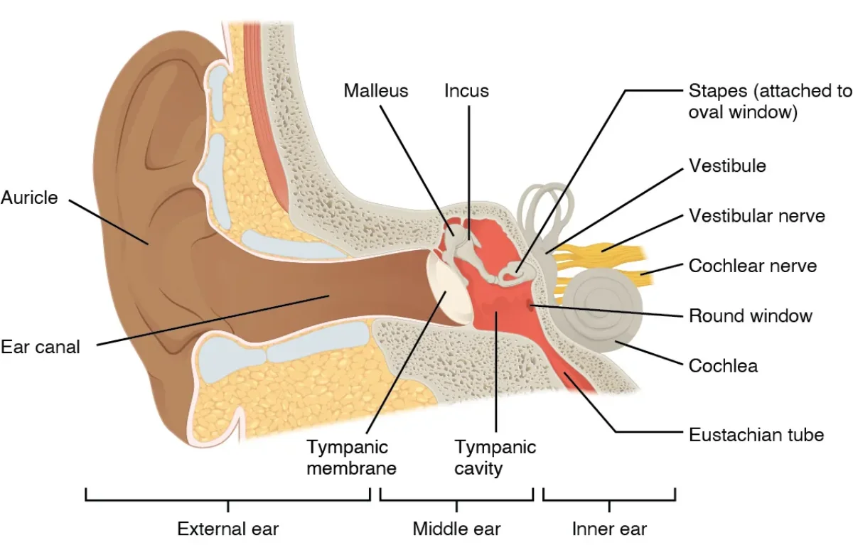
The Sound Pathway, Outside to Inside
Sound begins as pressurized air. When a speaker cone pushes forward, it shoves air molecules into a zone of high pressure; when it pulls back, air thins into low pressure. Those alternating pressure waves are what your ear is trying to detect.
The sound path:
Pinna. The floppy outer ear. Its curves collect air pressure waves and funnel them inward, like a satellite dish.
Auditory canal (external auditory meatus). The tube from the pinna toward your skull. Narrows the wave, concentrating it.
Tympanic membrane (eardrum). A thin, tense sheet at the end of the auditory canal. Pressure waves make it vibrate.
Ossicles. Three tiny bones in the middle ear - the smallest bones in your body - that amplify the vibration:
Malleus (hammer) is attached to the eardrum.
Incus (anvil) connects malleus to stapes.
Stapes (stirrup) presses on the oval window.
Mnemonic: M-I-S.
Oval window. A membrane on the cochlea that transmits the ossicle vibration into the cochlear fluid.
Cochlea. A snail-shaped, fluid-filled tube. The waves travel through the cochlea, bend hair cells, and then exit via the round window, which flexes outward to relieve pressure.
Auditory nerve. The axons from hair-cell synapses carry action potentials to the brain.
The three regions of the ear. Sound travels pinna → canal → tympanic membrane → ossicles (M-I-S) → oval window → cochlea. Semicircular canals branching from the inner ear handle balance. Credit: OpenStax Anatomy & Physiology 2e, CC BY 4.0.
The Three Regions of the Ear
Grouping the parts makes the anatomy easier to retain:
Outer ear: pinna through tympanic membrane.
Middle ear: ossicles (malleus, incus, stapes), sitting in an air-filled cavity.
Inner ear: cochlea and semicircular canals (the semicircular canals handle balance, not hearing - we will see them in the next section).
Inside the Cochlea: the Organ of Corti
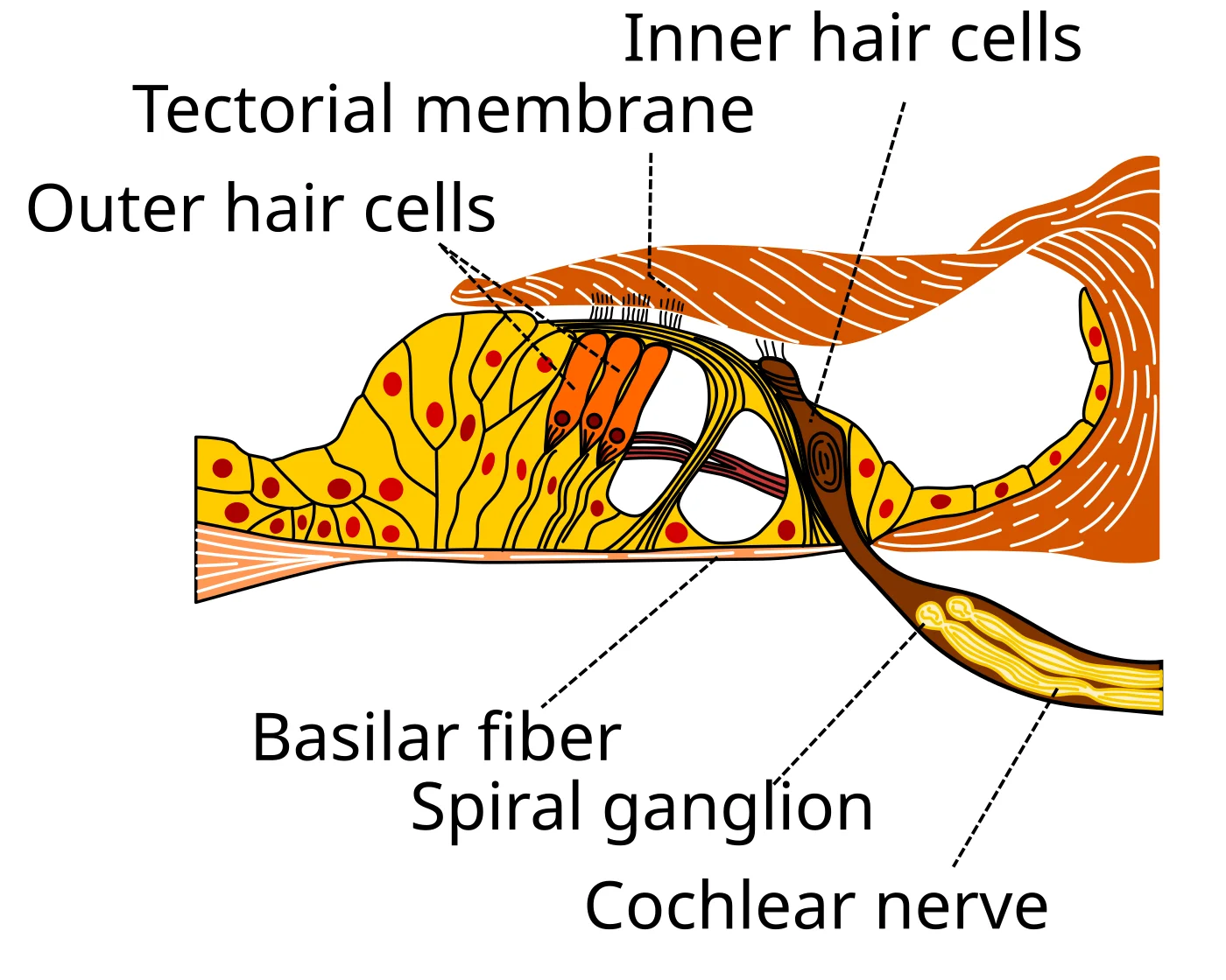
The cochlea is not a simple tube. A membrane runs down the middle called the basilar membrane, and sitting on top of it is the organ of Corti, which contains the actual transducers: hair cells.
A single hair cell has a bundle of stereocilia - tiny projections - whose tips are linked together by filaments called tip links. Each tip link is physically attached to a K⁺ channel. When the basilar membrane moves relative to the overhanging tectorial membrane, the stereocilia bend. Bending yanks the tip links, which opens the K⁺ channels. K⁺ flows in, the hair cell depolarizes, and voltage-gated Ca²⁺ channels open. Ca²⁺ triggers neurotransmitter release onto the auditory nerve.
That is auditory transduction in one paragraph. Mechanical motion → ion channels open → depolarization → action potential.
The Organ of Corti sits on the basilar membrane inside the cochlea. When the basilar membrane moves relative to the tectorial membrane, the stereocilia of the hair cells bend, opening K⁺ channels and triggering neurotransmitter release onto the cochlear (auditory) nerve. Credit: Madhero88 via Wikimedia Commons, CC BY-SA 3.0.
Place Theory: Where On the Basilar Membrane?
Here is the central fact for auditory processing: different frequencies shake different parts of the basilar membrane.
The base of the cochlea (nearest the oval window) is stiff and narrow. It resonates best with high-frequency sounds (short wavelengths).
The apex of the cochlea (farthest inside the coil) is floppy and wide. It resonates best with low-frequency sounds (long wavelengths).
So a high-frequency mosquito wail bends the hair cells at the base, while a low-frequency bass drum bends the hair cells at the apex. The brain knows what pitch you heard by which hair cells fired. This is called place theory or tonotopic mapping. It also persists all the way to the primary auditory cortex in the temporal lobe, where neurons are arranged by preferred frequency - a 500 Hz tone activates a different patch of cortex than a 5000 Hz tone.
Frequency Theory
Place theory handles high frequencies beautifully but struggles with very low frequencies. Below about 200 Hz, the whole basilar membrane vibrates at once and place becomes a poor code. The brain uses a second trick: frequency theory (or temporal coding). The auditory nerve fires action potentials at the same rate as the incoming sound wave. A 100 Hz tone → neuron fires 100 times per second.
For really fast frequencies, individual neurons cannot fire fast enough, so groups of neurons take turns (the volley principle): neuron A fires on wave 1, neuron B on wave 2, and so on, producing a combined firing rate that tracks the stimulus.
Practical split. Place theory for high pitches (about 2000 Hz and up), frequency theory for low pitches, both mechanisms together for the middle range.
Conductive vs. Sensorineural Hearing Loss
Conductive hearing loss comes from a problem in the pathway that conducts sound from outside to the cochlea: wax in the canal, a torn eardrum, stiff ossicles. Sound cannot physically reach the hair cells. Fixing conductive loss often means hearing aids that simply make sounds louder or bone-conduction devices.
Sensorineural hearing loss comes from damage downstream of the conduction pathway - typically to the hair cells themselves or the auditory nerve. Loud concerts kill hair cells, and they do not grow back. Age-related hearing loss (presbycusis) is largely sensorineural. A cochlear implant restores some hearing in severe sensorineural cases: a microphone outside the skull feeds a processor, which feeds electrodes threaded into the cochlea. The electrodes stimulate the auditory nerve directly, bypassing the dead hair cells.
What are the three ossicles, in order from eardrum to cochlea?
Click to reveal answer
Malleus, Incus, Stapes (M-I-S). Hammer, anvil, stirrup. They amplify the vibration of the eardrum by roughly 20× before it enters the denser fluid of the cochlea.
Which end of the basilar membrane detects high frequencies?
Click to reveal answer
The base (near the oval window) - stiff, narrow, resonates with high frequencies. The apex (far end) handles low frequencies.
What is the difference between place theory and frequency theory of hearing?
Click to reveal answer
Place theory: the basilar membrane location of maximum vibration encodes pitch (good for high frequencies). Frequency theory: the rate of neuron firing encodes pitch (good for low frequencies). The ear uses both.
What distinguishes conductive from sensorineural hearing loss?
Click to reveal answer
Conductive: problem in the sound-conducting pathway (outer/middle ear - wax, ruptured eardrum, fused ossicles). Sensorineural: damage to hair cells or auditory nerve. Cochlear implants help sensorineural loss by bypassing dead hair cells.
Vision and hearing get most of the air time, but four more senses ride on the MCAT: touch, balance, smell, and taste. They all use the same translate-energy-into-a-neural-signal game, but each has its own receptors and quirks.
Somatosensation: Touch, Pressure, Heat, Pain
Somatosensation is the “body sense.” It picks up four kinds of information:
Mechanoreception (pressure, vibration, stretch)
Thermoreception (temperature)
Nociception (pain)
Proprioception (body position)
Each has specialized receptors, but they all ultimately feed into the somatosensory cortex of the parietal lobe. That cortex contains the famous somatosensory homunculus: a distorted body map where each body part’s size reflects how much cortical real estate it has. Lips, hands, and tongue take up huge patches; torso and back, tiny ones. It is why a papercut on your finger is agony while a scratch on your back is background noise.
Nociception and the Gate Control Theory
Pain signals travel on three types of nerve fibers:
A-beta fibers - thick, heavily myelinated, fastest. Carry touch/pressure (not pain directly) but contribute to pain modulation.
C fibers - small diameter, unmyelinated, slow. Dull, lingering, aching pain (“second pain”).
The mnemonic is “fast to slow = A-beta, A-delta, C” (alphabetical).
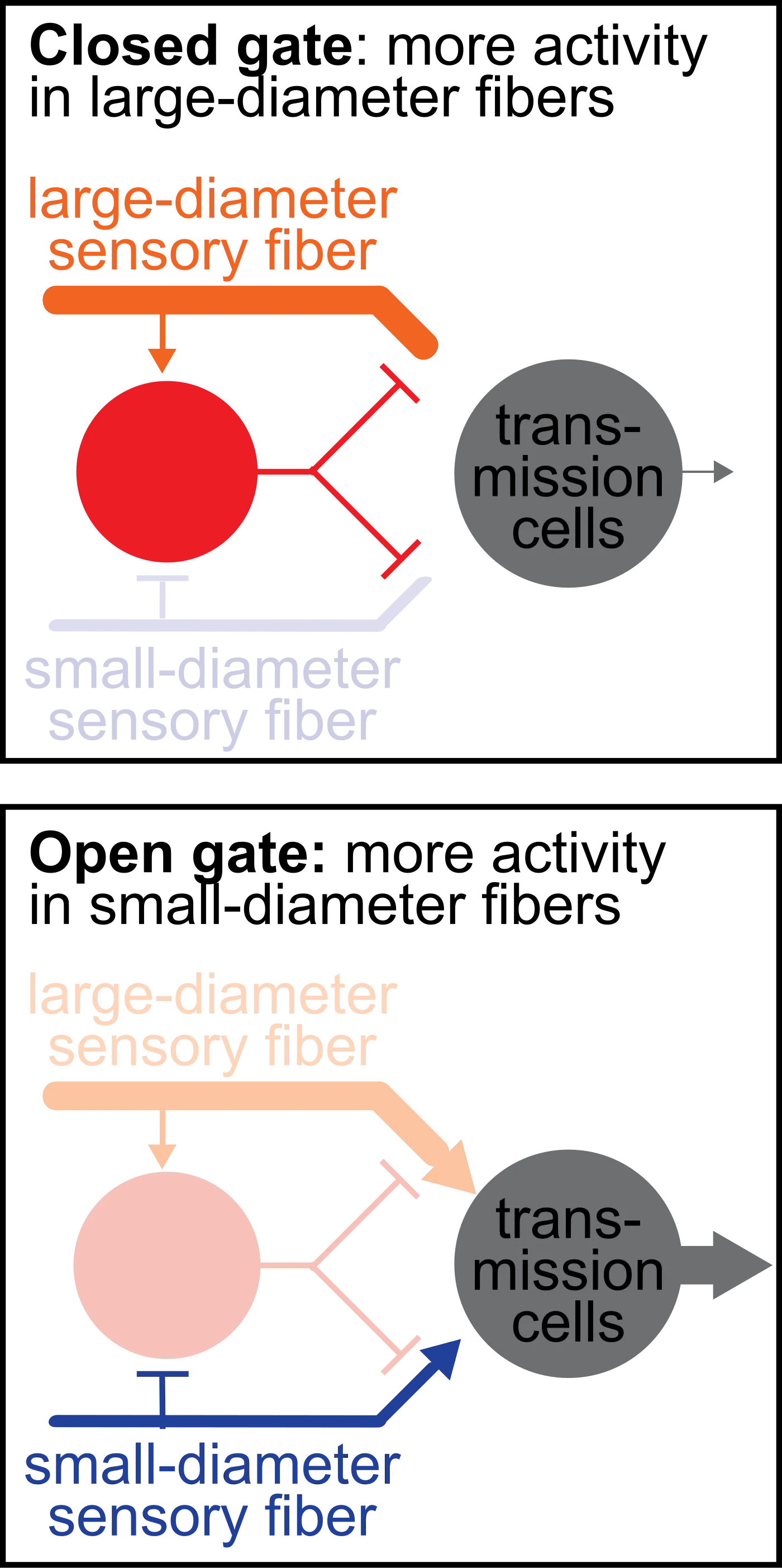
Gate control theory (Melzack and Wall) explains why rubbing a stubbed toe helps. Non-painful mechanical input (A-beta fibers) activates inhibitory interneurons in the spinal cord that close the “gate” on pain signals (C fibers) traveling to the brain. “Fast blocks slow.” This is the same principle behind TENS units and why mothers instinctively rub a child’s scraped knee.
Gate control theory. Top: small-diameter pain fibers alone activate the transmission cell — the gate is open and pain reaches the brain. Bottom: large-diameter touch fibers engage an inhibitory interneuron that shuts the gate, blocking pain. Rubbing an injury recruits large fibers to close the gate. Credit: John Tuthill via Wikimedia Commons, CC BY-SA 4.0.
Proprioception vs. Kinesthesia
Proprioception = sense of where your body parts are in space right now. It relies on muscle spindles (stretch-sensitive sensors inside muscles) and joint receptors. It lets you touch your nose with your eyes closed.
Kinesthesia = sense of how your body is moving. More about active motion than static position. Proprioception is cognitive (knowing); kinesthesia is behavioral (doing).
The Vestibular System: Balance
Inside your inner ear, right next to the cochlea, are the semicircular canals (posterior, anterior, lateral - each oriented in a different plane, like a 3D gyroscope) and the otolithic organs (utricle and saccule).
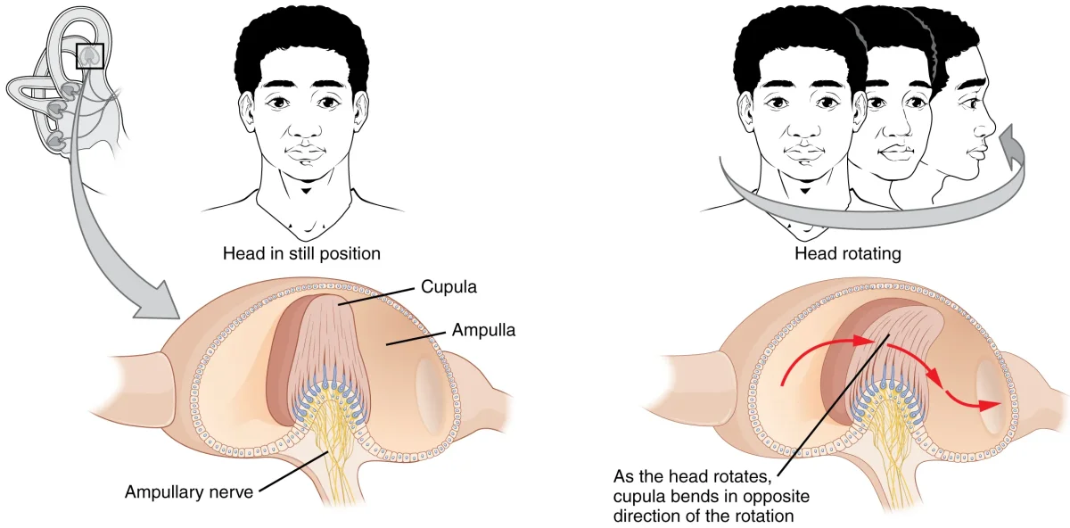
Semicircular canals detect rotational acceleration (your head is spinning). Fluid called endolymph inside the canals sloshes against hair cells when your head rotates. The direction and speed of the slosh tells the brain how you are turning.
Utricle and saccule detect linear acceleration and head tilt. They contain otoliths (calcium carbonate crystals) sitting on a bed of hair cells. When you tilt or accelerate, gravity and inertia drag the crystals, which bends the hair cells.
The merry-go-round problem: when you stop spinning, your head stops, but the endolymph keeps sloshing for a few seconds. Your inner ear says “still spinning” while your eyes say “stopped.” That sensory mismatch is vertigo and motion sickness.
The vestibular apparatus. Three semicircular canals (oriented roughly orthogonally) detect rotational acceleration; the otolithic organs (utricle, saccule) detect linear acceleration and head tilt using calcium-carbonate crystals pressing on hair cells. Credit: OpenStax Anatomy & Physiology 2e, CC BY 4.0.
Olfaction: Smell
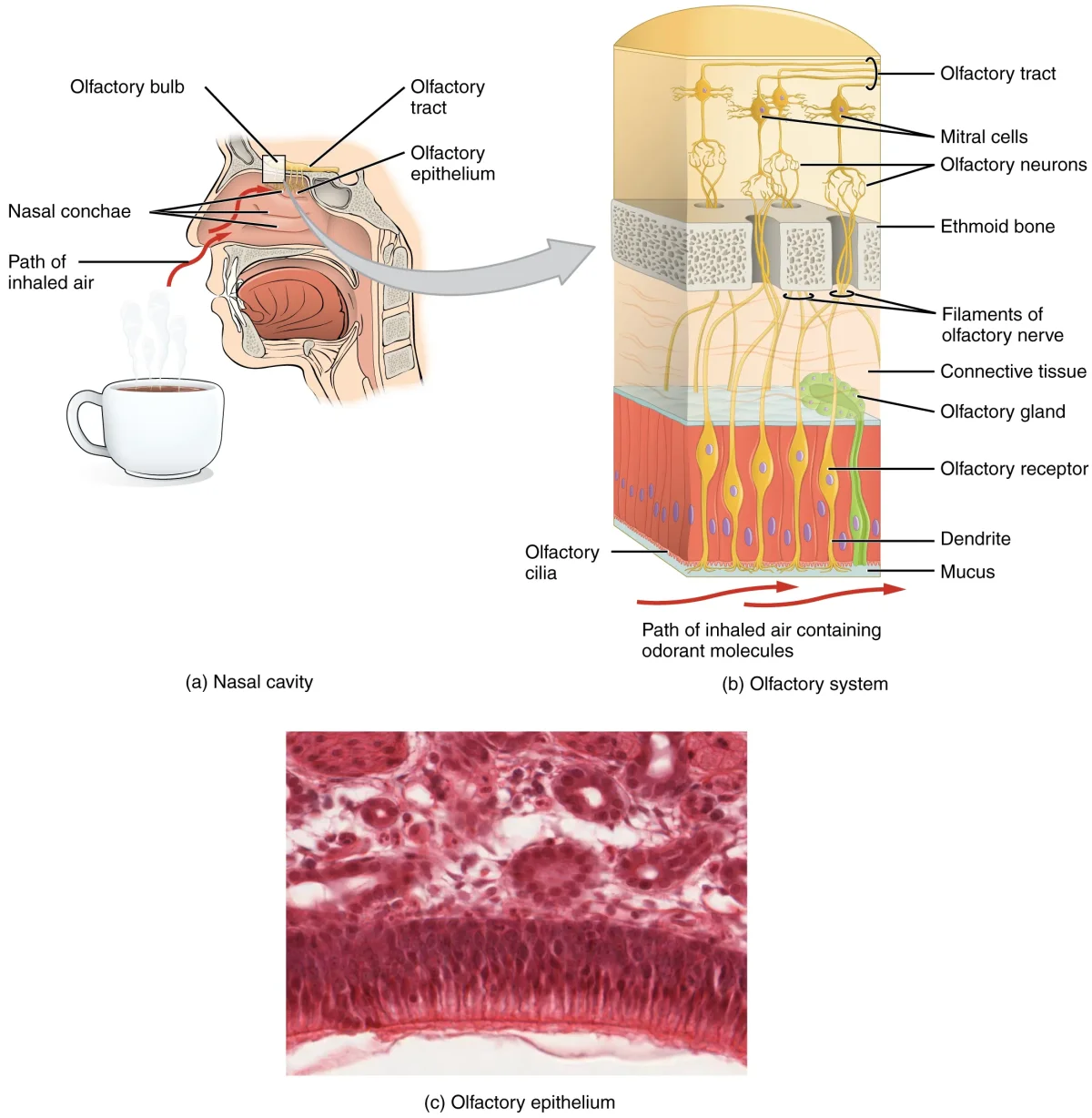
Smell is unique among the senses in one load-bearing way: it does not synapse in the thalamus first. It goes almost directly from the nose to the limbic system, which is why a whiff of a scent can trigger a vivid emotional memory - the signal hits the amygdala and hippocampus before your conscious brain catches up.
The pathway:
Odorant molecules dissolve in mucus in the olfactory epithelium at the top of the nasal cavity.
They bind G-protein-coupled receptors on specialized olfactory sensory neurons. Each neuron expresses one receptor type, sensitive to one class of molecules.
The neurons send axons through holes in the cribriform plate (part of the ethmoid bone) up to the olfactory bulb.
In the olfactory bulb, all the neurons expressing the same receptor converge on a single glomerulus. That glomerulus synapses onto a mitral/tufted cell.
Mitral/tufted cells project to the amygdala, piriform cortex, and eventually to the orbitofrontal cortex - all without passing through the thalamus.
The olfactory pathway. Odorant molecules bind receptors on olfactory sensory neurons in the nasal epithelium; axons pass through the cribriform plate to the olfactory bulb, then project to the limbic system without passing through the thalamus. Credit: OpenStax Anatomy & Physiology 2e, CC BY 4.0.
Head trauma that shears the delicate olfactory neurons through the cribriform plate causes anosmia (loss of smell). Anosmia is worth memorizing: “aNOSEmia = no smell.”
Competing Theories of Olfaction
Shape theory (steric theory) - odor molecules fit receptors like a lock and key based on molecular shape.
Vibrational theory - odor perception depends on the vibrational frequency of the molecule.
Labeled-line theory - each receptor type sends a dedicated line to the brain, like a separate phone line for each scent category.
Shape theory is dominant. The MCAT wants you to recognize the names, not pick a winner.
Pheromones
Pheromones are chemical signals released by one individual that trigger an innate response in another member of the same species - mating, territorial marking, alarm. Other mammals use a specialized vomeronasal organ with its own accessory olfactory bulb. Humans have a vestigial vomeronasal organ and minimal pheromone sensitivity.
Gustation: Taste
Five basic tastes, each with a dedicated receptor:
Sweet - GPCR receptors detect sugars.
Salty - Na⁺ enters directly through sodium channels.
Sour - H⁺ ions block K⁺ channels.
Bitter - GPCR receptors detect many unrelated compounds (defensive).
Taste receptor cells cluster into taste buds, which sit in structures called papillae on the tongue:
Fungiform papillae - mushroom-shaped, tip and sides.
Foliate papillae - folded, sides of tongue.
Circumvallate papillae - flat mounds, back of tongue.
Filiform papillae - thread-shaped, all over; no taste buds, just texture.
The traditional “tongue map” (sweet at the tip, bitter at the back) is wrong. Every taste can be detected everywhere taste buds exist. Each bud has cells for all five tastes.
Taste signals travel via three cranial nerves:
Front two-thirds of the tongue → facial nerve (CN VII) via the chorda tympani.
Back one-third of the tongue → glossopharyngeal nerve (CN IX).
Like smell, taste does not synapse in the thalamus before its first cortical integration - the first merge point is the orbitofrontal cortex.
What is the gate control theory of pain?
Click to reveal answer
Non-painful input (A-beta fibers) activates spinal inhibitory interneurons that "close the gate" on pain signals from C fibers. "Fast blocks slow." Why rubbing a stubbed toe eases the pain.
Which inner ear structures detect rotational vs. linear acceleration?
Click to reveal answer
Semicircular canals detect rotation (fluid sloshes against hair cells). Utricle and saccule (otolithic organs) detect linear acceleration and head tilt via calcium carbonate crystals on hair cells.
Why is smell unique among the senses in terms of brain pathway?
Click to reveal answer
Olfaction bypasses the thalamus. Signals go from the olfactory bulb directly to the amygdala, piriform cortex, and orbitofrontal cortex - explaining the potent emotional and memory triggers of smell.
What are the five basic tastes, and which one detects glutamate?
Click to reveal answer
Sweet, salty, sour, bitter, umami. Umami is the glutamate/savory taste (hence MSG, Parmesan, broth, ripe tomatoes).
What happens if the cribriform plate is fractured in a head injury?
Click to reveal answer
Olfactory nerve axons passing through the plate can be sheared, causing anosmia (loss of smell). "aNOSEmia."
Your retina catches photons. Your cochlea catches pressure waves. But “seeing a dog” and “hearing music” are not retinal or cochlear events - they are cortical ones. This section covers how your brain assembles sensory signals into a unified perception and why two people can look at the same drawing and see two different images.
Bottom-Up vs. Top-Down Processing
Bottom-up processing starts with the stimulus. Photons hit the retina, signals propagate upward through bipolar cells, ganglion cells, LGN, and visual cortex. Each step adds information. You “build” the percept from raw features. No prior expectation required. This is how a newborn, seeing a face for the very first time, figures out it is a face.
Top-down processing starts with your brain’s expectations. Your brain has a model of what is likely to be out there, and it uses that model to guide interpretation. “Where’s Waldo?” is top-down: you know what Waldo looks like before you start scanning, so your eyes lock onto red-and-white-striped patches. Without the expectation, you would see a chaotic crowd.
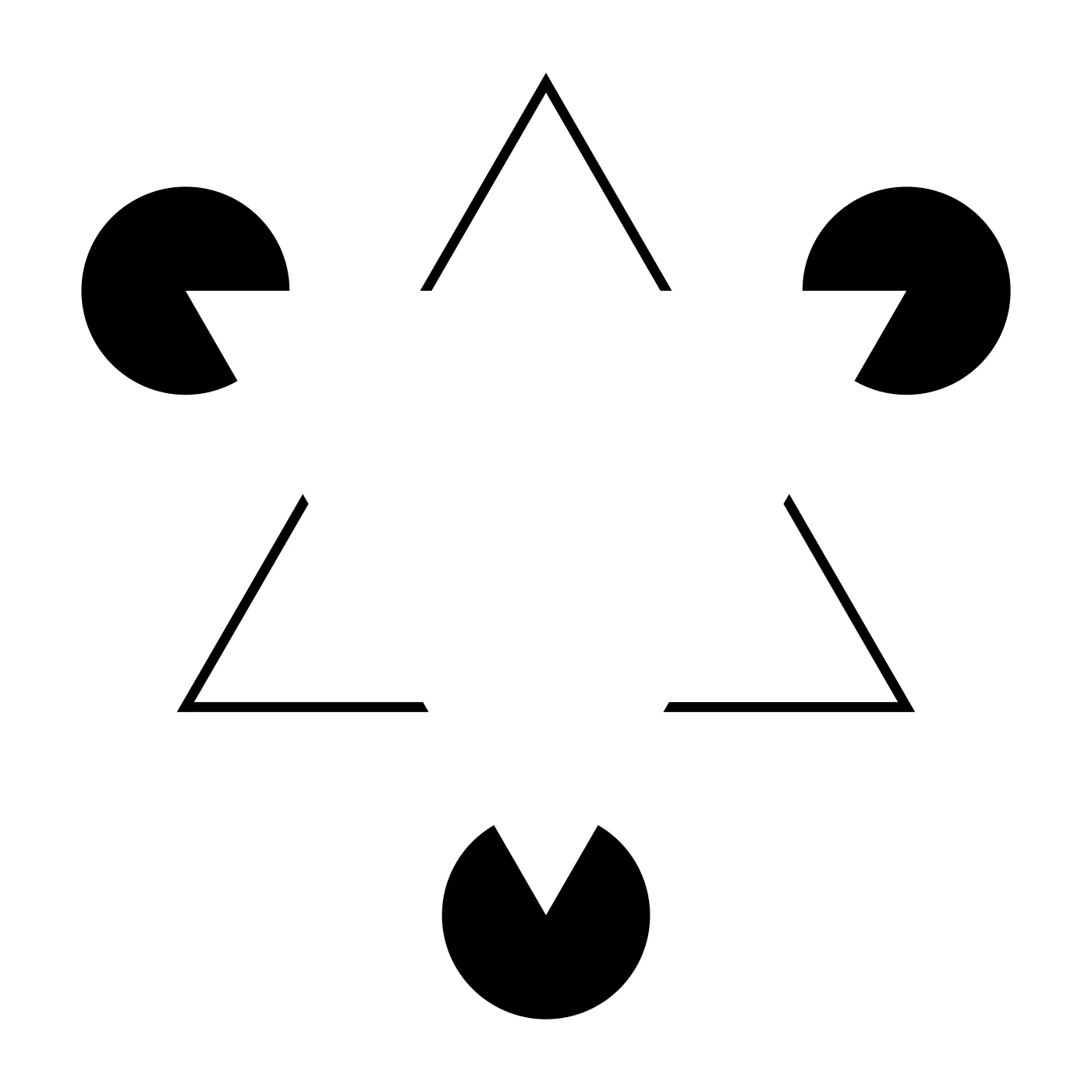
The MCAT loves to test one trap: top-down processing can make you perceive things that are not there. When you look at a Kanizsa triangle - three Pac-Man shapes arranged so their mouths form a triangle - you see a white triangle floating in front. There is no triangle. Your brain drew it based on expectation. Top-down is creative; bottom-up is data-driven.
The Kanizsa triangle. There is no white triangle in the image, yet most viewers see one floating in front. The brain closes the gaps between the Pac-Man cutouts using top-down expectation — a textbook demonstration of illusory contours. Credit: Fibonacci via Wikimedia Commons, CC BY-SA 3.0.
Gestalt Principles: The Whole Is More Than the Sum of the Parts
Gestalt (“form” in German) psychologists argued that our brains group sensory elements into coherent wholes using a handful of rules. Know each one by name, the MCAT will ask.
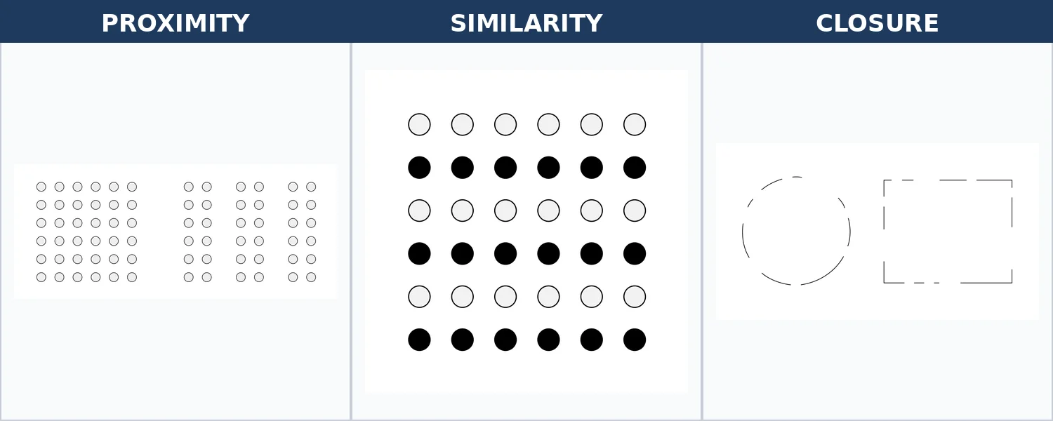
Proximity. Objects near each other are grouped together. Rows of dots spaced closer vertically than horizontally look like columns, not rows.
Similarity. Objects that look alike (same shape, color, size) are grouped. A mix of red and blue dots forms two groups even when they are interspersed.
Continuity (good continuation). Lines are perceived as following the smoothest path. An X is seen as two crossing lines, not four separate corners.
Closure. Incomplete figures are mentally completed. Three Pac-Men suggest a triangle; a broken circle looks like a whole circle.
Symmetry. The mind prefers symmetrical groupings, seeing two brackets facing each other as a pair.
Figure-ground. Any scene is split into a figure (the object of attention) and a ground (the background). The classic vase/faces illusion is a figure-ground flip.
Rubin's vase. Depending on which region your brain assigns as the figure and which as the ground, you see either a vase or two facing profiles — but never both at once. Credit: Nevit Dilmen via Wikimedia Commons, CC BY-SA 3.0.
- **Common fate.** Things moving together are grouped together. A flock of birds looks like a single swirling unit rather than dozens of separate birds.
- **Pragnanz (good form / simplicity).** Reality is organized into the simplest form possible. The Olympic rings are seen as five interlocked circles, not a dozen odd curved shapes. This is the overarching principle - every other Gestalt rule is a special case of pragnanz.
- **Past experience.** Prior exposure biases grouping. Reading "L" "I" next to each other as two letters rather than the uppercase "U" they could form.
Three of the most testable Gestalt principles. Proximity: dots spaced closer together are grouped as one cluster. Similarity: identical-looking dots form rows even when they interleave. Closure: the brain fills in gaps to complete familiar shapes. Credit: Assembled from Wikimedia Commons files by Kasufcgslfguhvsne et al. (Public Domain).
Depth Perception: Binocular and Monocular Cues
How does a 2D retinal image become a 3D percept? Your brain uses two kinds of cues.
Binocular cues require two eyes.
Retinal disparity. Your eyes are about 2.5 inches apart, so they see slightly different images. The bigger the disparity, the closer the object. This is how 3D movies work - they deliver different images to each eye to force disparity.
Convergence. When an object is close, your eyes turn inward (eye muscles contract). For a far object, the muscles relax. The brain uses the muscle signal as a distance cue.
Monocular cues work with only one eye.
Relative size. If two objects are the same known size but one looks bigger, it must be closer.
Interposition (overlap). An object that covers another is in front of it.
Relative height. Things higher in the visual field look farther away.
Shading and contour. Highlights and shadows suggest 3D form (crater vs. mountain illusion).
Motion parallax. When you move, near objects seem to move fast while distant objects drift slowly. Watch power poles streak past the train window while a distant mountain barely budges.
Linear perspective. Parallel lines converge at the horizon.
Your retinal image changes constantly, but your perception of objects does not. This is perceptual constancy.
Size constancy. A friend walking toward you grows on your retina, but you perceive them as the same size.
Shape constancy. An opening door casts an increasingly flat trapezoid on your retina, but you still perceive it as rectangular.
Color constancy. A white paper looks white under blue sky and warm incandescent bulb even though the light reflected off it differs dramatically. Your brain discounts the illuminant.
Without constancy, objects would seem to change size, shape, and color every time you turned your head. Constancy is a top-down correction that keeps the world stable.
Sensory Adaptation (One More Time)
We covered it briefly in section 1.1, but it is worth closing the chapter on: sensory adaptation is the down-regulation of receptor firing to a constant, unchanging stimulus. You stop feeling your socks after a minute. You stop smelling your own perfume after an hour. You stop hearing a humming air conditioner. The stimulus is still there; your receptors have simply muted themselves so the brain can focus on changes.
Adaptation is why detecting a new stimulus matters more than detecting a steady one. A predator sneaking up behind you is new - motion. A rock behind you is steady and irrelevant.
What is the difference between bottom-up and top-down processing?
Click to reveal answer
Bottom-up starts with raw stimulus data and builds upward. Top-down starts with expectations and interprets ambiguous data through prior knowledge. Top-down can generate percepts that don't exist (illusions).
Name three binocular depth cues.
Click to reveal answer
Retinal disparity (slight difference between images in the two eyes) and convergence (inward rotation of the eyes on near objects). Those are the two primary ones; most "depth cues" lists have only these two as binocular, with everything else monocular.
What is pragnanz (the law of good form)?
Click to reveal answer
The Gestalt principle that the brain perceives ambiguous or complex images in the simplest way possible. The Olympic rings look like five circles rather than a dozen separate curves.
What is perceptual constancy?
Click to reveal answer
The brain perceives objects as having stable properties (size, shape, color) despite continuous changes in the retinal image. Size, shape, and color constancy are the classic three.