
Visual Processing and Color Vision
The raw photon-to-neuron step happens in the retina. From there, the signal takes a highly organized trip to your visual cortex, and different features of the image get routed down different wires. This section is about what happens after the retinal ganglion cells fire.
The Visual Field Crosses Over
Your visual field splits into a left half and a right half. Here is the rule to memorize: the right half of the visual field is processed by the left brain, and the left half by the right brain. It is the same crossover you see with motor and somatosensory cortex.
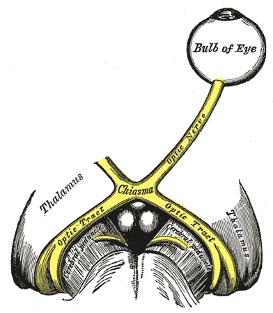
How does the crossover actually happen? By the optic chiasm.
- Each retina has a nasal side (closer to the nose) and a temporal side (closer to the temple).
- Light from the right visual field lands on the nasal side of the right eye and the temporal side of the left eye.
- At the optic chiasm, the axons from the nasal retinas cross to the opposite hemisphere; axons from the temporal retinas stay on the same side.
- The net effect: all signals carrying the right visual field end up in the left occipital lobe, and vice versa.
Feature Detection: Color, Form, Motion
After the optic chiasm, signals travel through the lateral geniculate nucleus (LGN) and up to the primary visual cortex in the occipital lobe. Along the way, different features of what you are seeing ride on different “wires”:
The parvocellular pathway handles color and fine spatial detail (form). It has good spatial resolution but poor temporal resolution - it cannot track fast movement, only stationary detail. Driven mostly by cones.
The magnocellular pathway handles motion and temporal detail. It is color-blind and low-resolution but excellent at tracking things that move. Driven mostly by rods and the larger ganglion cells.
Parallel processing means your brain handles color, form, and motion simultaneously on different tracks, then stitches them back together upstream. You do not see color first, then motion. You see them at once.
The Two Theories of Color Vision
The MCAT will absolutely ask you which theory explains which phenomenon. They are not competing - modern neuroscience says both are right at different stages.
Trichromatic Theory (Young-Helmholtz)
Three cone types exist: red-sensitive, green-sensitive, and blue-sensitive. Any perceived color is a mixture of the activations of these three. Purple? Red + blue cones firing. Orange? Red + green. White? All three.
This explains how a TV screen with only red, green, and blue subpixels can produce any color you see on it. Trichromatic theory lives at the level of the retina (the cones themselves).
Red-green color blindness - the most common form - usually comes from a missing or defective red or green cone. It is X-linked, which is why it hits men more often. Those individuals still distinguish blue from red or green; they just cannot tell red from green.
Opponent Process Theory (Hering)
Three opposing pairs of colors are processed together: red vs. green, blue vs. yellow, and black vs. white. In any pair, one color inhibits the other. You can have reddish-blue (magenta/purple) or yellowish-red (orange), but you can never have reddish-green or bluish-yellow. The pairs are mutually exclusive.
This is why after staring at a green square for 30 seconds and then looking at a white wall, you see a red afterimage. The green channel became fatigued; its opposing channel (red) rebounds. Opponent process theory explains afterimages, simultaneous color contrast, and why certain color combinations look “impossible.” It lives in the retinal ganglion cells and beyond - downstream of the cones.
Bonus: Color Constancy
Take a white sheet of paper outside at noon (blue-sky lighting) and then inside under a warm incandescent bulb (orange-ish lighting). The light hitting your retina is wildly different, but you still perceive the paper as white. That is color constancy - your brain accounts for the ambient lighting and recovers the object’s “true” color. Your phone camera does this too, clumsily; it is called “white balance.” Your visual system does it automatically and often without your noticing. The only time it fails conspicuously is in weird lighting like sodium-vapor streetlights, which can make red cars look black.