
The Eye and Phototransduction
Your phone camera has a lens that focuses light, a sensor that converts light into voltage, and a processor that interprets those voltages as an image. Your eye is the same architecture, assembled by biology instead of engineers. Understand the camera and the eye follows.
The Anatomy: Front to Back
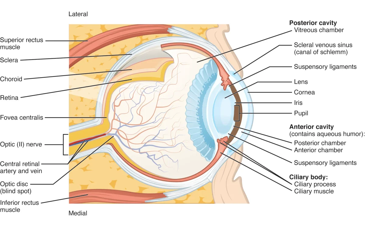
Light takes a short journey from the outside world to your retina. Memorize the pathway in this order and you have the structure locked down.
- Cornea. The clear, thick dome at the front of the eye. Does about two thirds of all light focusing. Covered by a thin mucus membrane called the conjunctiva.
- Aqueous humor. Watery fluid in the anterior chamber (in front of the lens). Nourishes the cornea and lens, which have no blood vessels of their own.
- Pupil. The hole in the middle of the iris. Dilates in the dark (let more light in), constricts in the sun (let less light in).
- Iris. The colored ring. A muscle that changes the size of the pupil.
- Lens. Fine-tunes the focus. The ciliary muscles attached to the lens via suspensory ligaments change the lens’s shape (accommodation) to focus on near vs. far objects.
- Vitreous humor. A jelly that fills most of the eyeball and keeps it inflated.
- Retina. The back wall of the eye, packed with photoreceptors (rods and cones). This is where transduction happens.
- Macula. A central patch of retina specialized for high-detail vision.
- Fovea. The very center of the macula. Packed with cones, no rods. This is where you see the word you are reading right now.
- Optic nerve. Bundles the output of all retinal ganglion cells and ships it to the brain.
Between the retina and the tough outer sclera (the white of your eye) is the choroid, a layer of blood vessels that feeds the retina and absorbs stray light so it does not bounce around.
Rods and Cones: Two Sensors in One Retina
Your retina has two kinds of photoreceptors, and they have almost opposite jobs.
| Feature | Rods | Cones |
|---|---|---|
| How many? | ~120 million | ~6 million |
| Location | Periphery | Fovea / macula |
| Sensitivity | ~1000× more sensitive | Less sensitive |
| Color? | No (black and white only) | Yes (R, G, B) |
| Detail? | Low resolution | High resolution |
| Recovery | Slow | Fast |
| Best for | Night vision, motion | Daylight, color, detail |
This is why you cannot see color clearly in the dark: cones need bright light, and at night only your rods are firing. It is also why motion catches your eye in peripheral vision: rods dominate the periphery and are wired for rapid, transient changes. To actually read something, you have to point your fovea at it - one reason reading a wall clock is easy but reading peripheral text is not.
The three cone types are not evenly distributed. Roughly 60% are red-sensitive, 30% green-sensitive, and 10% blue-sensitive. This uneven mix is why we can resolve red and green subtleties better than blue ones, and it becomes load-bearing in the color-vision theories covered in the next section.
Phototransduction: How a Photon Becomes a Nerve Signal
This is the single highest-yield mechanism in the whole sensory chapter. Get it right and you pick up multiple MCAT points.
The big counterintuitive twist: in the dark, photoreceptors are depolarized and constantly releasing neurotransmitter. Light turns them OFF. Your retina reports light by going silent. Every other sense reports a stimulus by firing more; photoreceptors are the exception.
Here is the cascade, in the dark and in the light.
In the Dark (Baseline, Depolarized)
- cGMP (cyclic GMP) is abundant inside the rod.
- cGMP binds sodium channels and keeps them open.
- Na⁺ flows in. The rod stays depolarized.
- The rod continuously releases glutamate onto bipolar cells.
When Light Hits a Rod
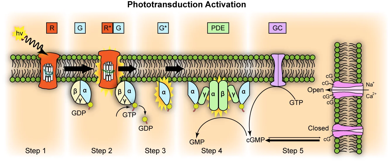
- A photon strikes rhodopsin (the photopigment in rods; cones use photopsin). Rhodopsin is a G-protein-coupled receptor with a small molecule called 11-cis retinal tucked inside.
- Light converts 11-cis retinal into all-trans retinal (the shape changes). This activates rhodopsin.
- Activated rhodopsin triggers transducin, a G-protein. The alpha subunit of transducin splits off.
- The alpha subunit activates phosphodiesterase (PDE).
- PDE converts cGMP → GMP. cGMP levels plummet.
- With cGMP gone, the Na⁺ channels close. Less Na⁺ enters.
- The rod hyperpolarizes and stops releasing glutamate.
- Downstream bipolar cells sense the drop in glutamate. ON-center bipolar cells (which are normally inhibited by glutamate) become more active.
- Active ON-center bipolar cells excite retinal ganglion cells, which fire action potentials down the optic nerve. The brain now knows a photon arrived.
Light Adaptation: Dark Room, Bright Sunlight
When you walk from a sunny sidewalk into a dark theater, it takes minutes to see. That delay is the cascade running in reverse: rhodopsin was mostly bleached (11-cis retinal all used up) in bright light, and your rods need time to regenerate it. Once rhodopsin is restocked, rods become sensitive again.
Going the other way - dark room to bright hallway - is fast because cones adjust quickly (high-activity pathways recover faster), and because pupil constriction happens in seconds. This is part of why rods take longer to adapt than cones: rods have to resynthesize more photopigment before they can fire again.