
The Ear and Auditory Processing
A bass drum thumps at 60 Hz. A mosquito whines at 15,000 Hz. Your ear turns both into electrical signals the same brain can read, using nothing but tiny bones, a snail-shaped tube of saltwater, and a row of microscopic hair-cell “sensors.” This section walks you through the conversion, piece by piece, and then unpacks the two theories the MCAT loves to test.
The Sound Pathway, Outside to Inside
Sound begins as pressurized air. When a speaker cone pushes forward, it shoves air molecules into a zone of high pressure; when it pulls back, air thins into low pressure. Those alternating pressure waves are what your ear is trying to detect.
The sound path:
-
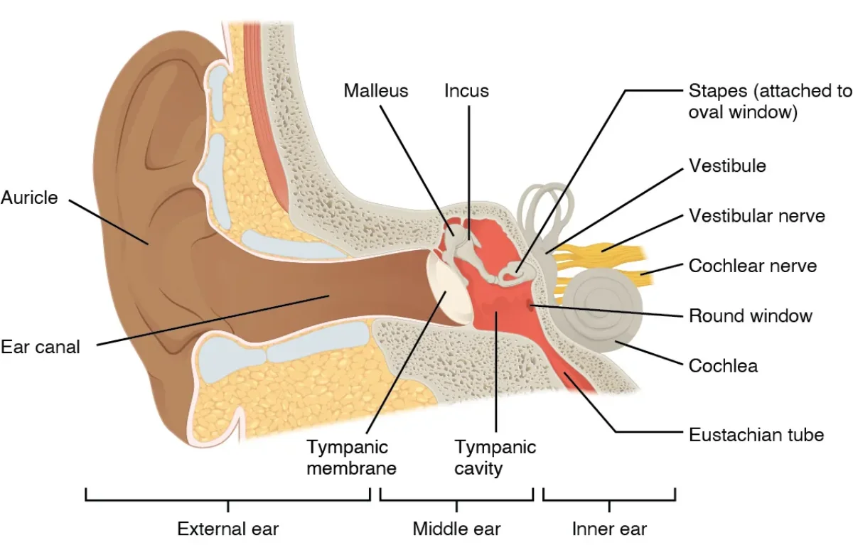
Pinna. The floppy outer ear. Its curves collect air pressure waves and funnel them inward, like a satellite dish.
-
Auditory canal (external auditory meatus). The tube from the pinna toward your skull. Narrows the wave, concentrating it.
-
Tympanic membrane (eardrum). A thin, tense sheet at the end of the auditory canal. Pressure waves make it vibrate.
-
Ossicles. Three tiny bones in the middle ear - the smallest bones in your body - that amplify the vibration:
- Malleus (hammer) is attached to the eardrum.
- Incus (anvil) connects malleus to stapes.
- Stapes (stirrup) presses on the oval window.
Mnemonic: M-I-S.
-
Oval window. A membrane on the cochlea that transmits the ossicle vibration into the cochlear fluid.
-
Cochlea. A snail-shaped, fluid-filled tube. The waves travel through the cochlea, bend hair cells, and then exit via the round window, which flexes outward to relieve pressure.
-
Auditory nerve. The axons from hair-cell synapses carry action potentials to the brain.
The Three Regions of the Ear
Grouping the parts makes the anatomy easier to retain:
- Outer ear: pinna through tympanic membrane.
- Middle ear: ossicles (malleus, incus, stapes), sitting in an air-filled cavity.
- Inner ear: cochlea and semicircular canals (the semicircular canals handle balance, not hearing - we will see them in the next section).
Inside the Cochlea: the Organ of Corti
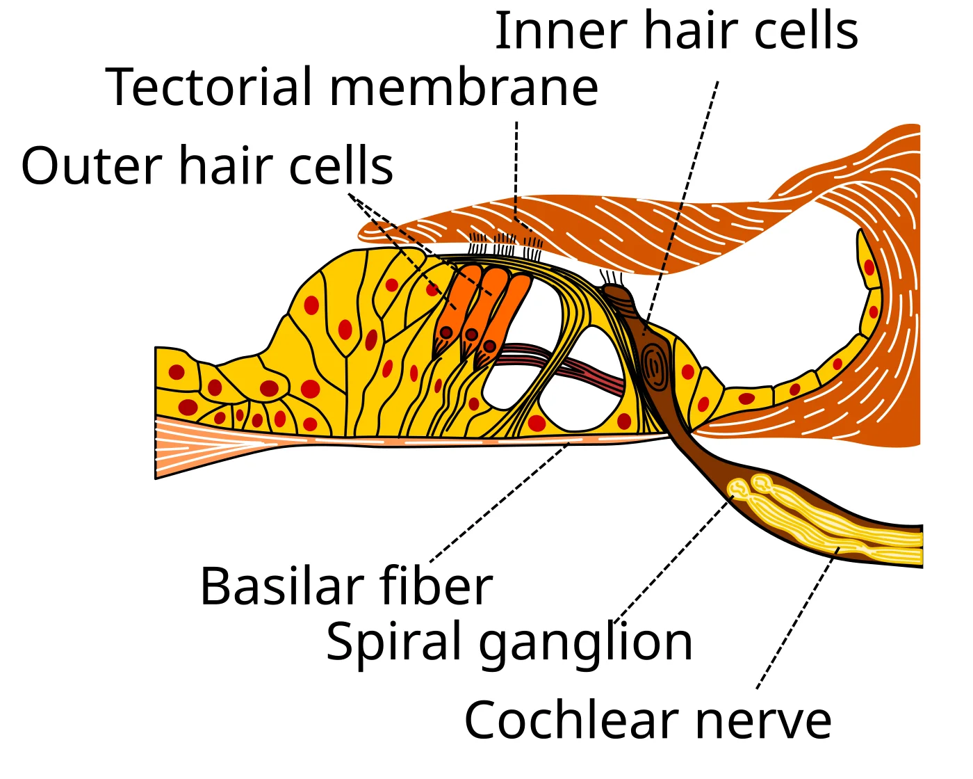
The cochlea is not a simple tube. A membrane runs down the middle called the basilar membrane, and sitting on top of it is the organ of Corti, which contains the actual transducers: hair cells.
A single hair cell has a bundle of stereocilia - tiny projections - whose tips are linked together by filaments called tip links. Each tip link is physically attached to a K⁺ channel. When the basilar membrane moves relative to the overhanging tectorial membrane, the stereocilia bend. Bending yanks the tip links, which opens the K⁺ channels. K⁺ flows in, the hair cell depolarizes, and voltage-gated Ca²⁺ channels open. Ca²⁺ triggers neurotransmitter release onto the auditory nerve.
That is auditory transduction in one paragraph. Mechanical motion → ion channels open → depolarization → action potential.
Place Theory: Where On the Basilar Membrane?
Here is the central fact for auditory processing: different frequencies shake different parts of the basilar membrane.
- The base of the cochlea (nearest the oval window) is stiff and narrow. It resonates best with high-frequency sounds (short wavelengths).
- The apex of the cochlea (farthest inside the coil) is floppy and wide. It resonates best with low-frequency sounds (long wavelengths).
So a high-frequency mosquito wail bends the hair cells at the base, while a low-frequency bass drum bends the hair cells at the apex. The brain knows what pitch you heard by which hair cells fired. This is called place theory or tonotopic mapping. It also persists all the way to the primary auditory cortex in the temporal lobe, where neurons are arranged by preferred frequency - a 500 Hz tone activates a different patch of cortex than a 5000 Hz tone.
Frequency Theory
Place theory handles high frequencies beautifully but struggles with very low frequencies. Below about 200 Hz, the whole basilar membrane vibrates at once and place becomes a poor code. The brain uses a second trick: frequency theory (or temporal coding). The auditory nerve fires action potentials at the same rate as the incoming sound wave. A 100 Hz tone → neuron fires 100 times per second.
For really fast frequencies, individual neurons cannot fire fast enough, so groups of neurons take turns (the volley principle): neuron A fires on wave 1, neuron B on wave 2, and so on, producing a combined firing rate that tracks the stimulus.
Practical split. Place theory for high pitches (about 2000 Hz and up), frequency theory for low pitches, both mechanisms together for the middle range.
Conductive vs. Sensorineural Hearing Loss
Conductive hearing loss comes from a problem in the pathway that conducts sound from outside to the cochlea: wax in the canal, a torn eardrum, stiff ossicles. Sound cannot physically reach the hair cells. Fixing conductive loss often means hearing aids that simply make sounds louder or bone-conduction devices.
Sensorineural hearing loss comes from damage downstream of the conduction pathway - typically to the hair cells themselves or the auditory nerve. Loud concerts kill hair cells, and they do not grow back. Age-related hearing loss (presbycusis) is largely sensorineural. A cochlear implant restores some hearing in severe sensorineural cases: a microphone outside the skull feeds a processor, which feeds electrodes threaded into the cochlea. The electrodes stimulate the auditory nerve directly, bypassing the dead hair cells.