
Touch, Balance, Smell, and Taste
Vision and hearing get most of the air time, but four more senses ride on the MCAT: touch, balance, smell, and taste. They all use the same translate-energy-into-a-neural-signal game, but each has its own receptors and quirks.
Somatosensation: Touch, Pressure, Heat, Pain
Somatosensation is the “body sense.” It picks up four kinds of information:
- Mechanoreception (pressure, vibration, stretch)
- Thermoreception (temperature)
- Nociception (pain)
- Proprioception (body position)
Each has specialized receptors, but they all ultimately feed into the somatosensory cortex of the parietal lobe. That cortex contains the famous somatosensory homunculus: a distorted body map where each body part’s size reflects how much cortical real estate it has. Lips, hands, and tongue take up huge patches; torso and back, tiny ones. It is why a papercut on your finger is agony while a scratch on your back is background noise.
Nociception and the Gate Control Theory
Pain signals travel on three types of nerve fibers:
- A-beta fibers - thick, heavily myelinated, fastest. Carry touch/pressure (not pain directly) but contribute to pain modulation.
- A-delta fibers - thinner, lightly myelinated, medium speed. Sharp, localized pain (“first pain”).
- C fibers - small diameter, unmyelinated, slow. Dull, lingering, aching pain (“second pain”).
The mnemonic is “fast to slow = A-beta, A-delta, C” (alphabetical).
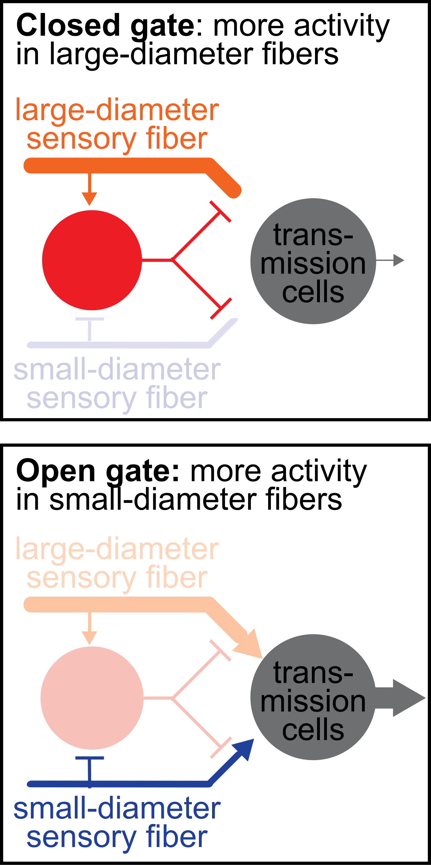
Gate control theory (Melzack and Wall) explains why rubbing a stubbed toe helps. Non-painful mechanical input (A-beta fibers) activates inhibitory interneurons in the spinal cord that close the “gate” on pain signals (C fibers) traveling to the brain. “Fast blocks slow.” This is the same principle behind TENS units and why mothers instinctively rub a child’s scraped knee.
Proprioception vs. Kinesthesia
Proprioception = sense of where your body parts are in space right now. It relies on muscle spindles (stretch-sensitive sensors inside muscles) and joint receptors. It lets you touch your nose with your eyes closed.
Kinesthesia = sense of how your body is moving. More about active motion than static position. Proprioception is cognitive (knowing); kinesthesia is behavioral (doing).
The Vestibular System: Balance
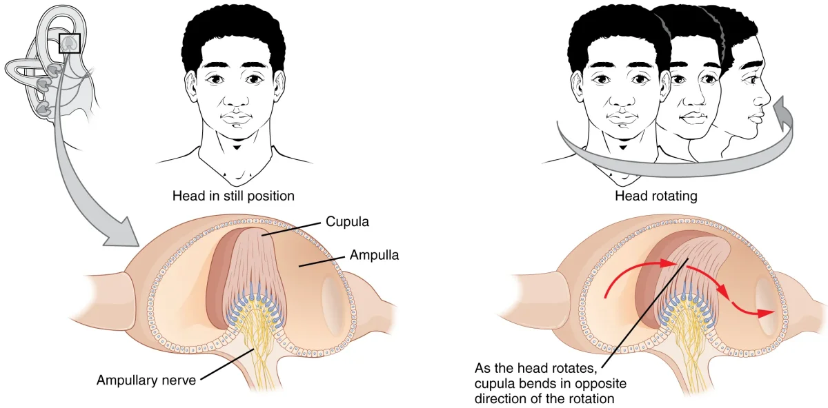
Inside your inner ear, right next to the cochlea, are the semicircular canals (posterior, anterior, lateral - each oriented in a different plane, like a 3D gyroscope) and the otolithic organs (utricle and saccule).
- Semicircular canals detect rotational acceleration (your head is spinning). Fluid called endolymph inside the canals sloshes against hair cells when your head rotates. The direction and speed of the slosh tells the brain how you are turning.
- Utricle and saccule detect linear acceleration and head tilt. They contain otoliths (calcium carbonate crystals) sitting on a bed of hair cells. When you tilt or accelerate, gravity and inertia drag the crystals, which bends the hair cells.
The merry-go-round problem: when you stop spinning, your head stops, but the endolymph keeps sloshing for a few seconds. Your inner ear says “still spinning” while your eyes say “stopped.” That sensory mismatch is vertigo and motion sickness.
Olfaction: Smell
Smell is unique among the senses in one load-bearing way: it does not synapse in the thalamus first. It goes almost directly from the nose to the limbic system, which is why a whiff of a scent can trigger a vivid emotional memory - the signal hits the amygdala and hippocampus before your conscious brain catches up.
The pathway:
- Odorant molecules dissolve in mucus in the olfactory epithelium at the top of the nasal cavity.
- They bind G-protein-coupled receptors on specialized olfactory sensory neurons. Each neuron expresses one receptor type, sensitive to one class of molecules.
- The neurons send axons through holes in the cribriform plate (part of the ethmoid bone) up to the olfactory bulb.
- In the olfactory bulb, all the neurons expressing the same receptor converge on a single glomerulus. That glomerulus synapses onto a mitral/tufted cell.
- Mitral/tufted cells project to the amygdala, piriform cortex, and eventually to the orbitofrontal cortex - all without passing through the thalamus.
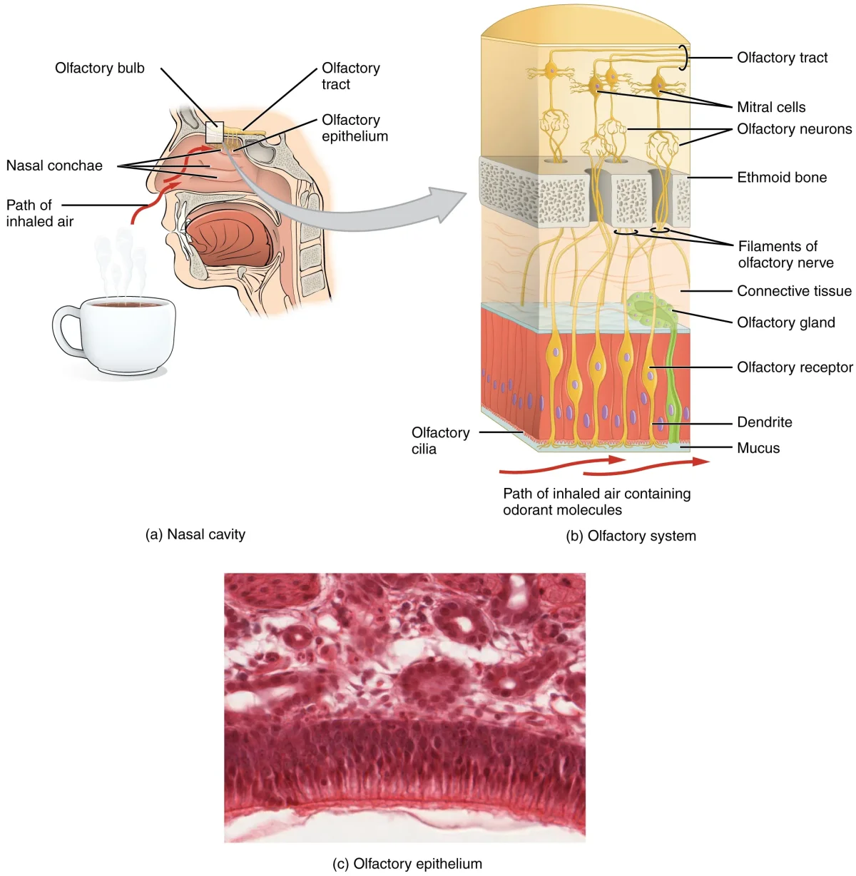
Head trauma that shears the delicate olfactory neurons through the cribriform plate causes anosmia (loss of smell). Anosmia is worth memorizing: “aNOSEmia = no smell.”
Competing Theories of Olfaction
- Shape theory (steric theory) - odor molecules fit receptors like a lock and key based on molecular shape.
- Vibrational theory - odor perception depends on the vibrational frequency of the molecule.
- Labeled-line theory - each receptor type sends a dedicated line to the brain, like a separate phone line for each scent category.
Shape theory is dominant. The MCAT wants you to recognize the names, not pick a winner.
Pheromones
Pheromones are chemical signals released by one individual that trigger an innate response in another member of the same species - mating, territorial marking, alarm. Other mammals use a specialized vomeronasal organ with its own accessory olfactory bulb. Humans have a vestigial vomeronasal organ and minimal pheromone sensitivity.
Gustation: Taste
Five basic tastes, each with a dedicated receptor:
- Sweet - GPCR receptors detect sugars.
- Salty - Na⁺ enters directly through sodium channels.
- Sour - H⁺ ions block K⁺ channels.
- Bitter - GPCR receptors detect many unrelated compounds (defensive).
- Umami - GPCR receptors detect glutamate (savory, meaty, Parmesan).
Taste receptor cells cluster into taste buds, which sit in structures called papillae on the tongue:
- Fungiform papillae - mushroom-shaped, tip and sides.
- Foliate papillae - folded, sides of tongue.
- Circumvallate papillae - flat mounds, back of tongue.
- Filiform papillae - thread-shaped, all over; no taste buds, just texture.
The traditional “tongue map” (sweet at the tip, bitter at the back) is wrong. Every taste can be detected everywhere taste buds exist. Each bud has cells for all five tastes.
Taste signals travel via three cranial nerves:
- Front two-thirds of the tongue → facial nerve (CN VII) via the chorda tympani.
- Back one-third of the tongue → glossopharyngeal nerve (CN IX).
- Posterior pharynx/epiglottis → vagus nerve (CN X).
Like smell, taste does not synapse in the thalamus before its first cortical integration - the first merge point is the orbitofrontal cortex.