A monosaccharide is a single sugar unit. Two pieces of information fully classify it: what functional group it carries and how many carbons it has.
Aldoses vs. Ketoses
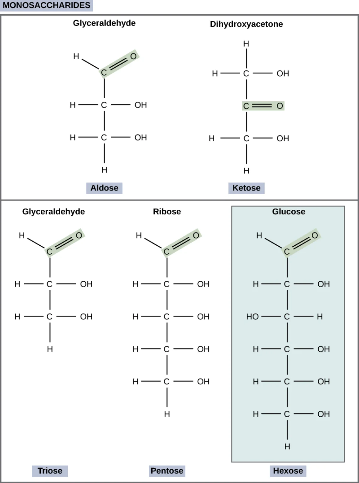
All monosaccharides have multiple hydroxyl groups and one carbonyl group (C=O). The position of the carbonyl splits them into two families.
Aldose: carbonyl is an aldehyde at the END of the chain (C1). Example: glucose.
Ketose: carbonyl is a ketone INSIDE the chain (usually C2). Example: fructose.
Aldose (left, glyceraldehyde) has the carbonyl at the end of the chain. Ketose (right, dihydroxyacetone) has the carbonyl internal to the chain. Credit: OpenStax Biology 2e, CC BY 4.0
Classification by Carbon Count
# Carbons
Name
Example aldose
Example ketose
3
Triose
Glyceraldehyde
Dihydroxyacetone (DHA)
4
Tetrose
Erythrose
Erythrulose
5
Pentose
Ribose
Ribulose
6
Hexose
Glucose, galactose
Fructose
Combining the Two Classifications
You can put the two descriptors together. Glucose is an aldohexose (aldose + hexose = 6-carbon sugar with aldehyde). Fructose is a ketohexose (ketose + hexose = 6-carbon sugar with ketone). Ribose is an aldopentose.
What is the difference between an aldose and a ketose?
Click to reveal answer
An aldose has its carbonyl group as an aldehyde at the end of the carbon chain (C1). A ketose has its carbonyl as a ketone inside the chain (usually C2). All aldoses can be written as the complementary ketose through enolization, but in biology each sugar typically exists as one form.
Classify glucose, fructose, ribose, and glyceraldehyde.
Click to reveal answer
Glucose = aldohexose (6C, aldehyde). Fructose = ketohexose (6C, ketone). Ribose = aldopentose (5C, aldehyde). Glyceraldehyde = aldotriose (3C, aldehyde). Every monosaccharide can be named this way.
Why does dihydroxyacetone lack a chiral center despite being a ketose with hydroxyl groups?
Click to reveal answer
Its three-carbon structure is HOCH2-CO-CH2OH. The central carbon bears a C=O (no H attached), and the two flanking carbons each bear two identical -CH2OH groups on either side. No carbon has four different substituents, so no carbon is chiral. DHA is the only achiral triose.
Sugars have lots of chiral centers and at least one ring. To avoid drawing 3D perspective every time, chemists use two conventional 2D shortcuts: Fischer projections (linear) and Haworth projections (cyclic).
Fischer Projections
Fischer projection rules:
Carbon chain is drawn vertically, with the most oxidized carbon (usually the aldehyde or ketone) at the top.
Horizontal bonds come OUT of the page toward you.
Vertical bonds go BEHIND the page.
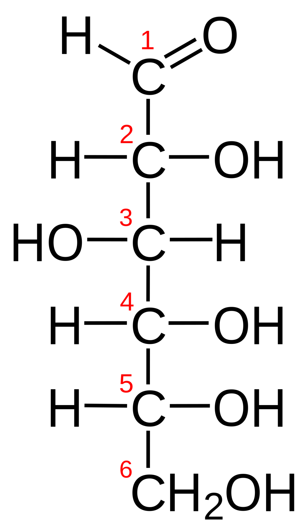
Fischer projection of D-glucose. The C1 aldehyde is at the top; the OH on the bottom-most chiral center (C5) points right, which defines D-sugars. Credit: Wikimedia Commons, CC BY-SA
The “D” or “L” label is set by the bottom chiral center. If its -OH is on the right, the sugar is D. If on the left, L. Almost all naturally occurring sugars are D.
Haworth Projections
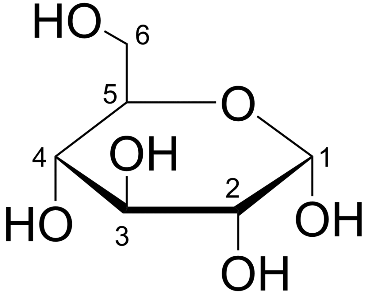
Most sugars exist as rings in solution. A Haworth projection depicts the ring structure as a flat hexagon (six-membered ring = pyranose) or pentagon (five-membered ring = furanose) with substituents drawn up or down.
Haworth projection of glucose. The ring is a flat hexagon; OH groups project up or down. The oxygen in the ring is the bridge between C5 (or C4 for furanoses) and the carbonyl carbon. Credit: Wikimedia Commons, CC BY-SA
Fischer to Haworth Conversion Rule
To convert a Fischer projection to Haworth:
Groups on the right in Fischer go DOWN in Haworth.
Groups on the left in Fischer go UP in Haworth.
The terminal -CH2OH (C6 for hexoses) goes UP for D-sugars.
Pyranose vs. Furanose
Pyranose = 6-membered ring (5 carbons + 1 oxygen). Named after pyran. Most hexoses (glucose, galactose, mannose) prefer the pyranose form.
Furanose = 5-membered ring (4 carbons + 1 oxygen). Named after furan. Fructose (a hexose) and ribose (a pentose, in nucleic acids) exist as furanoses.
The Chair Conformation
For a more realistic 3D view, pyranose rings are sometimes drawn in chair conformation (the six-membered ring looks like a lawn chair). In the chair of β-D-glucopyranose, every large substituent sits equatorial (sticking out horizontally), which is maximally stable. This is part of why glucose is the most common monosaccharide: it is unusually stable in its ring form.
In a Fischer projection, how do you tell whether a sugar is D or L?
Click to reveal answer
Look at the bottom-most chiral center (for a hexose, C5). If the hydroxyl group points to the right, the sugar is D. If it points to the left, L. The designation only cares about that single chiral center, regardless of orientations at other positions.
A hydroxyl group is drawn on the right in a Fischer projection. Where does it appear in the Haworth projection?
Click to reveal answer
Below the ring (pointing DOWN). The rule is "right in Fischer becomes down in Haworth, left in Fischer becomes up in Haworth." For D-sugars, the terminal -CH2OH (C6 in a hexose) points UP.
Why does glucose form a pyranose while fructose forms a furanose?
Click to reveal answer
Glucose's aldehyde carbonyl is at C1. When C5's hydroxyl attacks C1, the resulting ring has 6 members (5C + 1 O): a pyranose. Fructose's ketone carbonyl is at C2. When C5's hydroxyl attacks C2, the resulting ring has 5 members (4C + 1 O): a furanose. The ring size follows directly from which carbon bears the carbonyl.
Sugar stereochemistry is intimidating until you learn the three-word vocabulary: enantiomer, epimer, anomer. Each word describes a specific kind of stereochemical relationship.
D and L, epimers, anomers, mutarotation
Sugar stereochemistry
1
Scroll sideways to see the whole map.
Set at the last chiral carbon Differ at one carbon Differ at the anomeric carbon Interconverting in solution
Why the ring forms at allThe carbonyl carbon is attacked by one of the sugar's own hydroxyls, forming an internal hemiacetal (from an aldose) or hemiketal (from a ketose). That is why the reaction needs no reagent: the sugar closes on itself in water.
What makes a sugar reducingA sugar is reducing if its anomeric carbon is free, because only then can the ring open to expose an aldehyde that can be oxidized. Lock that carbon into a glycosidic bond, as sucrose does with both of its anomeric carbons, and the sugar is non-reducing.
Counting stereoisomersA sugar with n chiral centres has 2ⁿ stereoisomers. Glucose has four, so there are sixteen aldohexoses, of which glucose is one. Ring closure adds a new chiral centre and therefore doubles the count again into α and β.
Every one of these terms is a question about a single carbon. Identify which carbon the question is asking about and the term names itself: the last chiral one gives D or L, any one gives an epimer, and the anomeric one gives α or β.
Enantiomers
Two molecules that are mirror images and non-superimposable. For monosaccharides, this means every chiral center is flipped. D-glucose and L-glucose are enantiomers.
A single pair of enantiomers has identical physical properties (melting point, solubility, etc.) except for the direction of plane-polarized light rotation and their interactions with other chiral molecules (like enzymes).
Epimers
Two sugars that differ at exactly one chiral center. Everything else is identical.
D-glucose and D-galactose are C4 epimers (differ only at C4).
D-glucose and D-mannose are C2 epimers (differ only at C2).
D-galactose and D-mannose are NOT epimers (they differ at two centers - C2 and C4), so they are just diastereomers.
Anomers
A special subset of epimers. Anomers differ only at the anomeric carbon - the carbon that was the carbonyl (C1 for aldoses, C2 for ketoses) and is now the one with the new -OH after ring closure.
Alpha (α) anomer: the new -OH at the anomeric carbon points DOWN in Haworth (opposite side from C6 -CH2OH in D-sugars).
Beta (β) anomer: the new -OH at the anomeric carbon points UP in Haworth (same side as C6 -CH2OH).
Why Alpha vs. Beta Matters
This tiny difference determines whether humans can digest a polymer.
Starch (alpha-1,4 glucose chain) → helical, our amylase can break it.
Both are pure glucose polymers. Only the anomeric configuration at every link differs.
Diastereomers
Any stereoisomers that are NOT mirror images. Epimers and anomers are special types of diastereomers. Enantiomers are NOT diastereomers.
So the hierarchy is:
Stereoisomers (anything with the same connectivity but different 3D arrangement)
Enantiomers (mirror image)
Diastereomers (not mirror image)
Epimers (differ at one chiral center)
Anomers (epimers at the anomeric carbon specifically)
Counting Stereoisomers
An aldohexose has 4 chiral centers (C2, C3, C4, C5), giving 24 = 16 total stereoisomers (8 D-sugars and 8 L-sugars). A ketohexose has 3 chiral centers, giving 23 = 8 stereoisomers (4 D and 4 L). A pentose aldose has 3 chiral centers and also has 8 stereoisomers.
What is the difference between an epimer and an anomer?
Click to reveal answer
Both are diastereomers differing at exactly one chiral center. Epimers differ at any one chiral center. Anomers specifically differ at the anomeric carbon (C1 in aldoses, C2 in ketoses) - the carbon that was the carbonyl before ring closure. Every anomer is an epimer, but not every epimer is an anomer.
D-glucose and D-mannose differ only at one carbon. Which one?
Click to reveal answer
C2. D-mannose is the C2 epimer of D-glucose. The OH group at C2 points right in glucose (Fischer) and left in mannose. D-galactose, in contrast, is the C4 epimer of D-glucose. Galactose and mannose differ at two positions, so they are diastereomers but not epimers.
How do you tell alpha from beta anomers in a D-sugar Haworth projection?
Click to reveal answer
In a D-pyranose Haworth projection, the C6 -CH2OH points UP. Alpha has the anomeric -OH pointing DOWN (opposite the C6 -CH2OH). Beta has the anomeric -OH pointing UP (same side as C6). That is the only difference between the two anomers.
In solution, sugars exist mostly as rings. The ring forms by an intramolecular reaction between a hydroxyl group and the carbonyl group of the same molecule. Understanding this step is the key to understanding anomers and mutarotation.
Hemiacetals and Hemiketals
Hemiacetal: formed when an alcohol attacks an aldehyde. The resulting carbon has -OR and -OH on the same carbon.
Hemiketal: formed when an alcohol attacks a ketone. Same idea, but starting from a ketone.
For sugars, the attack is intramolecular - a hydroxyl from farther down the chain swings around and adds to the carbonyl.
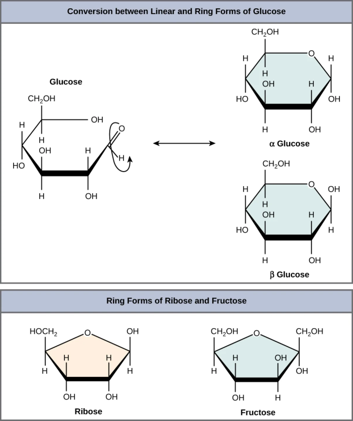
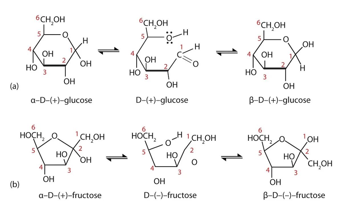
Glucose ring formation. The aldehyde at C1 is attacked by the hydroxyl oxygen at C5, closing a six-membered pyranose ring. The new -OH at C1 defines the anomeric carbon. Credit: OpenStax Biology 2e, CC BY 4.0
The hydroxyl can only reach the carbonyl because of how the chain folds. The specific carbons involved determine ring size.
The Anomeric Carbon Is Special
In the linear form, C1 of glucose is an aldehyde - planar, sp2, no chirality. When it cyclizes to the hemiacetal, C1 becomes a chiral carbon. The new hydroxyl can be on either face (top or bottom), giving two possible stereochemistries. These are the alpha and beta anomers.
The anomeric carbon is always the only carbon with both -OR (ring oxygen) and -OH groups. You can always spot it in a ring structure.
Mutarotation
Pure alpha-D-glucose dissolved in water slowly equilibrates between the alpha and beta anomers. The linear (open-chain) form is the intermediate - the ring opens briefly, the C1 stereochemistry is lost, and re-closure can produce either anomer. After a few hours at room temperature, the solution reaches equilibrium at roughly 64 percent beta, 36 percent alpha, and less than 1 percent open chain.
This is mutarotation. It can be measured by optical rotation (specific rotation changes over time because the two anomers have different rotations).
Mutarotation. Pure alpha- or beta-glucose in solution slowly equilibrates via the linear open-chain aldehyde, reaching about 64% beta and 36% alpha at equilibrium. Credit: Wikimedia Commons, CC BY-SA
Reducing Sugars
Any sugar with a free (non-glycosidically-bonded) anomeric carbon can open into its linear form and expose the aldehyde (or alpha-hydroxy ketone), which can be oxidized. Such sugars are called reducing sugars and react positively with Benedict’s, Tollens’, or Fehling’s reagents.
Glucose, galactose, maltose, lactose: reducing (free anomeric carbon somewhere in the molecule).
Sucrose: non-reducing - both anomeric carbons are locked in the glycosidic bond.
What is a hemiacetal, and how does it form in glucose?
Click to reveal answer
A hemiacetal is a carbon bearing both an -OR group and an -OH group. In glucose, the C5 hydroxyl attacks the C1 aldehyde carbonyl, forming a six-membered ring with a new -OH at C1 (the anomeric hydroxyl) and an -O- bridge to C5. That C1 is a hemiacetal. The corresponding reaction in fructose gives a hemiketal because it starts from a ketone.
What is mutarotation and why does pure alpha-D-glucose show it in water?
Click to reveal answer
Mutarotation is the spontaneous equilibration of alpha and beta anomers in solution. The ring briefly opens into the linear (open-chain) form, losing the C1 stereochemistry, and then recloses to either anomer. Over time, a pure anomer reaches the equilibrium ratio (about 64% beta, 36% alpha for glucose). The changing mix is detectable as a change in optical rotation.
Why is sucrose not a reducing sugar even though it is made from glucose and fructose?
Click to reveal answer
Both anomeric carbons (C1 of glucose and C2 of fructose) are locked in the glycosidic bond. Neither has a free -OH that can equilibrate with an open-chain aldehyde or alpha-hydroxy ketone. Without that free anomeric hydroxyl, sucrose cannot be oxidized and gives a negative Benedict's/Fehling's test.
A handful of monosaccharides do most of the physiologically important work. Memorize these by shape, by epimer relationship, and by food source.
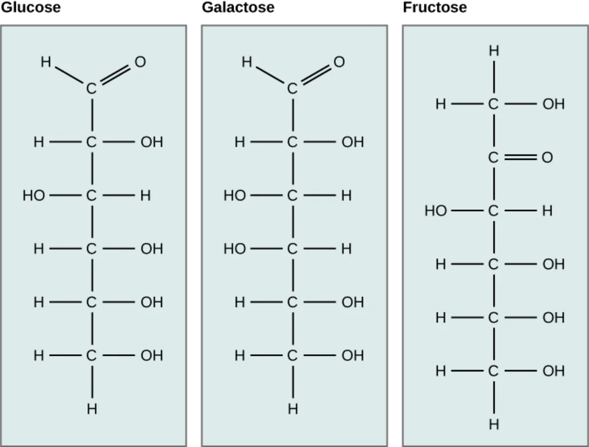
Linear structures of the three most common hexoses. Glucose and galactose are C4 epimers. Fructose differs from glucose by its C2 ketone (ketohexose) vs. C1 aldehyde (aldohexose). Credit: OpenStax Biology 2e, CC BY 4.0
Glucose
The universal monosaccharide. Blood sugar. Every cell in your body can use glucose for energy. It is an aldohexose. In solution, it exists mostly as beta-D-glucopyranose (64%) and alpha-D-glucopyranose (36%). Blood-glucose levels are tightly regulated around 80-100 mg/dL in the fasting state (about 4-5 mM).
Glucose enters cells through GLUT transporters, then gets phosphorylated by hexokinase (or glucokinase in the liver). Once phosphorylated, it cannot diffuse back out.
Fructose
Fruit sugar. A ketohexose and a C2-C5 structural rearrangement of glucose. In solution, fructose favors the furanose (five-membered ring) form. Sweeter than sucrose.
Fructose bypasses the usual glucose regulatory steps. It enters hepatocytes independently of insulin and gets phosphorylated by fructokinase at C1 - skipping phosphofructokinase-1 regulation in glycolysis. This is why very high fructose intake (e.g., from high-fructose corn syrup) can overload the liver faster than an equivalent amount of glucose.
Galactose
Galactose is the C4 epimer of glucose. It is half of lactose (the sugar in milk). After digestion of lactose, galactose is converted back to glucose-6-phosphate by a series of enzymes (the Leloir pathway), the most important of which is galactose-1-phosphate uridyltransferase. Deficiency of this enzyme causes classic galactosemia - a serious neonatal disease.
Fructose and galactose: the side doors into glycolysis
Pathway map
1
Scroll sideways to see the whole map.
Junction metabolite Step that causes disease when missing Cytosol of the liver cell Intermediate
Why fructose is not just another sugarGalactose joins the trunk at glucose-6-phosphate, upstream of PFK-1, so it is subject to the same brake as glucose. Fructose in the liver joins as DHAP and glyceraldehyde-3-phosphate, downstream of PFK-1, so it pours in past the gate. That is why a large fructose load feeds straight into pyruvate, acetyl-CoA, and fatty acid synthesis without waiting for permission.
The enzyme deficienciesEssential fructosuria (fructokinase) is harmless: fructose simply appears in urine. Hereditary fructose intolerance (aldolase B) is not: fructose-1-phosphate piles up and traps the cell's phosphate, which stalls glycogenolysis and gluconeogenesis and causes hypoglycemia after fruit or sucrose. The same pattern holds for galactose: galactokinase deficiency gives cataracts from galactitol, while GALT deficiency is classic galactosemia, which is severe.
Where the cataracts come fromWhen galactose backs up, aldose reductase converts it to galactitol, which cannot leave the lens and pulls in water osmotically. The same enzyme makes sorbitol from glucose, which is the mechanism behind diabetic cataracts and neuropathy.
Two side doors into glycolysis. Galactose enters above PFK-1 and is regulated like glucose. Liver fructose enters below PFK-1 and is not, which is why fructose is handled so much faster and pushes so readily toward fat. Every enzyme marked in red causes a named disease when it is missing.
Mannose
The C2 epimer of glucose. Important as a component of glycoproteins, particularly on the surface of many microorganisms and on newly synthesized eukaryotic proteins in the ER. You do not need to memorize mannose metabolism, but recognize that it is a C2-glucose epimer.
Ribose and Deoxyribose
Both are pentoses (5 carbons).
Ribose is the sugar in RNA and in many cofactors (ATP, NAD+, FAD, CoA). In ring form, it is a furanose.
2-Deoxyribose is the sugar in DNA - the only difference is that C2 has no -OH (just -H).
The missing -OH on deoxyribose makes DNA more chemically stable than RNA, which is why genetic information is stored in DNA rather than RNA.
Why can fructose overload the liver faster than the same amount of glucose?
Click to reveal answer
Fructose is phosphorylated by fructokinase at C1, skipping the main regulatory step of glycolysis (PFK-1). Glucose metabolism is tightly controlled at PFK-1 by ATP, citrate, AMP, and fructose-2,6-BP. Fructose enters glycolysis downstream of this regulation and is poured into the liver’s metabolism with little control, contributing to lipogenesis with high intake.
What is the single structural difference between glucose and galactose?
Click to reveal answer
The orientation of the -OH at C4. Glucose has it on one side; galactose has it flipped. Everything else - the aldehyde at C1, the other -OH groups, the ring closure - is identical. They are C4 epimers.
Why is DNA more chemically stable than RNA?
Click to reveal answer
DNA’s sugar (2-deoxyribose) lacks the C2 hydroxyl that ribose has. The 2’-OH in RNA can attack the phosphodiester backbone intramolecularly, leading to hydrolysis. The absence of that 2’-OH in DNA makes the backbone far less prone to spontaneous cleavage, which is why evolution stores genetic information in DNA rather than RNA.
Two monosaccharides joined by a glycosidic bond form a disaccharide. Three disaccharides dominate the MCAT: maltose, lactose, and sucrose. Each has a specific pair of sugar monomers and a specific bond type.
The Big Three
Disaccharide
Composition
Bond type
Reducing?
Everyday source
Maltose
Glucose + glucose
α-1,4
Yes
Malt, beer, barley
Lactose
Galactose + glucose
β-1,4
Yes
Milk
Sucrose
Glucose + fructose
α-1,2 glycosidic (anomeric-to-anomeric)
No
Table sugar, sugar cane
Maltose
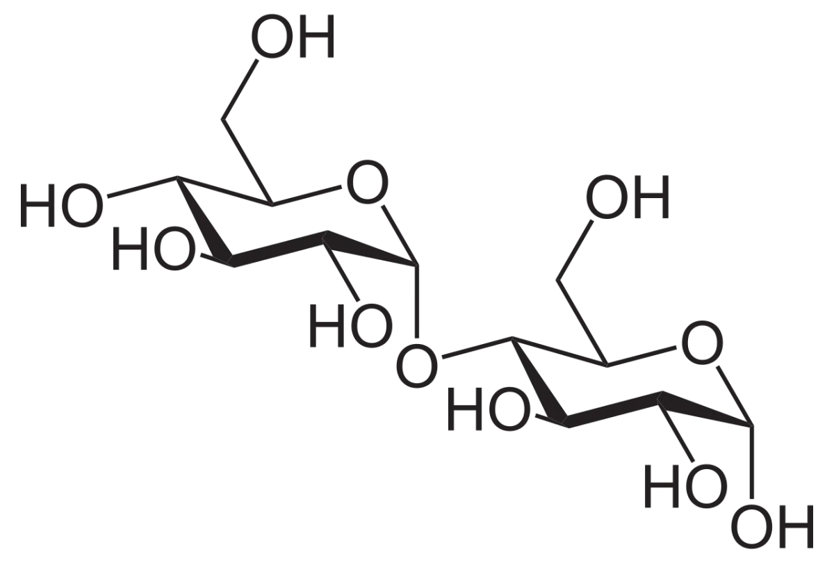
Maltose: two glucose units connected by an alpha-1,4 glycosidic bond. The right-side C1 is free (reducing). Credit: Wikimedia Commons, CC BY-SA
Maltose is a glucose-glucose disaccharide with an alpha-1,4 bond. It is a digestion product of starch - amylase breaks starch into maltose fragments, and maltase (in the intestinal brush border) splits maltose into two glucoses.
Malted barley has high maltose content, hence the name. Maltose is a reducing sugar because one anomeric carbon (on the right-hand glucose) is still free.
Lactose
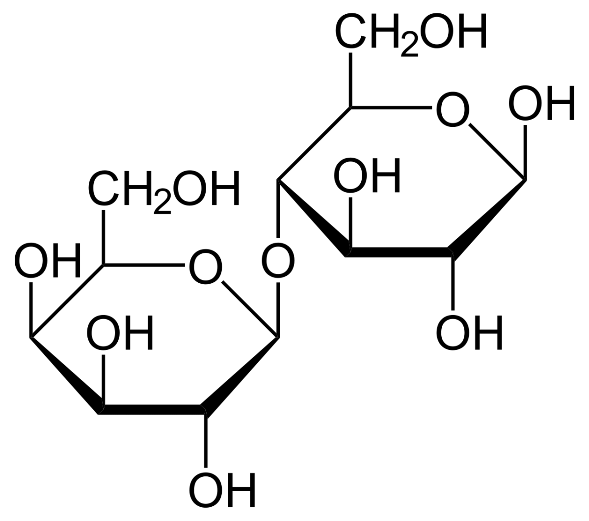
Lactose is the sugar in milk. It is galactose (on the left) linked to glucose (on the right) via a beta-1,4 glycosidic bond. Both units are in their pyranose forms.
Lactose: galactose-beta-1,4-glucose. The beta linkage requires lactase (in humans, only infants and some adults express lactase fully into adulthood). Credit: Wikimedia Commons, CC BY-SA
Digestion requires lactase (a beta-galactosidase) in the brush border. Adults who stop expressing lactase develop lactose intolerance - undigested lactose reaches the colon, bacteria ferment it, and gas and diarrhea result.
Sucrose
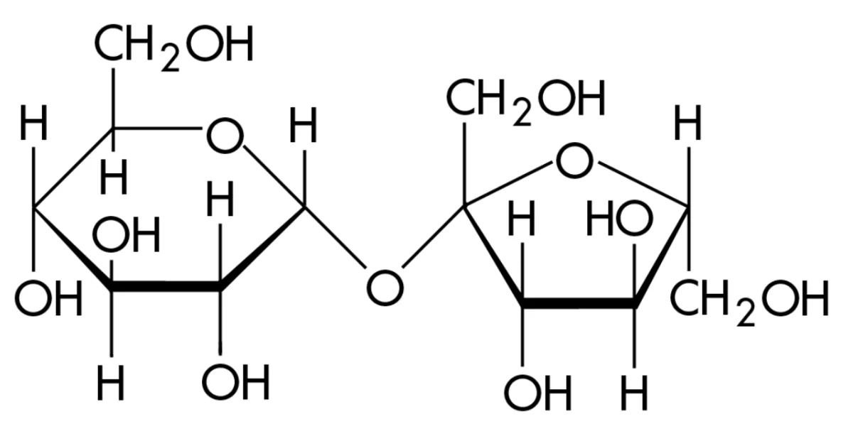
Sucrose is glucose + fructose. The bond is unusual: it is formed between the anomeric C1 of glucose and the anomeric C2 of fructose. Because BOTH anomeric carbons are locked in the glycosidic bond, sucrose has no free anomeric -OH. It is a non-reducing sugar.
Sucrose: glucose-α-1,2-β-fructose. Both anomeric carbons are in the bond, so there is no free -OH available for oxidation. Sucrose is non-reducing. Credit: Wikimedia Commons, CC BY-SA
Digestion requires sucrase in the brush border, which yields glucose + fructose.
Disaccharidases
All disaccharides are broken down by specific brush-border enzymes:
Maltase splits maltose into 2 glucose.
Lactase splits lactose into galactose + glucose.
Sucrase (part of sucrase-isomaltase) splits sucrose into glucose + fructose.
Isomaltase cleaves alpha-1,6 branch points from starch digestion products.
The monosaccharide products are absorbed by enterocytes via specific transporters (SGLT1 for glucose/galactose, GLUT5 for fructose).
What monosaccharides make up each of maltose, lactose, and sucrose, and what is each bond type?
Click to reveal answer
Maltose = glucose + glucose via α-1,4. Lactose = galactose + glucose via β-1,4. Sucrose = glucose + fructose via α-1,2 glycosidic (both anomeric carbons in the bond).
Why is sucrose a non-reducing sugar?
Click to reveal answer
Because both component anomeric carbons (C1 of glucose and C2 of fructose) are in the glycosidic bond. Neither monomer has a free anomeric -OH available to open and expose an aldehyde or alpha-hydroxy ketone. Without that, sucrose cannot be oxidized by Benedict's/Tollens'/Fehling's reagent.
What enzyme digests lactose, and why do many adults stop making it?
Click to reveal answer
Lactase, a brush-border beta-galactosidase, cleaves lactose into galactose + glucose. In many adults, lactase expression decreases after weaning (the ancestral human state). Undigested lactose reaches the colon where bacteria ferment it, producing gas, bloating, and diarrhea - lactose intolerance. Lactase persistence into adulthood is a relatively recent mutation enriched in historically dairy-dependent populations.
A glycosidic bond links an anomeric carbon of one sugar to a group on another molecule (usually another sugar). It is formed by a condensation reaction (water is released) and broken by hydrolysis.
O-Glycosidic vs. N-Glycosidic
O-glycosidic bond: anomeric carbon links to an -OH group. Most sugar-sugar bonds. Most glycoproteins (Ser/Thr -OH) and glycolipids.
N-glycosidic bond: anomeric carbon links to an -NH group. Nucleotides (base-to-ribose) and N-linked glycoproteins (sugar-to-Asn).
Alpha vs. Beta
The critical question for any glycosidic bond is: which anomeric configuration is the bond in?
Alpha linkage: the anomeric -O- is in the alpha position (on the opposite face from C6 -CH2OH in D-sugars). Creates a bend/helix.
Beta linkage: the anomeric -O- is in the beta position (on the same face as C6 -CH2OH). Creates a flat, linear polymer.
Bond Naming
Glycosidic bonds are named by the carbons involved. An alpha-1,4 bond means:
The bond is to the alpha anomer.
C1 of the first sugar is linked to C4 of the second sugar.
Examples:
Starch amylose: alpha-1,4 bonds between glucoses.
Starch amylopectin: alpha-1,4 main chain with alpha-1,6 branches every ~24-30 residues.
Glycogen: alpha-1,4 with alpha-1,6 branches every ~8-12 residues (more branched than starch).
Cellulose: beta-1,4 between glucoses.
Maltose: alpha-1,4.
Lactose: beta-1,4.
Sucrose: alpha-1,2 (glucose C1 to fructose C2, both anomeric).
Formation and Hydrolysis
To form the bond, an -OH on one sugar attacks the anomeric -OH on another sugar. Water is released. This is a condensation reaction. In the body, glycosyltransferases (using activated sugar donors like UDP-glucose) catalyze this step.
To break the bond, a water molecule adds across the -O- between sugars. This is hydrolysis. Glycosidases (amylase, maltase, lactase, sucrase) catalyze this step.
What is the difference between an alpha and a beta glycosidic bond?
Click to reveal answer
The difference is the configuration at the anomeric carbon. Alpha points the linking oxygen on the opposite face from C6 -CH2OH (in D-sugars); beta points it on the same face. In glucose polymers, alpha bonds form helices (digestible starch, glycogen) while beta bonds form flat sheets (indigestible cellulose).
Why can you digest starch but not cellulose even though both are pure glucose polymers?
Click to reveal answer
Starch uses alpha-1,4 linkages; cellulose uses beta-1,4. Human amylases and glucosidases cleave alpha bonds only. We lack cellulase, the enzyme needed to cleave beta bonds. Herbivores like cows rely on gut bacteria that do have cellulase to digest cellulose on their behalf.
How does a glycosidic bond form and how is it broken?
Click to reveal answer
Formation is a condensation: the anomeric -OH of one sugar reacts with an -OH (or -NH) on another molecule, producing the glycosidic bond and releasing water. Breaking is hydrolysis: a water molecule adds across the -O- linkage, regenerating the two separate -OH groups. In the body, synthesis is driven by activated sugar donors (e.g., UDP-glucose) and hydrolysis is catalyzed by specific glycosidases.
Polysaccharides are long chains of monosaccharides. Four MCAT-critical polysaccharides: glycogen, starch, cellulose, and chitin. Each is built from a simple sugar but with distinctive linkages that give it a specific role.
The linkage decides everything
Polysaccharides
1
Scroll sideways to see the whole map.
Plant storage (starch) Animal storage (glycogen) Structural, and indigestible to us The linkage that decides everything
One bond, two worldsAmylose and cellulose are both unbranched chains of glucose. The only difference is α versus β at carbon 1. α gives a helix you can eat; β gives a flat ribbon that hydrogen bonds into fibers you cannot. Humans have no β-glucosidase, which is why cellulose is dietary fiber rather than food.
Why glycogen is so branchedPhosphorylase can only work from a non-reducing end. Branching every ten residues multiplies those ends, so a whole molecule can be dismantled at once when adrenaline arrives. Amylopectin branches every twenty-five, which is fine for a plant that is in no hurry.
Reducing or notA disaccharide is reducing if at least one anomeric carbon is still free. Maltose and lactose each keep one free. Sucrose joins glucose C1 to fructose C2, locking both, which is why it is the classic non-reducing sugar.
Read the linkage and you have the answer. α means digestible and helical; β means structural and, for us, indigestible. A 1,6 bond means a branch point, which means speed. Everything else on this map follows from those two facts.
Storage Polysaccharides
Glycogen (animals)
Glycogen is the animal storage form of glucose. It is stored mainly in liver (for regulating blood glucose) and muscle (for local energy during exercise). Structure: alpha-1,4 main chain with alpha-1,6 branches every 8-12 residues. The branching is extensive because:
Branches create many non-reducing ends where glycogen phosphorylase can simultaneously remove glucose - rapid mobilization.
Branches pack the polymer compactly and increase water-solubility.
Starch is the plant storage form of glucose. It has two components:
Amylose: unbranched alpha-1,4 chains of glucose. Forms a helix.
Amylopectin: alpha-1,4 main chains with alpha-1,6 branches every 24-30 residues. Less branched than glycogen.
Structural Polysaccharides
Cellulose
Cellulose is the structural polysaccharide of plant cell walls. It is a long chain of glucose units joined by beta-1,4 bonds. The beta linkage makes the chain flat and extended, and adjacent chains hydrogen-bond into rigid sheets and microfibrils.
Humans do not make cellulase and cannot digest cellulose. Cellulose is the main component of dietary fiber, which adds bulk to stool and affects gut transit time.
Chitin
Chitin is the structural polysaccharide of insect exoskeletons and fungal cell walls. It is like cellulose but the monomer is N-acetylglucosamine (GlcNAc, glucose with an -NHCOCH3 on C2) linked by beta-1,4 bonds. The extra N-acetyl groups form additional hydrogen bonds, making chitin even tougher than cellulose.
Why Store Glucose as Glycogen Instead of Free Glucose
Free glucose is osmotically active - every millimolar of dissolved glucose contributes to cellular osmotic pressure. A liver cell storing 100 mM worth of free glucose would draw in so much water that it would burst. Glycogen is one giant polymer: each glycogen particle bundles tens of thousands of glucose residues into a single molecule, so the same 100 mM of glucose residues counts as only a few micromolar glycogen particles. Osmotic pressure drops by roughly the branching factor.
Why is glycogen more extensively branched than starch, and why does that matter?
Click to reveal answer
Glycogen has alpha-1,6 branches every 8-12 residues; amylopectin every 24-30. More branches mean more non-reducing ends, so more glycogen phosphorylase molecules can chew off glucose at the same time during a rapid energy demand (sprint, fight-or-flight). Animals tend to need glucose faster than plants, so our storage polymer is built for fast mobilization.
What is the structural difference between cellulose and chitin?
Click to reveal answer
Cellulose is a polymer of glucose connected by beta-1,4 bonds. Chitin is a polymer of N-acetylglucosamine (glucose with an -NH-CO-CH3 group on C2) connected by beta-1,4 bonds. The N-acetyl groups in chitin allow extra hydrogen bonding, making chitin even more rigid than cellulose. Both are structural polysaccharides of beta-1,4 linkages.
Why does the body store glucose as glycogen rather than as free glucose?
Click to reveal answer
Osmotic pressure. Free glucose is osmotically active - millimolar concentrations pull water into the cell. Polymerizing thousands of glucoses into a single glycogen molecule reduces the particle count by thousands and thereby minimizes osmotic pull. The cell can store large amounts of glucose without drawing in destructive amounts of water.
Most proteins that reach the cell surface or the extracellular space are glycosylated - they have one or more sugar chains covalently attached. The same is true of many lipids in the outer leaflet of the cell membrane. These sugar decorations are not structural curiosities; they are critical for folding, stability, recognition, and signaling.
Glycoproteins
A glycoprotein is a protein with one or more covalently attached sugar chains. Glycosylation happens in the endoplasmic reticulum and Golgi apparatus.
Two main glycosylation types, named after the amino acid the sugar chain links to:
N-linked glycosylation: sugar attaches to the -NH2 of an asparagine (Asn) side chain. The first sugar is N-acetylglucosamine (GlcNAc).
O-linked glycosylation: sugar attaches to the -OH of a serine or threonine side chain.
What Glycans Do for a Protein
Folding assistance: N-linked glycans act as “quality control flags” in the ER. Chaperones (calnexin, calreticulin) recognize incompletely trimmed glycans and retain unfolded proteins until folding completes.
Stability: glycans shield protein surfaces from proteases and extend serum half-life. Engineered protein drugs are often glycosylated to last longer in circulation.
Cell-cell recognition: surface glycans are the “signature” of a cell. Immune cells read them to distinguish self from non-self, tissue from tissue.
Targeting: mannose-6-phosphate on lysosomal enzymes is the address tag that sends them to the lysosome. Defects in this tagging cause I-cell disease.
The Glycocalyx
Every eukaryotic cell is covered by a dense layer of sugars sticking out from glycoproteins and glycolipids on the outer leaflet of the plasma membrane. This is the glycocalyx. It protects the membrane, mediates interactions with other cells, and presents receptors for hormones, pathogens, and immune cells.
Glycolipids
Lipids (mostly sphingolipids) on the outer membrane leaflet can carry sugar chains. The result is a glycolipid. The sugar portion faces the extracellular space.
Major glycolipid classes:
Cerebrosides: one sugar (usually glucose or galactose) on a ceramide. Abundant in brain tissue.
Gangliosides: complex branched oligosaccharides containing sialic acid on a ceramide. Especially abundant in neurons; defects in their catabolism cause lysosomal storage diseases (Tay-Sachs, Gaucher).
Blood group antigens (ABO system) are partly carried by glycolipids on the red blood cell surface. This is covered in the next section.
Proteoglycans
A proteoglycan is a protein with one or more long, negatively charged carbohydrate chains called glycosaminoglycans (GAGs) attached. GAGs are repeating disaccharide units, most bearing sulfate groups. Examples of GAGs: chondroitin sulfate, heparan sulfate, dermatan sulfate, keratan sulfate, hyaluronic acid.
Proteoglycans dominate the extracellular matrix of connective tissues like cartilage. They act like sponges, binding water and giving cartilage its compressive strength.
What is the difference between N-linked and O-linked glycosylation?
Click to reveal answer
N-linked glycosylation attaches a sugar chain to the -NH2 side chain of an asparagine (Asn, N) residue, with N-acetylglucosamine as the first sugar. O-linked glycosylation attaches a chain to the -OH side chain of a serine or threonine. Both happen in the ER/Golgi and use specific glycosyltransferases.
What is the glycocalyx and why does it matter for cell-cell recognition?
Click to reveal answer
The glycocalyx is the dense layer of carbohydrate chains on the outer leaflet of a cell's plasma membrane, built from glycoproteins and glycolipids. Its distinctive sugar patterns serve as identifiers that immune cells, hormones, and other cells can recognize. Blood group antigens, for example, are glycocalyx components; viruses like influenza exploit specific glycocalyx sugars to dock onto host cells.
Why is cartilage so compressible yet resilient?
Click to reveal answer
Cartilage extracellular matrix is packed with proteoglycans - proteins decorated with long sulfated glycosaminoglycans. The negatively charged GAGs bind large amounts of water like a sponge. When compressed, water is squeezed out; when the load is removed, the negative charges pull water back in. The bulk water flow gives cartilage its resilience.
Cell surface sugars are not passive decoration. They are the fingerprints cells use to identify each other and to catch passing signals. The ABO blood group system is the cleanest MCAT example.
The ABO Blood Group System
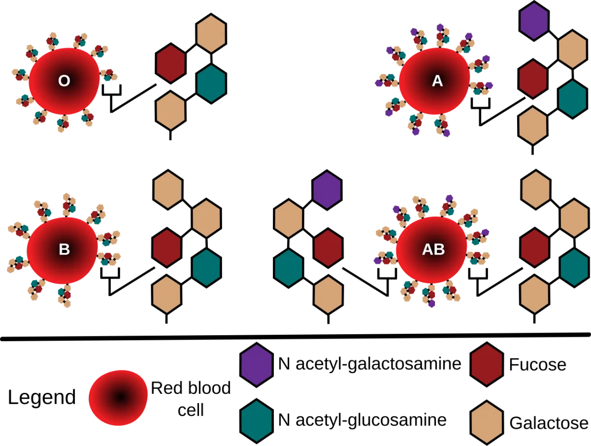
Red blood cells display a glycan on their surface built on a core called the H antigen. Different blood types differ in what single extra sugar (if any) is added to the H antigen:
Blood type
Antigen on RBC
Antibodies in plasma
Can receive from
A
H antigen + N-acetylgalactosamine (GalNAc)
Anti-B
A, O
B
H antigen + galactose
Anti-A
B, O
AB
Both A and B antigens
None
A, B, AB, O (universal recipient)
O
H antigen only
Anti-A and anti-B
O only (universal donor of RBCs)
ABO blood group antigens. Each blood type has a specific terminal sugar on the core H antigen: GalNAc for A, galactose for B, both for AB, and neither for O. Credit: Wikimedia Commons, CC BY-SA
Why Universal Donor and Recipient
The body makes antibodies against whichever A/B antigens it lacks. These anti-A and anti-B antibodies arise naturally in infancy from exposure to environmental sugars on bacteria.
Type A person has anti-B antibodies. If given type B blood, the antibodies clump B cells - a transfusion reaction.
Type O has anti-A and anti-B. Type O cannot receive A or B; Type O is the universal donor of red cells because O cells have neither A nor B to trigger attack.
Type AB has no anti-A or anti-B antibodies. Type AB is the universal recipient.
Rh System
Separate from ABO, the Rh antigen is a protein (not a carb). Someone who expresses Rh is Rh+; absent is Rh-. Critical clinically because an Rh- mother carrying an Rh+ fetus can develop anti-Rh antibodies that attack a subsequent Rh+ pregnancy (hemolytic disease of the newborn). Anti-Rh immunoglobulin (RhoGAM) given prophylactically prevents this sensitization.
Selectins and Lectins
Lectins are proteins that bind specific carbohydrate structures. They are not enzymes - they just grab sugars. Many cellular recognition events use lectins on one side and glycans on the other.
Selectins are a subfamily of lectins on the surface of white blood cells and endothelium. During inflammation, selectins on the endothelial surface grab sugar chains on leukocyte surfaces, causing the leukocytes to slow down and roll along the blood vessel wall. This is the first step of leukocyte extravasation - the process by which white blood cells leave the bloodstream to fight infection.
Integrins (protein-protein binders) later replace the selectin interaction with a firmer attachment, and the white cell squeezes through the endothelium into tissue.
Cell-Cell and Host-Pathogen Recognition
Many viruses and bacteria identify host cells by binding specific surface glycans:
Influenza virus binds sialic acid on respiratory epithelium.
Rotavirus binds histo-blood group antigens on gut epithelium.
H. pylori binds Lewis b antigens on stomach epithelium.
Blocking the sugar-receptor interaction can block infection. Some experimental drugs are sugar mimetics designed to saturate the viral binding protein and prevent attachment.
What is the molecular difference between type A and type B blood?
Click to reveal answer
Both types start from the H antigen, a core glycan on the red blood cell surface. Type A adds an N-acetylgalactosamine (GalNAc) to the H core. Type B adds a galactose. Type AB has both. Type O leaves the H antigen untouched. Type A people have natural anti-B antibodies; type B have anti-A; type O have both; type AB have neither.
Why is type O the universal RBC donor but type AB the universal plasma donor?
Click to reveal answer
Universal donor depends on what is being transfused. Type O red cells carry no A or B antigens, so they cannot trigger anti-A or anti-B antibodies in any recipient. But type O plasma contains both anti-A and anti-B antibodies, which would attack a non-O recipient. Type AB plasma has neither anti-A nor anti-B antibodies, so it can be given to any recipient without causing immune attack, making AB the universal plasma donor.
What do selectins do during inflammation?
Click to reveal answer
Selectins are lectins (sugar-binding proteins) expressed on activated endothelium that bind carbohydrate structures on circulating leukocytes. The binding is transient and triggers "rolling" - the leukocyte slows down and rolls along the vessel wall. This is step 1 of leukocyte extravasation; integrin-mediated firm adhesion and transmigration follow.