
Structure Determination
Knowing a protein’s amino acid sequence is a start, but function comes from 3D shape. Three techniques dominate structural biology. The MCAT focuses mostly on X-ray crystallography; NMR and cryo-EM often appear as passage context.
X-Ray Crystallography
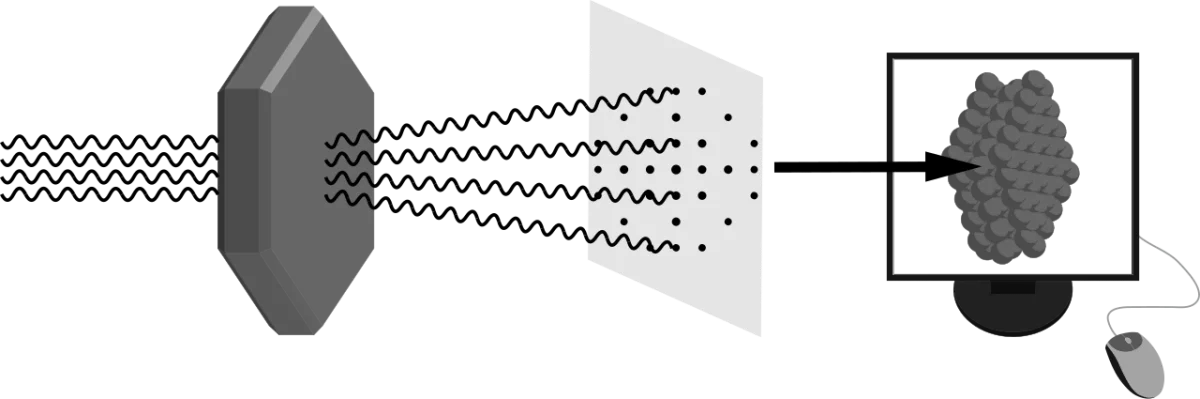
The gold-standard method for atomic-resolution protein structures since the 1960s. The workflow:
- Grow a crystal of the purified protein. This is often the hardest step - proteins do not always crystallize cleanly.
- Diffract X-rays through the crystal. The regular lattice of protein molecules scatters X-rays into a characteristic spot pattern.
- Compute an electron density map from the diffraction pattern (mathematically, an inverse Fourier transform).
- Fit the amino acid sequence into the electron density to build the 3D model.
Strengths and Limits
- Strength: atomic resolution (~1-2 Å) for well-ordered crystals. Shows every backbone and side-chain atom.
- Limit: requires a crystal. Membrane proteins and flexible proteins often refuse to crystallize. The crystal also locks the protein in one conformation, so you may miss dynamic motion.
Nuclear Magnetic Resonance (NMR) Spectroscopy
NMR uses the magnetic properties of atomic nuclei (primarily 1H, 13C, 15N) in strong magnetic fields. The chemical environment of each nucleus shifts its resonance frequency slightly, and interactions between neighboring nuclei produce characteristic peaks. Computational methods convert NMR spectra into 3D structure constraints.
NMR works on proteins in solution, not crystals. This is a major advantage - you see the protein in a more natural environment and can also measure dynamics. The major limit is size: NMR becomes very difficult above ~30 kDa because peaks overlap and relaxation effects distort spectra.
Cryo-Electron Microscopy (Cryo-EM)
Cryo-EM captures thousands of pictures of individual protein molecules frozen in a thin layer of vitreous ice. Each picture is a different angle on the same molecule. Computer software aligns and averages the pictures to reconstruct a 3D density map, from which a model is built.
Cryo-EM does not need a crystal. It excels at large complexes (ribosomes, whole viruses, membrane proteins in their natural environment). Resolution has improved dramatically, and modern cryo-EM routinely achieves 2-3 Å, competitive with crystallography. The 2017 Nobel Prize in Chemistry recognized this “resolution revolution.”
Quick Comparison
| Method | Sample state | Size range | Resolution | Sees dynamics? |
|---|---|---|---|---|
| X-ray crystallography | Crystalline solid | ~5 kDa to megadalton complexes | ~1-3 Å | No (static snapshot) |
| NMR | Solution | Usually < 30 kDa | ~2-4 Å | Yes (time-resolved) |
| Cryo-EM | Vitreous ice (flash-frozen) | Large complexes (>150 kDa typical) | ~2-4 Å modern | Partial (can capture different states) |