
Signaling & Blood Typing
Cell surface sugars are not passive decoration. They are the fingerprints cells use to identify each other and to catch passing signals. The ABO blood group system is the cleanest MCAT example.
The ABO Blood Group System
Red blood cells display a glycan on their surface built on a core called the H antigen. Different blood types differ in what single extra sugar (if any) is added to the H antigen:
| Blood type | Antigen on RBC | Antibodies in plasma | Can receive from |
|---|---|---|---|
| A | H antigen + N-acetylgalactosamine (GalNAc) | Anti-B | A, O |
| B | H antigen + galactose | Anti-A | B, O |
| AB | Both A and B antigens | None | A, B, AB, O (universal recipient) |
| O | H antigen only | Anti-A and anti-B | O only (universal donor of RBCs) |
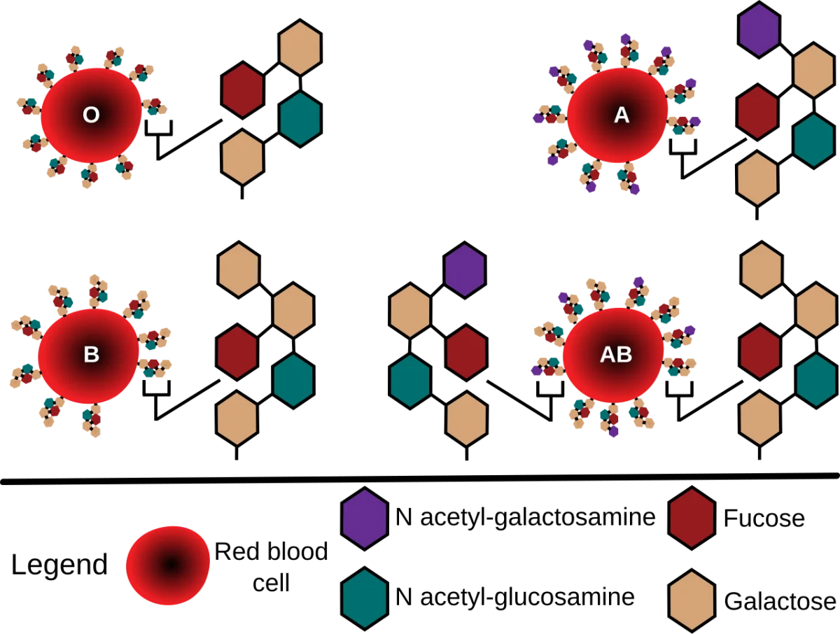
Why Universal Donor and Recipient
The body makes antibodies against whichever A/B antigens it lacks. These anti-A and anti-B antibodies arise naturally in infancy from exposure to environmental sugars on bacteria.
- Type A person has anti-B antibodies. If given type B blood, the antibodies clump B cells - a transfusion reaction.
- Type O has anti-A and anti-B. Type O cannot receive A or B; Type O is the universal donor of red cells because O cells have neither A nor B to trigger attack.
- Type AB has no anti-A or anti-B antibodies. Type AB is the universal recipient.
Rh System
Separate from ABO, the Rh antigen is a protein (not a carb). Someone who expresses Rh is Rh+; absent is Rh-. Critical clinically because an Rh- mother carrying an Rh+ fetus can develop anti-Rh antibodies that attack a subsequent Rh+ pregnancy (hemolytic disease of the newborn). Anti-Rh immunoglobulin (RhoGAM) given prophylactically prevents this sensitization.
Selectins and Lectins
Lectins are proteins that bind specific carbohydrate structures. They are not enzymes - they just grab sugars. Many cellular recognition events use lectins on one side and glycans on the other.
Selectins are a subfamily of lectins on the surface of white blood cells and endothelium. During inflammation, selectins on the endothelial surface grab sugar chains on leukocyte surfaces, causing the leukocytes to slow down and roll along the blood vessel wall. This is the first step of leukocyte extravasation - the process by which white blood cells leave the bloodstream to fight infection.
Integrins (protein-protein binders) later replace the selectin interaction with a firmer attachment, and the white cell squeezes through the endothelium into tissue.
Cell-Cell and Host-Pathogen Recognition
Many viruses and bacteria identify host cells by binding specific surface glycans:
- Influenza virus binds sialic acid on respiratory epithelium.
- Rotavirus binds histo-blood group antigens on gut epithelium.
- H. pylori binds Lewis b antigens on stomach epithelium.
Blocking the sugar-receptor interaction can block infection. Some experimental drugs are sugar mimetics designed to saturate the viral binding protein and prevent attachment.