An enzyme is a biological catalyst - a protein (or in rare cases an RNA) that speeds up a chemical reaction without being consumed. The rate increase is enormous. A typical enzyme accelerates its reaction by 106 to 1017 fold. Without enzymes, digesting a sandwich would take years.
Crucially, enzymes do not change whether a reaction can happen. A reaction with positive Gibbs free energy (ΔG > 0) stays non-spontaneous whether or not an enzyme is present. Enzymes only change how quickly an already-possible reaction reaches equilibrium.
Lowering Activation Energy
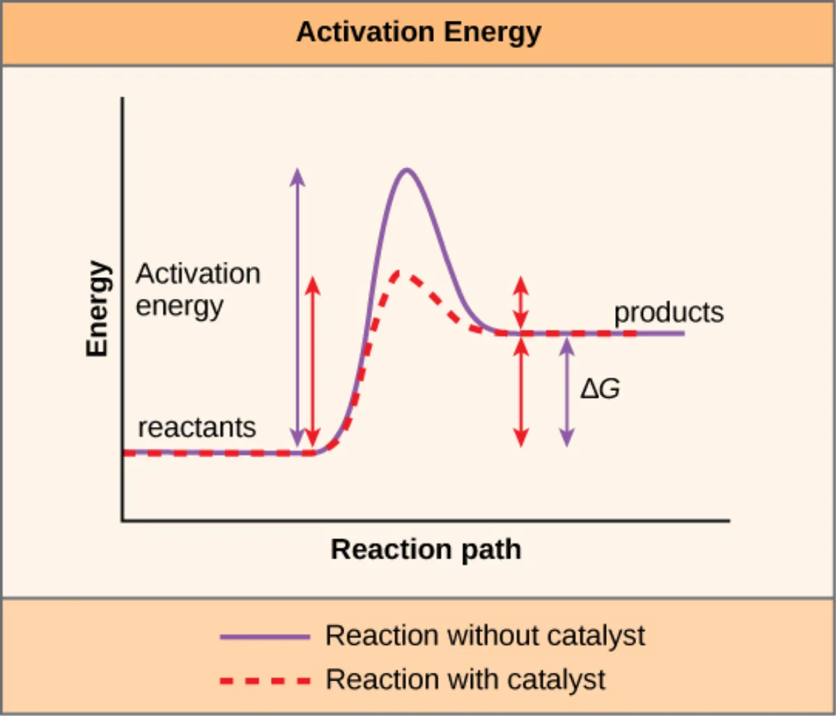
Every reaction has to climb an energy hill called the activation energy (Ea) before it can proceed. Substrates must twist, bend, or partially break bonds into a strained intermediate called the transition state before new bonds can form. Enzymes stabilize that transition state. A lower energy hill means more molecules have enough energy to make it over at body temperature, so the reaction runs faster.
A catalyzed reaction (lower curve) has a smaller activation energy but the overall ΔG between reactants and products is unchanged. Credit: OpenStax Biology 2e, CC BY 4.0
The Active Site
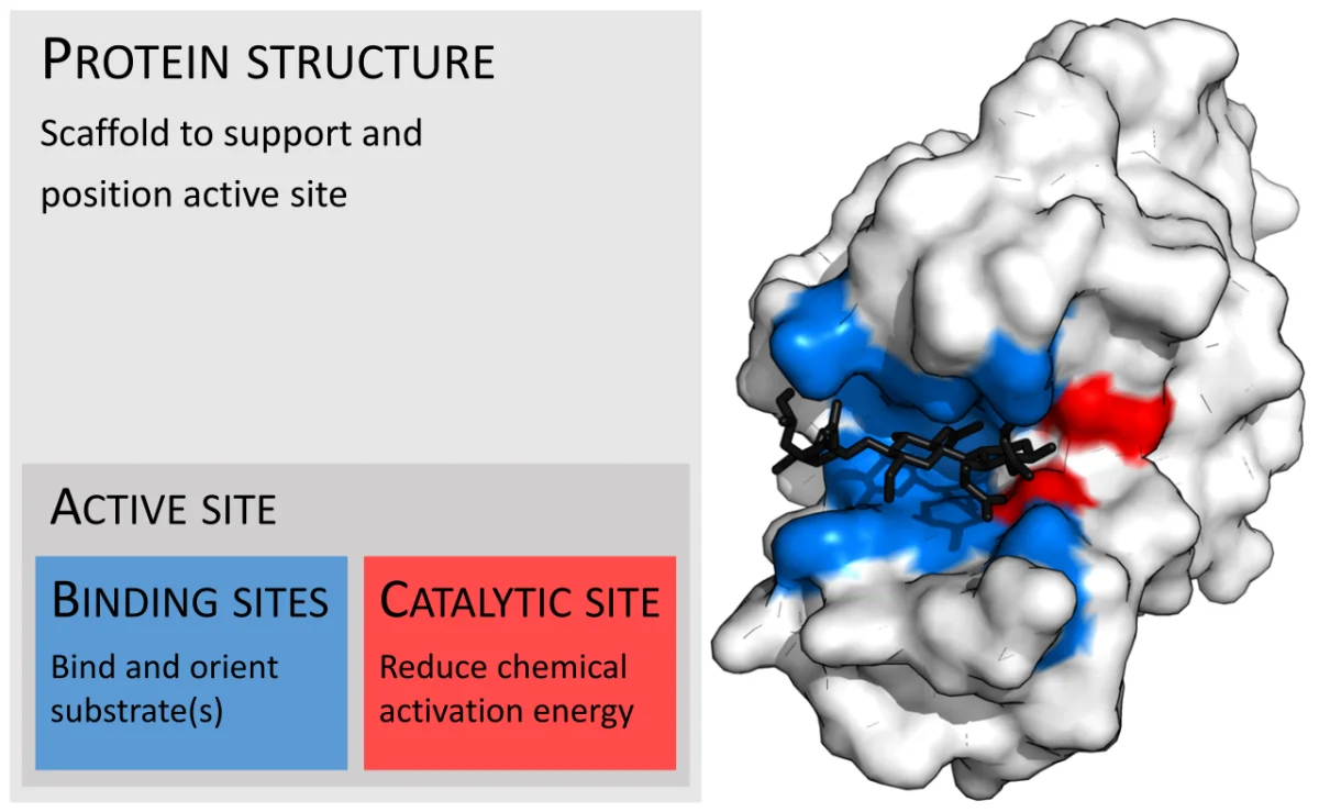
The active site is a small pocket or cleft on the enzyme, usually only a few amino acids wide, where the substrate binds and catalysis happens. Two types of residues matter there:
Binding residues grip the substrate through hydrogen bonds, van der Waals forces, hydrophobic packing, and sometimes ionic interactions. They set the specificity.
Catalytic residues actually participate in the bond-making or bond-breaking. Common offenders are Ser, His, Asp, Cys, Lys, and Tyr - amino acids with reactive side chains.
The active site is a tiny fraction of the whole protein. Most of the enzyme is scaffolding that holds those critical residues in exactly the right 3D positions.
Substrate Specificity
Enzymes are dramatically more specific than non-biological catalysts. Hexokinase phosphorylates glucose but ignores nearly identical sugars like galactose. Specificity comes from the shape, charge, and hydrophobicity of the active site matching only a narrow set of substrates.
Some enzymes are absolutely specific (only one substrate, e.g., urease acts only on urea). Others have group specificity (any sugar with a free -OH, or any peptide bond adjacent to a lysine). Most fall somewhere in between.
Catalytic Mechanisms
Enzymes lower activation energy through a handful of recurring tricks. The MCAT expects you to recognize the four main mechanisms:
Acid-base catalysis: an active-site residue donates or accepts a proton at a key step (histidine’s pKa near 6 makes it the star).
Covalent catalysis: a nucleophilic residue (often serine, cysteine, or lysine) transiently forms a covalent bond with the substrate, creating a reactive intermediate. Serine proteases (chymotrypsin, trypsin) build an acyl-enzyme intermediate through Ser-OH.
Metal-ion catalysis: a metal cofactor (Zn2+, Fe2+/Fe3+, Mg2+) polarizes substrates, stabilizes negative charges, or performs redox. Carbonic anhydrase uses Zn2+; cytochromes use iron.
Electrostatic catalysis: charged residues in the active site stabilize oppositely charged transition-state intermediates (the “oxyanion hole” in serine proteases is the classic example).
The Catalytic Triad
The textbook case is the Ser-His-Asp triad of chymotrypsin. Asp polarizes His; His deprotonates Ser; Ser attacks the peptide bond’s carbonyl carbon. All three mechanisms appear simultaneously: acid-base (His shuttles protons), covalent (Ser forms acyl-enzyme), and electrostatic (Asp and the oxyanion hole stabilize charged intermediates).
Cofactors and Coenzymes
Many enzymes need a non-protein partner to function:
Term
What it is
Examples
Cofactor
Any non-protein helper
Zn2+, Mg2+, Fe2+, a coenzyme
Coenzyme
An organic cofactor, often vitamin-derived
NAD+, FAD, CoA, PLP, biotin
Prosthetic group
A cofactor covalently or tightly bound to the enzyme
Heme in catalase, biotin in pyruvate carboxylase
Apoenzyme
The protein part alone - inactive
Hexokinase minus Mg2+
Holoenzyme
Apoenzyme + cofactor, fully active
Working hexokinase
The B Vitamin Cofactor Map
The MCAT loves “vitamin → coenzyme → reaction → deficiency disease” chains. Memorize the vitamin-to-coenzyme link; the rest is context.
Methyl transfer (homocysteine → methionine); methylmalonyl-CoA mutase
C
Ascorbate
Ascorbic acid
Reducing agent for prolyl hydroxylase (collagen synthesis)
Does an enzyme change the ΔG or the equilibrium constant of a reaction?
Click to reveal answer
No. Enzymes only lower the activation energy (Ea), speeding up the rate at which equilibrium is reached. The ΔG, Keq, and overall thermodynamic favorability are identical with or without the enzyme.
What is the difference between an apoenzyme and a holoenzyme?
Click to reveal answer
The apoenzyme is the protein portion alone, which is catalytically inactive. The holoenzyme is the apoenzyme plus its required cofactor (metal ion, coenzyme, or prosthetic group), and is the fully functional form.
Which amino acid side chains most often serve as catalytic residues at active sites?
Click to reveal answer
Ser, His, Asp (and Glu), Cys, Lys, and Tyr. These have functional groups capable of acting as nucleophiles, proton donors/acceptors, or stabilizers of charged intermediates. The classic example is the Ser-His-Asp "catalytic triad" of serine proteases like chymotrypsin.
Biologists have used two models to describe how a substrate fits into an active site. The MCAT expects you to know both, to know which is modern and correct, and to know why the distinction matters.
Lock-and-Key Model (Emil Fischer, 1894)
In the lock-and-key model, the active site has a fixed, rigid shape. Only a substrate with the exact complementary shape can fit - like a key fitting a particular lock. Binding does not change either partner; the shapes are pre-formed.
This model correctly predicts enzyme specificity but is too simple. It implies the enzyme is a static sculpture, which it is not.
Induced Fit Model (Daniel Koshland, 1958)
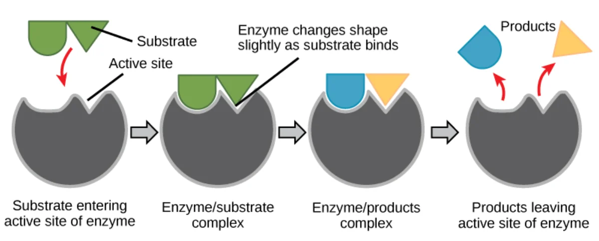
In induced fit, the active site is flexible. When the substrate begins to bind, the enzyme changes shape to wrap around the substrate more snugly. The substrate may also deform slightly. The resulting “mutual adjustment” creates the catalytically active arrangement of residues that was not quite right in the empty enzyme.
Induced fit: both enzyme and substrate adjust their shapes when they come together. The empty active site is not the same shape as the bound active site. Credit: OpenStax Biology 2e, CC BY 4.0
Induced fit is the modern, correct model for most enzymes. It explains why enzymes are so good at stabilizing the transition state - the post-binding conformation is complementary to the transition state, not the ground-state substrate.
Hexokinase - The Classic Example
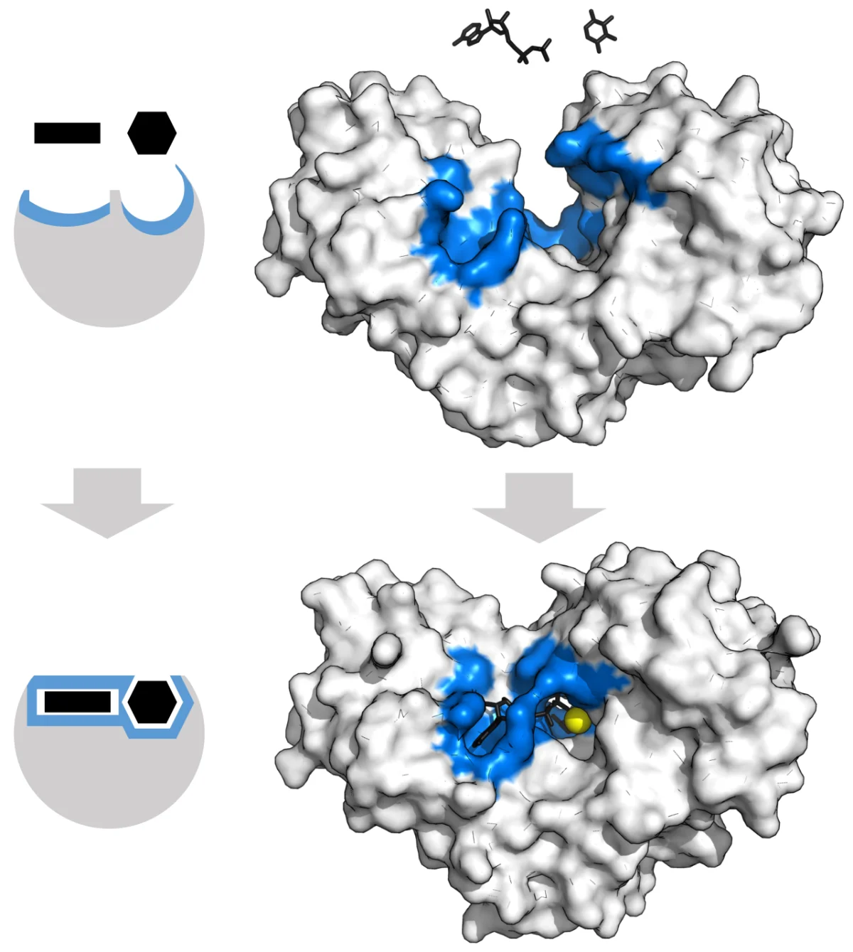
Hexokinase is the first enzyme of glycolysis. It transfers a phosphate from ATP onto glucose. When only the enzyme is present, the two lobes of hexokinase are open. When glucose binds, the lobes clamp down like a Venus flytrap, surrounding the sugar and excluding water.
Why the drama? Water would ruin everything. If water got into the active site, the ATP phosphate would be transferred onto water instead of glucose (ATP hydrolysis, a wasted reaction). By slamming shut on glucose, hexokinase guarantees that only glucose - not water - gets phosphorylated.
Hexokinase open vs. closed states. Glucose binding triggers a large conformational change that closes the active site and excludes water. Credit: Wikimedia Commons, CC BY-SA
What is the key difference between lock-and-key and induced fit models?
Click to reveal answer
Lock-and-key says the active site is rigid and pre-shaped to fit only one exact substrate. Induced fit says the active site (and sometimes the substrate) changes shape during binding, producing the active conformation only after contact. Induced fit is the modern, correct model.
Why does hexokinase undergo such a large conformational change on binding glucose?
Click to reveal answer
Closing around glucose excludes water from the active site. If water were present, the enzyme would wastefully transfer the phosphate from ATP onto water (ATP hydrolysis) instead of glucose. Induced fit enforces substrate specificity by geometrically shielding the chemistry.
The International Union of Biochemistry and Molecular Biology sorts every enzyme into one of six classes based on the reaction it catalyzes. The MCAT will not ask for EC numbers, but you absolutely must recognize the six classes and what each one does.
The Six Classes
Class
What it does
Common suffix / name clue
Example
1. Oxidoreductases
Transfer electrons (redox)
“dehydrogenase”, “oxidase”, “reductase”
Lactate dehydrogenase
2. Transferases
Move a functional group from one molecule to another
”kinase” (moves phosphate), “transaminase”
Hexokinase
3. Hydrolases
Break bonds using water
”ase” on a substrate name (lipase, protease, amylase)
Trypsin
4. Lyases
Break bonds without water, often leaving a double bond
”synthase”, “decarboxylase”, “aldolase”
Pyruvate decarboxylase
5. Isomerases
Rearrange atoms within a molecule (no net change in formula)
“isomerase”, “mutase”
Phosphoglucose isomerase
6. Ligases
Join two molecules using ATP hydrolysis
”synthetase”, “ligase”
DNA ligase, pyruvate carboxylase
How to Tell Which Class from the Name
Enzyme names are often transparent once you know the suffix rules.
-ligase or -carboxylase (with ATP) joins things. Ligase. Example: pyruvate carboxylase, DNA ligase.
One-Sentence Examples per Class
Oxidoreductase: lactate dehydrogenase moves H (and electrons) between lactate and pyruvate, paired with NAD+/NADH.
Transferase: hexokinase transfers a phosphate from ATP onto glucose, producing glucose-6-phosphate.
Hydrolase: trypsin adds water across a peptide bond, cleaving a protein into fragments.
Lyase: fructose-1,6-bisphosphate aldolase cleaves a six-carbon sugar into two three-carbon pieces without water.
Isomerase: triose phosphate isomerase interconverts dihydroxyacetone phosphate and glyceraldehyde-3-phosphate.
Ligase: DNA ligase seals a nick in the DNA backbone, using ATP to drive the joining.
Which enzyme class uses water to break a bond, and which uses ATP to form a bond?
Click to reveal answer
Hydrolases (class 3) use water to cleave bonds. Ligases (class 6) use ATP to form bonds between two substrates. The two are, in effect, thermodynamic opposites.
An enzyme rearranges glucose-6-phosphate into fructose-6-phosphate without changing the atomic formula. What class is it?
Click to reveal answer
Isomerase (class 5). This particular enzyme is phosphoglucose isomerase. No atoms are added or removed - the molecule is simply rearranged.
Is glycogen synthase a lyase or a ligase?
Click to reveal answer
Neither exactly as named - the "synthase" suffix suggests a lyase, but glycogen synthase actually uses UDP-glucose (an activated sugar) and is classified as a glycosyltransferase. The rule "synthase = lyase, synthetase = ligase" is a good starting point, but not every enzyme obeys it perfectly.
Enzyme kinetics is the study of how fast enzymes work and how that speed changes when you vary conditions. The core equation is Michaelis-Menten, published by Leonor Michaelis and Maud Menten in 1913. Every MCAT kinetics question ultimately comes back to it.
The Scenario
Mix one substrate (S) with an enzyme (E). The enzyme grabs the substrate to form an enzyme-substrate complex (ES), does its chemistry, then releases the product (P) and returns free.
E + S ⇌ ES → E + P
If you keep adding substrate and measure how fast product appears, you get the classic Michaelis-Menten curve.
AnimationEnzyme Kinetics (Michaelis–Menten)
Key idea
As substrate rises, rate climbs then plateaus at Vmax — every enzyme is busy. Km is the [S] giving half-Vmax (lower Km = tighter binding). Competitive inhibitors raise Km (more substrate beats them); noncompetitive inhibitors lower Vmax (substrate can't).
Slide [S] to move along the curve; switch the inhibitor to see Km and Vmax shift.
Three features jump out:
At low [S], the curve is nearly linear. Doubling substrate doubles the rate.
At high [S], the curve flattens. Enzyme is saturated - nearly every molecule is already working on a substrate. Adding more does not help.
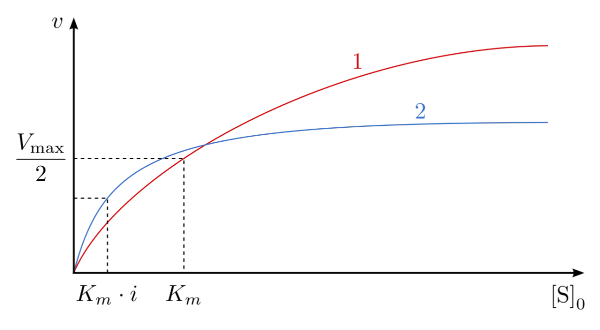
The asymptote is called Vmax. The substrate concentration at half-Vmax is called Km.
The Equation
V0=Km+[S]Vmax[S]
V0 = initial velocity (rate of product formation at t near zero, before product builds up enough to matter)
Vmax = maximum rate achievable, when enzyme is fully saturated
Km = Michaelis constant, the [S] at which V0 = Vmax / 2
[S] = substrate concentration
What Km Actually Means
Km has two equivalent interpretations:
The substrate concentration that gives half the maximum reaction rate.
A rough measure of the affinity of the enzyme for the substrate. A small Km means high affinity (the enzyme is satisfied at low [S]). A large Km means low affinity (the enzyme needs a lot of substrate before it hits half-Vmax).
Assumptions of Michaelis-Menten
The derivation relies on a few assumptions that matter for the MCAT:
Steady state: the concentration of ES is approximately constant while you measure. Formation and breakdown of ES are balanced.
[S] >> [E]: you add much more substrate than enzyme, so free [S] is not measurably depleted by forming ES.
V0 is measured before the reverse reaction matters. Product concentration is near zero, so P → S is negligible.
One substrate, one active site. Simple Michaelis-Menten kinetics do not apply directly to allosteric enzymes with multiple binding sites (those give sigmoidal curves - covered later).
What does a low Km tell you about an enzyme?
Click to reveal answer
A low Km means high affinity for the substrate. The enzyme reaches half-maximal velocity at very low substrate concentrations, so it can run effectively even when substrate is scarce.
At what substrate concentration does V0 equal half of Vmax?
Click to reveal answer
When [S] equals Km. This is essentially the definition of Km - it is the substrate concentration that produces half the maximum rate.
Why do we measure initial velocity (V0) rather than rate later in the reaction?
Click to reveal answer
At t near zero, [S] is still essentially the starting concentration and product has not yet built up enough to drive the reverse reaction. Measuring V0 isolates the forward reaction at a known, controlled [S], which is what the equation requires.
Km and Vmax are the two parameters you will be asked to read from kinetics plots and interpret in passages. They carry different information, and the MCAT will test whether you can tell them apart.
Vmax is About Enzyme Amount
Vmax is the ceiling - the maximum velocity the enzyme can achieve when substrate is so abundant that every active site is occupied at all times. Double the amount of enzyme and Vmax doubles. Half the enzyme and Vmax is halved. Vmax is proportional to [E]total.
Vmax is also set by how fast one enzyme molecule can turn over substrates - the turnover number kcat.
Vmax=kcat×[E]total
kcat = number of substrate molecules converted per active site per second, at saturation
[E]total = total enzyme concentration
Km is About Affinity, Not Amount
Km depends only on the identity of the enzyme and the substrate. It does NOT change when you add more enzyme. A passage that says “doubling [E] increased both Km and Vmax” is wrong - only Vmax responds to enzyme concentration.
Km changes only when:
You change the enzyme (mutation, isoform).
You change the substrate.
You add something that changes the effective affinity, like a competitive inhibitor (which raises apparent Km) or a noncompetitive inhibitor (which does not).
Reading the Curve
AnimationEnzyme Kinetics (Michaelis–Menten)
Key idea
As substrate rises, rate climbs then plateaus at Vmax — every enzyme is busy. Km is the [S] giving half-Vmax (lower Km = tighter binding). Competitive inhibitors raise Km (more substrate beats them); noncompetitive inhibitors lower Vmax (substrate can't).
Slide [S] to move along the curve; switch the inhibitor to see Km and Vmax shift.
To estimate Vmax by eye, look for where the curve flattens - that horizontal asymptote is Vmax. Drop down to half that value, then read left to the x-axis - that substrate concentration is Km.
Hexokinase vs. Glucokinase - A Classic Comparison
| Property | Hexokinase (muscle) | Glucokinase (liver) |
|----------|---------------------|---------------------|
| Km for glucose | ~0.1 mM (low, high affinity) | ~10 mM (high, low affinity) |
| Works at | Any blood glucose level | Only after a meal (high glucose) |
| Feedback | Inhibited by glucose-6-P (product) | Not inhibited by glucose-6-P |
| Role | Ensures muscle always has fuel | Buffers blood glucose, stores it |
kcat and the Specificity Constant
kcat / Km is called the specificity constant. It measures how well an enzyme distinguishes between two competing substrates.
A catalytically perfect enzyme has kcat/Km near 108 M-1 s-1, meaning the rate is limited only by how fast substrate and enzyme can bump into each other (diffusion limit).
You do not need to calculate kcat/Km on the MCAT, but you should recognize that a bigger kcat/Km means the enzyme strongly prefers that substrate.
You double the amount of enzyme in an assay. What happens to Vmax and Km?
Click to reveal answer
Vmax doubles (it is proportional to [E]total). Km is unchanged. Km is a property of the enzyme-substrate interaction, not of how much enzyme is present.
Enzyme A has Km = 0.01 mM for its substrate; enzyme B has Km = 10 mM. Which has higher affinity?
Click to reveal answer
Enzyme A. A lower Km corresponds to higher affinity. Enzyme A reaches half-maximal velocity at a 1000-fold lower substrate concentration than enzyme B, meaning it binds substrate more tightly.
Why does glucokinase (liver) have a much higher Km than hexokinase (muscle)?
Click to reveal answer
Physiologic role. Liver should only take up glucose when blood glucose is high (after a meal), so its kinase should “wake up” only at high substrate. Muscle needs glucose at all times, so its kinase must work at normal, low blood-glucose levels. Different Km values tune the tissues to different jobs.
Hyperbolas are annoying to analyze. Straight lines are easy. The Lineweaver-Burk plot linearizes the Michaelis-Menten equation by taking the reciprocal of both sides, which makes it easy to read Km and Vmax off a graph and diagnose inhibitor types at a glance.
The Linearization
Start with Michaelis-Menten.
V0=Km+[S]Vmax[S]
Flip both sides.
V01=Vmax[S]Km+[S]=VmaxKm⋅[S]1+Vmax1
That is the equation of a line when 1/V0 is plotted against 1/[S].
What to Read Off the Plot
Lineweaver-Burk plot. The y-intercept gives 1/Vmax. The x-intercept gives -1/Km. The slope is Km/Vmax. Credit: Wikimedia Commons, public domain
Feature
Meaning
y-intercept
1 / Vmax (so a higher y-intercept means a lower Vmax)
x-intercept
-1 / Km (so a more negative x-intercept, farther from origin, means a smaller Km and higher affinity)
Slope
Km / Vmax
Why the Plot Matters for Inhibitors
The real power of Lineweaver-Burk is distinguishing inhibitor types by how the lines shift.
Inhibitor type
What moves
Intuition
Competitive
x-intercept moves right (Km up), y-intercept unchanged (Vmax unchanged)
More substrate needed to win the competition, but top speed is still possible
Noncompetitive
y-intercept moves up (Vmax down), x-intercept unchanged (Km unchanged)
Broken enzyme molecules cannot go faster; remaining ones still bind normally
Uncompetitive
Both intercepts shift toward origin. Lines are parallel (same slope). Vmax and Km both decrease
Inhibitor traps ES complex, so effective affinity increases but top speed falls
Mixed
Both intercepts move. Slope changes. Vmax always drops; Km may rise or fall
Hybrid case - inhibitor binds both E and ES but with different affinities
We will unpack each of these in the next three sections. For now, memorize what to look for on the plot.
Common Trap: Direction of Change
A “higher” y-intercept on Lineweaver-Burk means a LOWER Vmax (because the y-intercept is 1/Vmax). A “more positive” x-intercept - closer to zero or right of it - means a HIGHER Km. Students constantly flip this sign. Practice with a dozen plots until it is automatic.
Worked Example: Reading Km and Vmax from a Plot
A passage hands you a Lineweaver-Burk plot with a y-intercept at 0.02 (min/µM) and an x-intercept at -0.1 (1/µM). Find Km and Vmax.
On a Lineweaver-Burk plot, where is Vmax read off and where is Km read off?
Click to reveal answer
Vmax is read off the y-intercept (y-intercept = 1/Vmax). Km is read off the x-intercept (x-intercept = -1/Km). A larger y-intercept value means smaller Vmax. A less negative x-intercept means larger Km.
Two Lineweaver-Burk lines cross exactly on the y-axis when an inhibitor is added. What type of inhibitor is it?
Click to reveal answer
Competitive. If the y-intercept is unchanged, Vmax is unchanged. The x-intercept shifts (Km rises), but at infinite substrate, the enzyme can still hit the same top speed. That is the signature of competitive inhibition.
The Lineweaver-Burk plot shows parallel lines before and after adding an inhibitor. What type is it?
Click to reveal answer
Uncompetitive. Parallel lines mean the slope (Km/Vmax) is unchanged, but both intercepts shift. Both Vmax and Km decrease by the same factor, so their ratio stays constant.
Competitive Noncompetitive Uncompetitive No inhibitor (dashed reference)
Read the intercepts, not the slopesOn a Lineweaver-Burk plot the y-intercept is 1/Vmax and the x-intercept is −1/Km. A line that meets the y-axis at the same point has an unchanged Vmax; a line that meets the x-axis at the same point has an unchanged Km. Two glances answer the question.
Mixed inhibitionA mixed inhibitor binds both free enzyme and the ES complex, but not equally. Vmax always falls; Km rises or falls depending on which form it prefers. Noncompetitive is simply the special case where it binds both exactly equally and Km does not move.
Why uncompetitive looks backwardsIt binds only the enzyme-substrate complex, so it removes ES from the equilibrium and pulls more enzyme into binding substrate. Apparent affinity improves (Km falls) while activity drops, which is why it is the only pattern where Km and Vmax move in the same direction.
Only three numbers ever change: where the curve saturates, where it reaches half-saturation, and whether more substrate can rescue it. Competitive is the only one substrate can out-compete, which is why it is the only one that leaves Vmax alone.
A competitive inhibitor looks enough like the real substrate to bind the active site, but it cannot be catalyzed. When the inhibitor is sitting in the active site, the enzyme cannot do its job. When a real substrate arrives, it has to wait its turn - or, if there is a lot of substrate, it just outcompetes the inhibitor.
That word “outcompete” is the entire concept. Competitive inhibition can be reversed by adding more substrate. This is why Vmax does not change: with enough substrate, the enzyme still hits its full speed. Only the apparent Km goes up - it takes more substrate to reach half-Vmax because some of the substrate is being wasted fighting the inhibitor for active sites.
Effect on Km and Vmax
| Parameter | Change | Why |
|-----------|--------|-----|
| Km (apparent) | Increases | You need more substrate to reach half-Vmax because some substrate is wasted displacing inhibitor |
| Vmax | Unchanged | At infinite [S], substrate always wins; the enzyme still reaches its full speed |
| Binding site | Active site only | Inhibitor binds where substrate does |
Lineweaver-Burk Signature
On Lineweaver-Burk, competitive inhibition produces lines that all cross at the y-intercept. The y-intercept (1/Vmax) is fixed because Vmax is unchanged. The x-intercept (-1/Km) moves rightward (closer to zero), showing that Km has increased.
Real Drug Examples
Competitive inhibitors make excellent drugs because the body can flush them out, and more substrate restores normal function.
Statins (atorvastatin, simvastatin) competitively inhibit HMG-CoA reductase, the rate-limiting enzyme of cholesterol synthesis.
Methotrexate is a folate analog that competitively inhibits dihydrofolate reductase, used in cancer chemotherapy and autoimmune disease.
Ethanol is given as a treatment for methanol poisoning because it competes with methanol for alcohol dehydrogenase, slowing the production of toxic formaldehyde.
How does a competitive inhibitor affect Vmax and Km?
Click to reveal answer
Vmax is unchanged. Apparent Km increases. Because the inhibitor binds the active site but can be displaced by substrate, adding enough substrate still brings the enzyme to full speed - only more substrate is required to reach half-max velocity.
Methotrexate competes with folate at dihydrofolate reductase. On a Lineweaver-Burk plot with and without methotrexate, where will the two lines meet?
Click to reveal answer
They will meet on the y-axis at the same y-intercept (1/Vmax). This is the hallmark of competitive inhibition - Vmax is preserved, so the y-intercepts are identical, while the x-intercepts (reflecting Km) shift.
Why is ethanol given as a treatment for methanol poisoning?
Click to reveal answer
Ethanol competitively inhibits alcohol dehydrogenase, the same enzyme that converts methanol to toxic formaldehyde. By saturating the enzyme with ethanol, you slow methanol oxidation long enough for the body to clear it unchanged. It is a deliberate therapeutic competitive inhibition.
Not every inhibitor plays by the rules of competition. Some bind sites other than the active site and distort the enzyme. Some only bind after the substrate has already arrived. These inhibitors produce distinctly different kinetic signatures, and the MCAT will test whether you can tell them apart.
Noncompetitive Inhibition
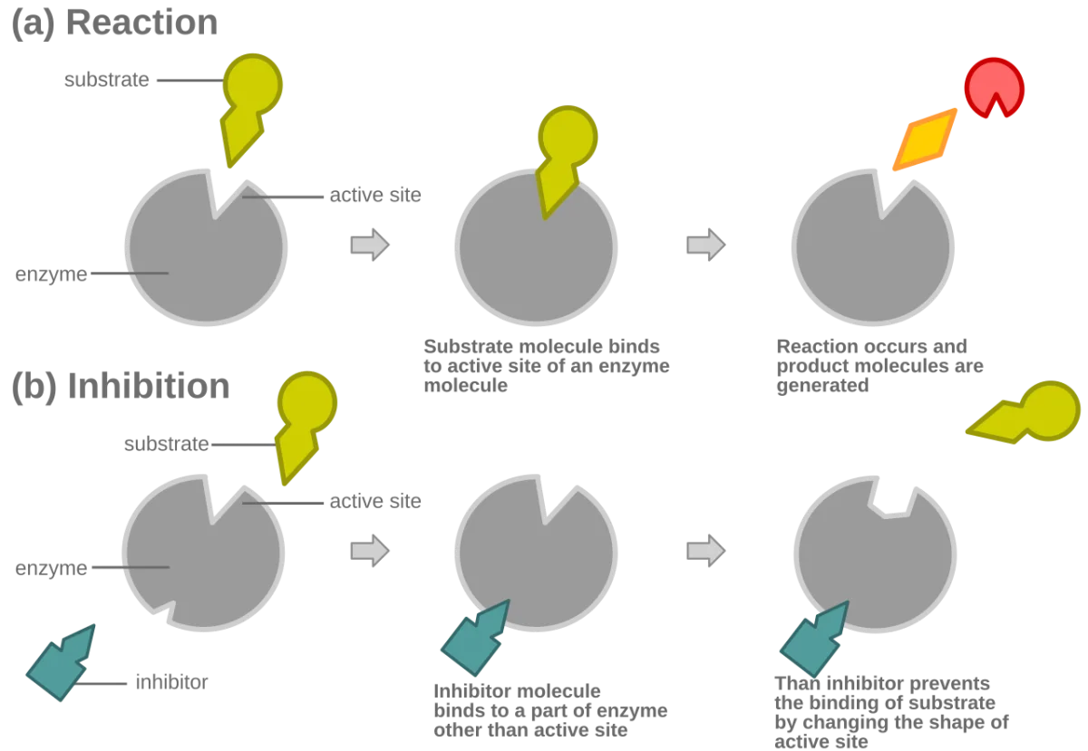
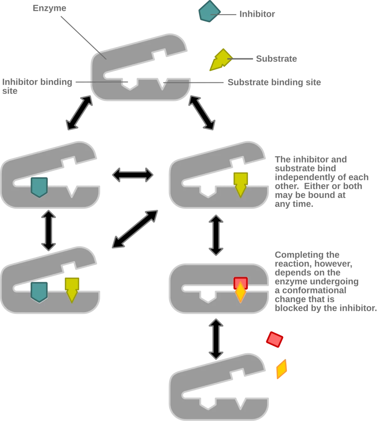
A noncompetitive inhibitor binds at a site other than the active site (an allosteric site) and bends the enzyme into an inactive shape. It can bind the free enzyme (E) or the enzyme-substrate complex (ES) with equal affinity. Adding more substrate does NOT help - the substrate can still slip into the active site, but the catalytic step is broken because the enzyme is distorted.
Noncompetitive inhibitor binds an allosteric site, changing the enzyme's shape. Substrate can still bind, but the enzyme can no longer catalyze the reaction properly. Credit: Wikimedia Commons, CC BY-SA
Kinetic Effect
Parameter
Change
Why
Vmax
Decreases
Some enzyme molecules are permanently broken (effectively [E] decreases)
Km
Unchanged
Substrate still binds the remaining functional enzymes the same way
Binding site
Allosteric (non-active)
Inhibitor does not compete with substrate for position
On Lineweaver-Burk, noncompetitive inhibition shifts lines up (higher y-intercept, because 1/Vmax is larger) but the x-intercept stays the same (Km unchanged). The two lines cross on the x-axis.
Uncompetitive Inhibition
An uncompetitive inhibitor binds ONLY the enzyme-substrate complex (ES), not the free enzyme. The inhibitor needs the substrate to be in place first - it binds a shape that only exists after substrate binding triggers a conformational change.
The result is counterintuitive: both Vmax AND Km decrease.
Vmax decreases because some ES complexes are trapped and cannot release product.
Km decreases because trapping ES pulls the E + S ⇌ ES equilibrium to the right (Le Chatelier), making it look as if the enzyme has higher affinity. More substrate is captured, not less.
Uncompetitive inhibition. Both Vmax and Km decrease by the same factor, keeping their ratio (and therefore the slope on a double-reciprocal plot) constant. Credit: Wikimedia Commons, CC BY-SA
Because both Vmax and Km decrease by the same factor, Km/Vmax (the slope on Lineweaver-Burk) is unchanged. The result is parallel lines on a double-reciprocal plot - different intercepts, same slope.
Real Examples
Lithium (for bipolar disorder) uncompetitively inhibits inositol monophosphatase. Because lithium only binds when substrate is already there, its effect is stronger where the substrate is abundant - a neat clinical quirk.
Some antibiotics targeting bacterial enzymes are uncompetitive to avoid complete shutdown of related human enzymes that lack the ES conformation the drug recognizes.
Side-by-Side Comparison
Inhibitor type
Binds
Km
Vmax
LB lines
Competitive
E only (active site)
Up
Same
Cross on y-axis
Noncompetitive
E and ES equally (allosteric)
Same
Down
Cross on x-axis
Uncompetitive
Only ES
Down
Down
Parallel
Mixed (next section)
E and ES, different affinity
Up or down
Down
Cross off-axis
How does a noncompetitive inhibitor affect Vmax and Km?
Click to reveal answer
Vmax decreases. Km is unchanged. Substrate still binds the functional enzyme the same way, but a fraction of the enzyme is permanently nonfunctional, so the top speed drops.
Uncompetitive inhibitors lower Km. Why does Km decrease instead of increase?
Click to reveal answer
The inhibitor binds only ES, effectively pulling E + S into ES by Le Chatelier's principle. More substrate gets bound at a given [S], which makes the enzyme look like it has higher affinity - so apparent Km drops. Vmax still falls because the trapped ES cannot release product.
You see parallel lines on a Lineweaver-Burk plot for inhibited vs. uninhibited enzyme. What type of inhibition is it?
Click to reveal answer
Uncompetitive. Parallel lines mean both Vmax and Km changed by the same factor, leaving the slope (Km/Vmax) constant. This is the classic signature of uncompetitive inhibition.
Pure noncompetitive inhibition (inhibitor binds E and ES with identical affinity) is mathematically tidy but biologically rare. Real-world “noncompetitive” inhibitors usually have unequal affinities for E vs. ES. That is mixed inhibition. It is the most common real form of allosteric inhibition.
The Big Idea
A mixed inhibitor binds both free enzyme (E) and the enzyme-substrate complex (ES), but not with the same strength. Let Ki be the dissociation constant for binding E, and Ki’ be the dissociation constant for binding ES.
If Ki < Ki’ (inhibitor prefers the free enzyme), behavior looks competitive-ish: Km appears to rise.
If Ki > Ki’ (inhibitor prefers the enzyme-substrate complex), behavior looks uncompetitive-ish: Km appears to fall.
In both cases, Vmax drops because some enzyme is always in an inhibitor-bound state.
Mixed inhibition on Lineweaver-Burk. The two lines intersect at a point that is neither on the x-axis nor the y-axis - an "off-axis" intersection. Credit: Wikimedia Commons, CC BY-SA
Kinetic Effect
Parameter
Change
Why
Vmax
Always decreases
Some enzyme is always inactive in an inhibitor complex
Km
Can go up or down
Depends on which form (E or ES) the inhibitor prefers
Binding site
Allosteric
Same site, different affinities for E and ES
LB signature
Off-axis intersection
The classic “neither purely competitive nor purely noncompetitive” pattern
Lineweaver-Burk Decision Tree
When you see an unknown inhibitor on a double-reciprocal plot, use this decision tree:
Do both lines share the y-intercept? Yes → competitive. No → go to step 2.
Do both lines share the x-intercept? Yes → noncompetitive. No → go to step 3.
Are the lines parallel? Yes → uncompetitive. No → mixed.
Why This Category Exists
Mixed inhibition is the honest description of what most allosteric regulators actually do. The MCAT used to lump everything non-competitive into “noncompetitive.” Modern biochem courses and the AAMC outline now distinguish mixed from pure noncompetitive. You should recognize both.
How does mixed inhibition differ from pure noncompetitive inhibition?
Click to reveal answer
Both bind allosteric sites and both lower Vmax. Pure noncompetitive requires identical affinity for E and ES, leaving Km unchanged. Mixed inhibition has different affinities for E and ES, so Km shifts (up or down depending on which form is preferred).
On a Lineweaver-Burk plot showing mixed inhibition, where do the two lines intersect?
Click to reveal answer
At an off-axis point - neither on the y-axis (which would indicate competitive) nor on the x-axis (which would indicate noncompetitive). The position of that intersection depends on whether Km rises or falls.
A mixed inhibitor binds the free enzyme more tightly than the ES complex. What happens to the apparent Km?
Click to reveal answer
Apparent Km increases. Because the inhibitor preferentially grabs free enzyme, substrate must compete for the remaining free E, so it takes more substrate to reach half-Vmax. Vmax still drops because no matter how much substrate you add, some enzyme is always inhibitor-bound.
Some enzymes do not follow a simple Michaelis-Menten curve. Their velocity-vs-substrate plot is not a hyperbola but an S-shape (sigmoid). These enzymes are allosteric - they have multiple subunits and multiple binding sites that talk to each other. The conversation between subunits produces cooperativity, which is how your cells do precise, switch-like regulation of critical metabolic steps.
Four ways to change enzyme activity, fastest first
Why zymogens exist at allSome enzymes are too dangerous to build in working form. A protease made active inside the cell that produced it would digest that cell, so it is made as an inactive zymogen and only cut open where it is meant to work. Acute pancreatitis is what happens when that cutting starts in the wrong place.
Feedback inhibitionThe classic allosteric arrangement: the end product of a pathway inhibits the enzyme that catalyzed its first committed step. It is the cheapest possible control, because the cell measures the thing it actually cares about and acts on the one step that commits resources.
Cooperativity is allostery tooIn a multi-subunit enzyme the substrate itself is the allosteric regulator: binding at one site changes the shape of the others. That converts the hyperbolic Michaelis-Menten curve into a sigmoid, which makes the enzyme behave like a switch rather than a dial.
Read the timescale in the question and the mechanism is already chosen. Something that responds within a heartbeat is allosteric. Something that responds to a hormone is covalent modification. Something that only happens once is a zymogen. Something that takes hours is a change in how much enzyme exists.
What “Allosteric” Means
Allos (other) + steric (place) = “binds at another place.” An allosteric site is any site on an enzyme other than the active site where a regulator can bind. Binding there triggers a conformational change that ripples through the protein, changing activity at the distant active site.
Allosteric activators stabilize the high-activity shape (R state, “relaxed”).
Allosteric inhibitors stabilize the low-activity shape (T state, “tense”).
Cooperativity and Sigmoidal Kinetics
Most allosteric enzymes have multiple substrate-binding sites. When one subunit binds substrate, it changes shape - and nudges the other subunits into a higher-affinity shape. The second substrate binds more easily, the third even more easily, and so on. That is positive cooperativity.
Plotting velocity vs. [S] for a cooperative enzyme gives a sigmoidal curve instead of a hyperbola. At low [S], the enzyme is sluggish. Once a critical [S] is reached, activity ramps up sharply. Past that threshold, the enzyme is essentially maxed out. Sigmoidal kinetics turn an enzyme into a switch.
Positive cooperativity: binding one substrate makes subsequent binding easier. Sigmoidal curve. Classic example: hemoglobin binding oxygen.
Negative cooperativity: binding one substrate makes subsequent binding harder. Less common but real (insulin receptor kinase).
Quantifying Cooperativity: The Hill Coefficient
Cooperativity is not just qualitative. You can put a number on it. The Hill coefficient (n or nH) is a measure of how strongly the subunits “talk” to each other.
You do not need to calculate Hill coefficients on the MCAT, but you should recognize a number like “n = 2.8” as “this protein shows strong positive cooperativity.” A passage that gives you a Hill plot or Hill value is testing whether you know which direction the cooperativity runs.
T State vs. R State
Two conformations interconvert:
T state (tense) - low affinity, low activity. Promoted by allosteric inhibitors.
R state (relaxed) - high affinity, high activity. Promoted by allosteric activators and by substrate itself.
Substrate binding pulls the equilibrium toward R. Inhibitors lock the enzyme in T. Activators lock it in R. The sigmoidal curve emerges from this T-to-R transition, which happens in a concerted all-or-nothing fashion in some models (MWC / concerted model) or one subunit at a time (KNF / sequential model). The MCAT does not require naming these models, but the concept of shifting between two states is essential.
Feedback Inhibition
Most metabolic pathways end in a product that allosterically inhibits the first committed step of its own pathway. This is feedback inhibition, and it is the main way cells avoid overproducing things they already have plenty of.
Canonical MCAT examples:
ATP is an allosteric inhibitor of phosphofructokinase-1 (PFK-1), the rate-limiting enzyme of glycolysis. When the cell has enough energy (high ATP), glycolysis slows.
AMP is an allosteric activator of PFK-1. Low energy (high AMP) accelerates glycolysis.
Citrate is an allosteric inhibitor of PFK-1. A full TCA cycle signals no need for more fuel.
CTP feedback-inhibits aspartate transcarbamoylase (ATCase), the classic textbook allosteric enzyme of pyrimidine synthesis.
Hemoglobin Is the MCAT’s Favorite Allosteric Protein
Hemoglobin is not an enzyme, but it is the textbook cooperative protein. Four subunits, each binding one O2. Binding the first O2 is hard (T state). Once bound, the subunit shifts to R, pulling the others with it. The second, third, and fourth O2 molecules bind much more easily. The result is a sigmoidal O2 saturation curve that allows hemoglobin to load O2 efficiently in the lungs (high O2) and unload it in tissues (low O2).
Negative regulators that stabilize T (2,3-BPG, H+, CO2) cause hemoglobin to release more O2 in tissues - the Bohr effect. Know this example cold; it shows up constantly. Full coverage of hemoglobin is in Chapter 3.
Why is a sigmoidal velocity-substrate curve a signature of cooperative allosteric enzymes?
Click to reveal answer
The sigmoidal shape comes from positive cooperativity - binding one substrate molecule increases the affinity of the remaining subunits. This produces a slow start at low [S] and a rapid jump once the first binding nudges the enzyme from T to R. Non-cooperative enzymes yield a simple hyperbolic curve.
What is feedback inhibition?
Click to reveal answer
A regulatory mechanism in which the end product of a pathway allosterically inhibits the first committed enzyme of the same pathway. It prevents overproduction when the product is abundant. A classic example is ATP and citrate inhibiting phosphofructokinase-1 in glycolysis.
What happens to an allosteric enzyme when an activator binds?
Click to reveal answer
The activator stabilizes the high-affinity R state, shifting the T-R equilibrium toward R. The sigmoidal curve shifts to the left (lower apparent K0.5, higher activity at a given [S]). Substrate binds more easily, and the enzyme approaches Vmax at lower substrate concentrations.
Cells switch enzymes on and off by covalently attaching small chemical groups to specific residues. This is the fastest way to tune activity without synthesizing or degrading the protein, which makes covalent modification the workhorse of cellular regulation.
Phosphorylation - The Master Switch
The most common and MCAT-tested modification is phosphorylation. A kinase transfers a phosphate from ATP onto a hydroxyl-containing residue (Ser, Thr, or Tyr). A phosphatase removes the phosphate by hydrolysis.
Enzyme-OH+ATPkinaseEnzyme-O-PO32−+ADP
Phosphate adds two negative charges and a bulky group. That changes local shape and often switches activity - sometimes activating, sometimes inhibiting, depending on the enzyme.
Enzymes are shaped proteins whose activity can be switched by adding or removing small chemical groups at specific residues. Credit: Wikimedia Commons, CC BY-SA
Examples of Phosphorylation Switches
Enzyme
Phosphorylated form
Physiologic trigger
Glycogen phosphorylase
Active (breaks glycogen)
Glucagon / epinephrine (fasting, stress)
Glycogen synthase
Inactive (does not build glycogen)
Glucagon / epinephrine
Pyruvate kinase
Inactive (glycolysis slowed)
Glucagon (liver)
Acetyl-CoA carboxylase
Inactive (no fatty acid synthesis)
Glucagon / AMPK
Hormone-sensitive lipase
Active (releases fat)
Epinephrine, glucagon
Notice the pattern: in the fasted/stressed state, phosphorylation activates enzymes that release glucose and fat, and inactivates enzymes that store them. Insulin, the “fed” hormone, reverses each of these by activating phosphatases.
Glycosylation
Sugars (usually carbohydrate chains) are attached to specific residues - Asn (N-linked) or Ser/Thr (O-linked). Glycosylation happens in the ER and Golgi and is essential for:
Directing proteins to the correct destination (lysosomal enzymes carry mannose-6-phosphate tags).
Stabilizing secreted proteins in the harsh extracellular environment.
Cell-surface recognition (blood type antigens are carbohydrates on red blood cells).
Ubiquitination - The Destruction Signal
Ubiquitin is a small protein that gets covalently attached to lysine residues of target proteins. A single ubiquitin can regulate location or activity. A chain of ubiquitins (polyubiquitination) tags the protein for destruction by the 26S proteasome.
Acetylation and Methylation
Two more modifications worth recognizing, especially in the context of gene regulation:
Acetylation of lysine residues by HATs (histone acetyltransferases) neutralizes their positive charge. In histones, this loosens the DNA-histone interaction and activates transcription. HDACs (histone deacetylases) reverse the process.
Methylation of lysine or arginine (and of DNA cytosines) can activate or repress transcription depending on the residue and context. Methylation of CpG islands in DNA typically silences gene expression.
What does a kinase do, and what residues does it typically modify on a target protein?
Click to reveal answer
A kinase transfers a phosphate group from ATP onto a hydroxyl-containing amino acid - serine, threonine, or tyrosine - on a target protein. The added phosphate changes local conformation and activity. A phosphatase does the reverse.
Why does polyubiquitination usually lead to protein destruction?
Click to reveal answer
The 26S proteasome recognizes long ubiquitin chains as a degradation signal. It unfolds the tagged protein and cleaves it into small peptides. Single or short ubiquitin tags, by contrast, often signal localization or activity changes rather than destruction.
Glucagon is released during fasting. What happens to glycogen phosphorylase and glycogen synthase in response?
Click to reveal answer
Glucagon triggers a kinase cascade that phosphorylates both enzymes. Phosphorylation activates glycogen phosphorylase (breaks down glycogen, releasing glucose) and inactivates glycogen synthase (stops storage). The result: glucose is released into the blood, as expected in the fasted state.
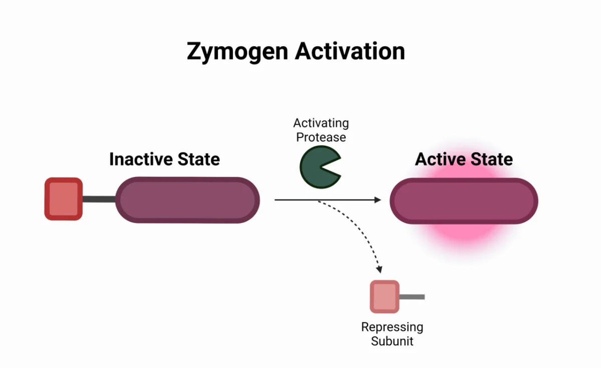
Some enzymes are dangerous if they are active in the wrong place at the wrong time. The cell synthesizes them in an inactive precursor form called a zymogen (or proenzyme) and activates them only when and where needed, usually by cleaving off a peptide. Once cut, the zymogen cannot go back to being inactive - activation is irreversible, which is why the cell is so careful about where it happens.
Digestive Zymogens
Pancreatic digestive enzymes are the textbook example. If pepsin, trypsin, chymotrypsin, or elastase were active in the pancreas, they would digest the pancreas itself (a real disease: acute pancreatitis). So the pancreas synthesizes them as zymogens and releases them into the small intestine for activation.
Zymogen
Where made
Where activated
Active enzyme
Activated by
Pepsinogen
Stomach (chief cells)
Stomach (low pH)
Pepsin
Autocatalysis at pH < 5, plus existing pepsin
Trypsinogen
Pancreas
Small intestine
Trypsin
Enteropeptidase (duodenum), then autocatalysis
Chymotrypsinogen
Pancreas
Small intestine
Chymotrypsin
Trypsin
Proelastase
Pancreas
Small intestine
Elastase
Trypsin
Procarboxypeptidase
Pancreas
Small intestine
Carboxypeptidase
Trypsin
Prolipase
Pancreas
Small intestine
Lipase
Trypsin
Notice the key node: trypsin activates all the others. Enteropeptidase (also called enterokinase) in the duodenal brush border cleaves trypsinogen to trypsin; trypsin then activates itself and every other pancreatic zymogen. This is called a proteolytic cascade.
Zymogen activation. Cleavage removes a small peptide, and the remaining chain folds into the active enzyme. The process is irreversible. Credit: Wikimedia Commons, CC BY-SA
The Blood Clotting Cascade
Clotting is a proteolytic cascade of zymogen activations. Each factor is a zymogen; when cut, it becomes an active protease that cuts the next factor in line. This amplifies the signal - a tiny initial trigger produces a large, localized clot within seconds.
Intrinsic pathway: triggered by contact with damaged surface (Factor XII → XI → IX → X).
Extrinsic pathway: triggered by tissue factor (VII → X).
Both pathways converge at Factor X → Xa, which activates prothrombin (II) to thrombin (IIa). Thrombin converts fibrinogen to fibrin, forming the clot.
Warfarin blocks vitamin K recycling, which is needed to gamma-carboxylate clotting factors II, VII, IX, and X - so warfarin indirectly inhibits clotting by producing non-functional zymogens.
Apoptosis - Caspase Cascade
Programmed cell death uses a similar cascade of zymogen activations. Caspases (cysteine-aspartate proteases) are synthesized as procaspases. Initiator caspases (e.g., caspase-8, -9) cleave and activate executioner caspases (caspase-3, -6, -7), which then dismantle the cell in an orderly way. Because the cascade is irreversible, the decision to die is committed once the first executioner caspase is activated.
Regulatory Enzymes in a Pathway
Beyond zymogens, most metabolic pathways have one or two “regulatory enzymes” that control flux. These are usually:
The rate-limiting enzyme (slowest step of the pathway at normal conditions).
An enzyme catalyzing an irreversible step, so regulation is meaningful.
These are expanded in the metabolism chapters. For now, just know that the cell concentrates regulation at a few key control points rather than controlling every step of every pathway.
Why are digestive proteases made as zymogens?
Click to reveal answer
So they do not digest the tissue that makes them. If trypsin or chymotrypsin were active in the pancreas, they would degrade pancreatic proteins (causing pancreatitis). By keeping them in inactive zymogen form until they reach the small intestine, the pancreas is protected.
What single enzyme activates nearly all pancreatic zymogens, and how is it itself activated?
Click to reveal answer
Trypsin. It activates chymotrypsinogen, procarboxypeptidase, proelastase, prolipase, and additional trypsinogen molecules. Trypsinogen itself is activated by enteropeptidase (enterokinase) in the duodenal brush border, and then by trypsin autocatalysis.
Why is proteolytic activation of zymogens called "irreversible"?
Click to reveal answer
Because the activating cut removes or rearranges a peptide segment in a way the cell cannot undo. Unlike phosphorylation (reversible by phosphatases) or allosteric regulation (reversible by removing the regulator), the cell cannot stitch the pro-peptide back on. Once cut, the enzyme is permanently active until it is degraded.

![Lineweaver-Burk double reciprocal plot showing 1/V0 on the y-axis and 1/[S] on the x-axis. The y-intercept is 1/Vmax, the x-intercept is -1/Km, and the slope is Km/Vmax](/images/books/biochemistry/ch2/wikimedia-lineweaver-burk.webp)