
Structure and Function
An enzyme is a biological catalyst - a protein (or in rare cases an RNA) that speeds up a chemical reaction without being consumed. The rate increase is enormous. A typical enzyme accelerates its reaction by to fold. Without enzymes, digesting a sandwich would take years.
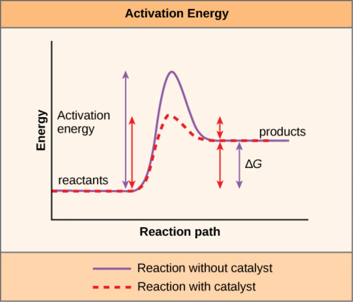
Crucially, enzymes do not change whether a reaction can happen. A reaction with positive Gibbs free energy (ΔG > 0) stays non-spontaneous whether or not an enzyme is present. Enzymes only change how quickly an already-possible reaction reaches equilibrium.
Lowering Activation Energy
Every reaction has to climb an energy hill called the activation energy (Ea) before it can proceed. Substrates must twist, bend, or partially break bonds into a strained intermediate called the transition state before new bonds can form. Enzymes stabilize that transition state. A lower energy hill means more molecules have enough energy to make it over at body temperature, so the reaction runs faster.
The Active Site
The active site is a small pocket or cleft on the enzyme, usually only a few amino acids wide, where the substrate binds and catalysis happens. Two types of residues matter there:
- Binding residues grip the substrate through hydrogen bonds, van der Waals forces, hydrophobic packing, and sometimes ionic interactions. They set the specificity.
- Catalytic residues actually participate in the bond-making or bond-breaking. Common offenders are Ser, His, Asp, Cys, Lys, and Tyr - amino acids with reactive side chains.
The active site is a tiny fraction of the whole protein. Most of the enzyme is scaffolding that holds those critical residues in exactly the right 3D positions.
Substrate Specificity
Enzymes are dramatically more specific than non-biological catalysts. Hexokinase phosphorylates glucose but ignores nearly identical sugars like galactose. Specificity comes from the shape, charge, and hydrophobicity of the active site matching only a narrow set of substrates.
Some enzymes are absolutely specific (only one substrate, e.g., urease acts only on urea). Others have group specificity (any sugar with a free -OH, or any peptide bond adjacent to a lysine). Most fall somewhere in between.
Catalytic Mechanisms
Enzymes lower activation energy through a handful of recurring tricks. The MCAT expects you to recognize the four main mechanisms:
- Acid-base catalysis: an active-site residue donates or accepts a proton at a key step (histidine’s pKa near 6 makes it the star).
- Covalent catalysis: a nucleophilic residue (often serine, cysteine, or lysine) transiently forms a covalent bond with the substrate, creating a reactive intermediate. Serine proteases (chymotrypsin, trypsin) build an acyl-enzyme intermediate through Ser-OH.
- Metal-ion catalysis: a metal cofactor (Zn2+, Fe2+/Fe3+, Mg2+) polarizes substrates, stabilizes negative charges, or performs redox. Carbonic anhydrase uses Zn2+; cytochromes use iron.
- Electrostatic catalysis: charged residues in the active site stabilize oppositely charged transition-state intermediates (the “oxyanion hole” in serine proteases is the classic example).
The Catalytic Triad
The textbook case is the Ser-His-Asp triad of chymotrypsin. Asp polarizes His; His deprotonates Ser; Ser attacks the peptide bond’s carbonyl carbon. All three mechanisms appear simultaneously: acid-base (His shuttles protons), covalent (Ser forms acyl-enzyme), and electrostatic (Asp and the oxyanion hole stabilize charged intermediates).
Cofactors and Coenzymes
Many enzymes need a non-protein partner to function:
| Term | What it is | Examples |
|---|---|---|
| Cofactor | Any non-protein helper | Zn2+, Mg2+, Fe2+, a coenzyme |
| Coenzyme | An organic cofactor, often vitamin-derived | NAD+, FAD, CoA, PLP, biotin |
| Prosthetic group | A cofactor covalently or tightly bound to the enzyme | Heme in catalase, biotin in pyruvate carboxylase |
| Apoenzyme | The protein part alone - inactive | Hexokinase minus Mg2+ |
| Holoenzyme | Apoenzyme + cofactor, fully active | Working hexokinase |
The B Vitamin Cofactor Map
The MCAT loves “vitamin → coenzyme → reaction → deficiency disease” chains. Memorize the vitamin-to-coenzyme link; the rest is context.
| Vitamin | Name | Coenzyme form | Role |
|---|---|---|---|
| B1 | Thiamine | Thiamine pyrophosphate (TPP) | Decarboxylations: pyruvate → acetyl-CoA (PDH), α-KG dehydrogenase |
| B2 | Riboflavin | FAD / FMN | One- or two-electron transfer in redox reactions |
| B3 | Niacin | NAD+ / NADP+ | Two-electron hydride transfer in redox reactions |
| B5 | Pantothenic acid | Coenzyme A (CoA) | Acyl-group carrier (acetyl-CoA, fatty-acyl-CoA) |
| B6 | Pyridoxine | Pyridoxal phosphate (PLP) | Amino-acid transamination, decarboxylation |
| B7 | Biotin | Biotin (covalent prosthetic group) | Carboxylations (pyruvate carboxylase, acetyl-CoA carboxylase) |
| B9 | Folate | Tetrahydrofolate (THF) | One-carbon transfers in nucleotide synthesis |
| B12 | Cobalamin | Methylcobalamin / adenosylcobalamin | Methyl transfer (homocysteine → methionine); methylmalonyl-CoA mutase |
| C | Ascorbate | Ascorbic acid | Reducing agent for prolyl hydroxylase (collagen synthesis) |