Cells switch enzymes on and off by covalently attaching small chemical groups to specific residues. This is the fastest way to tune activity without synthesizing or degrading the protein, which makes covalent modification the workhorse of cellular regulation.
Phosphorylation - The Master Switch
The most common and MCAT-tested modification is phosphorylation. A kinase transfers a phosphate from ATP onto a hydroxyl-containing residue (Ser, Thr, or Tyr). A phosphatase removes the phosphate by hydrolysis.
Enzyme-OH+ATPkinaseEnzyme-O-PO32−+ADP
Phosphate adds two negative charges and a bulky group. That changes local shape and often switches activity - sometimes activating, sometimes inhibiting, depending on the enzyme.
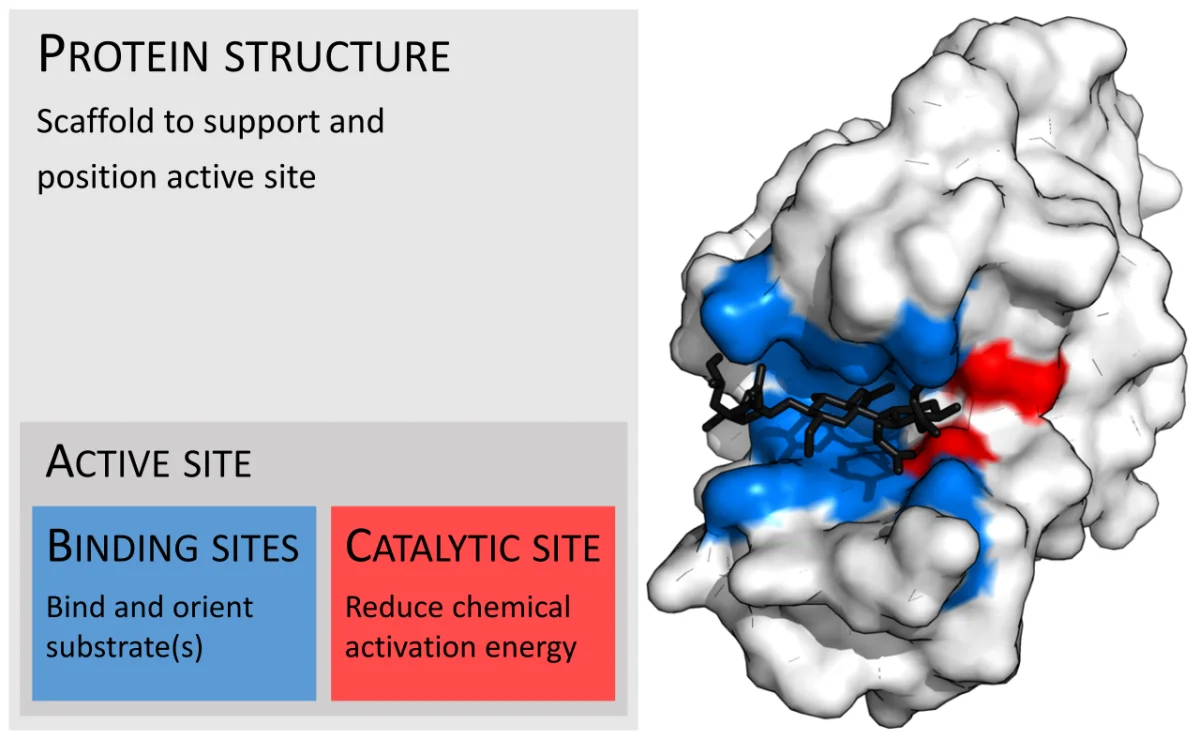
Enzymes are shaped proteins whose activity can be switched by adding or removing small chemical groups at specific residues. Credit: Wikimedia Commons, CC BY-SA
Examples of Phosphorylation Switches
Enzyme
Phosphorylated form
Physiologic trigger
Glycogen phosphorylase
Active (breaks glycogen)
Glucagon / epinephrine (fasting, stress)
Glycogen synthase
Inactive (does not build glycogen)
Glucagon / epinephrine
Pyruvate kinase
Inactive (glycolysis slowed)
Glucagon (liver)
Acetyl-CoA carboxylase
Inactive (no fatty acid synthesis)
Glucagon / AMPK
Hormone-sensitive lipase
Active (releases fat)
Epinephrine, glucagon
Notice the pattern: in the fasted/stressed state, phosphorylation activates enzymes that release glucose and fat, and inactivates enzymes that store them. Insulin, the “fed” hormone, reverses each of these by activating phosphatases.
Glycosylation
Sugars (usually carbohydrate chains) are attached to specific residues - Asn (N-linked) or Ser/Thr (O-linked). Glycosylation happens in the ER and Golgi and is essential for:
Directing proteins to the correct destination (lysosomal enzymes carry mannose-6-phosphate tags).
Stabilizing secreted proteins in the harsh extracellular environment.
Cell-surface recognition (blood type antigens are carbohydrates on red blood cells).
Ubiquitination - The Destruction Signal
Ubiquitin is a small protein that gets covalently attached to lysine residues of target proteins. A single ubiquitin can regulate location or activity. A chain of ubiquitins (polyubiquitination) tags the protein for destruction by the 26S proteasome.
Acetylation and Methylation
Two more modifications worth recognizing, especially in the context of gene regulation:
Acetylation of lysine residues by HATs (histone acetyltransferases) neutralizes their positive charge. In histones, this loosens the DNA-histone interaction and activates transcription. HDACs (histone deacetylases) reverse the process.
Methylation of lysine or arginine (and of DNA cytosines) can activate or repress transcription depending on the residue and context. Methylation of CpG islands in DNA typically silences gene expression.
What does a kinase do, and what residues does it typically modify on a target protein?
Click to reveal answer
A kinase transfers a phosphate group from ATP onto a hydroxyl-containing amino acid - serine, threonine, or tyrosine - on a target protein. The added phosphate changes local conformation and activity. A phosphatase does the reverse.
Why does polyubiquitination usually lead to protein destruction?
Click to reveal answer
The 26S proteasome recognizes long ubiquitin chains as a degradation signal. It unfolds the tagged protein and cleaves it into small peptides. Single or short ubiquitin tags, by contrast, often signal localization or activity changes rather than destruction.
Glucagon is released during fasting. What happens to glycogen phosphorylase and glycogen synthase in response?
Click to reveal answer
Glucagon triggers a kinase cascade that phosphorylates both enzymes. Phosphorylation activates glycogen phosphorylase (breaks down glycogen, releasing glucose) and inactivates glycogen synthase (stops storage). The result: glucose is released into the blood, as expected in the fasted state.