An amine is a nitrogen atom with one or more alkyl or aryl substituents. Classification depends on how many carbons are directly bonded to the nitrogen - a classification scheme DIFFERENT from alcohols (where you count carbons attached to the C-OH carbon).
Classification
Primary (1°): RNH₂. One C on N; two H’s.
Secondary (2°): R₂NH. Two C’s on N; one H.
Tertiary (3°): R₃N. Three C’s on N; zero H’s.
Quaternary ammonium (4°): R₄N⁺. Four C’s on N; positive charge; no lone pair available.
Geometry and Hybridization
Amine nitrogen is sp³ hybridized with a pyramidal geometry. Three sp³ sigma bonds (to R or H groups) and one sp³ orbital containing the lone pair. The lone pair is essential: it makes amines basic (donor) and nucleophilic (attack electrophiles).
Because of rapid pyramidal inversion (nitrogen flipping through a planar transition state), chiral amines with three different R groups usually do NOT have a fixed configuration - they racemize spontaneously. Quaternary ammoniums with four different R groups CAN be chiral (no lone pair to invert through).
Nomenclature
IUPAC names amines by replacing alkane “-e” with “-amine”:
Aniline and pyridine are retained common names that dominate usage.
Quaternary Ammonium Compounds
Quaternary ammoniums (R₄N⁺) have four R groups and a positive charge. They have no lone pair and are NOT basic. They act as permanent cations (always charged regardless of pH). Examples: choline (HOCH₂CH₂N⁺(CH₃)₃), acetylcholine, benzalkonium chloride (disinfectant). Biologically important in membrane phospholipid head groups.
Amines vs Amides
Amines (RNH₂, R₂NH, R₃N) have sp³ nitrogen with an available lone pair - basic and nucleophilic. Amides (RCONR’₂) have sp² nitrogen with the lone pair delocalized into the carbonyl - NOT basic, NOT nucleophilic at N under physiological conditions.
Students often confuse these. The C=O adjacent to N changes everything: it makes amide N flat, unreactive, and part of protein backbones that resist hydrolysis.
Classify each as 1°, 2°, 3° amine, or quaternary ammonium: (a) N,N-dimethylaniline, (b) diethylamine, (c) tetramethylammonium chloride, (d) benzylamine.
Click to reveal answer
(a) 3° tertiary - nitrogen has three C substituents (two methyls + one phenyl). (b) 2° secondary - nitrogen has two ethyl groups and one H. (c) Quaternary ammonium - four methyls on N with positive charge. (d) 1° primary - nitrogen has one benzyl group and two H's. Count the C's directly on nitrogen, not on adjacent atoms.
Amines have physical properties sitting between alcohols (strong H-bonding) and ethers (no H-bonding). The N-H bond is polar but not as polar as O-H, so hydrogen bonding is moderate. This places amine boiling points between those of comparable alcohols and alkanes.
Boiling Points
For MW ~30:
Compound
MW
Boiling point
H-bonding
Methane (CH₄)
16
-162°C
None
Methylamine (CH₃NH₂)
31
-6°C
Moderate (N-H donor + acceptor)
Methanol (CH₃OH)
32
65°C
Strong (O-H donor + acceptor)
Methylamine boils about 70°C higher than methane but 70°C lower than methanol - an intermediate H-bonding situation.
Tertiary Amines Cannot Donate H-Bonds
Tertiary amines (R₃N) have no N-H bonds, so they cannot DONATE hydrogen bonds. They can still accept H-bonds (via the nitrogen lone pair), but the overall intermolecular forces are weaker than with primary or secondary amines:
Trimethylamine ((CH₃)₃N): b.p. 3°C.
Triethylamine ((CH₃CH₂)₃N): b.p. 89°C.
Compare dimethylamine ((CH₃)₂NH, MW 45, b.p. 7°C) with trimethylamine ((CH₃)₃N, MW 59, b.p. 3°C). Despite a higher MW, trimethylamine boils lower because it cannot H-bond with itself.
Water Solubility
Small amines (1-4 carbons) are miscible with water or highly soluble. N lone pair and N-H bonds H-bond to water molecules. Larger amines (>5 carbons) have diminishing water solubility as the hydrophobic chain grows, following the same pattern as alcohols.
Protonated amines (in acidic solution, RNH₃⁺) are always water-soluble due to their positive charge. This is why “amine hydrochloride” salts (RNH₃⁺Cl⁻) are used in pharmaceuticals - they are dramatically more water-soluble than the free amine.
Odor
Amines have distinctive odors, often unpleasant:
Trimethylamine: rotten fish smell (found in spoiling seafood, vaginal secretions in bacterial vaginosis).
Putrescine (1,4-butanediamine): decomposing meat.
Cadaverine (1,5-pentanediamine): rotting flesh.
These are the compounds that give decomposition its characteristic smell - all amines.
Rank by boiling point (highest first): trimethylamine (MW 59), propanol (MW 60), propane (MW 44), ethylamine (MW 45).
Click to reveal answer
Propanol (97°C) > ethylamine (17°C) > trimethylamine (3°C) > propane (-42°C). Propanol has strong O-H hydrogen bonding. Ethylamine has moderate N-H H-bonding (primary amine, can donate and accept). Trimethylamine has NO N-H so cannot donate H-bonds - surprisingly low for a 59-MW molecule. Propane has only London forces. Hydrogen-bonding ability + donor/acceptor status dominates boiling point ranking.
Amines are weak Brønsted bases. When protonated, they form ammonium ions (RNH₃⁺, R₂NH₂⁺, R₃NH⁺) with characteristic pKa values around 9-11. The specific basicity depends on electron availability at nitrogen, modulated by alkyl donation, aromatic delocalization, and steric effects.
Amine basicity trends: alkyl amines > ammonia > aryl amines (anilines). Alkyl donation pushes electron density onto N; aromatic ring withdraws electron density via resonance. Credit: Wikimedia Commons, CC BY-SA
Key pKa Values
Species
Conjugate acid pKa
Notes
Aniline (C₆H₅NH₂)
4.6
Aryl - weak base
Ammonia (NH₃)
9.2
Reference
Methylamine (CH₃NH₂)
10.6
Alkyl - stronger base than ammonia
Dimethylamine ((CH₃)₂NH)
10.8
Two alkyl donors; slightly more basic
Trimethylamine ((CH₃)₃N)
9.8
Three alkyl donors BUT steric solvation effects reduce basicity slightly
Pyridine (C₅H₅N)
5.2
Aromatic but sp² lone pair not in pi system
Imidazole
7.0
Biologically important (histidine)
Amide (R-CO-NH₂)
~-0.5
Essentially not basic (lone pair delocalized)
Higher pKa of conjugate acid = stronger base (harder to remove the H⁺ from the ammonium = ammonium is less willing to lose H = amine is more willing to accept H = stronger base).
Alkyl Amines: More Basic Than Ammonia
Alkyl groups donate electron density to nitrogen (induction + hyperconjugation), making the lone pair more electron-rich and more available to accept a proton. Trend in gas phase: (CH₃)₃N > (CH₃)₂NH > CH₃NH₂ > NH₃.
In water, the trend is blurred by solvation effects: the ammonium cation is stabilized by H-bonding to water, and more H-N bonds on the ammonium mean more H-bond donors to water. This extra solvation stabilizes primary ammoniums more than tertiary. The net result in water: R₂NH (secondary) ≈ RNH₂ (primary) ≈ R₃N (tertiary) > NH₃. The differences are small (pKa varies by only ~1 unit).
Aryl Amines: Much Less Basic
Aniline (C₆H₅NH₂) has pKa 4.6 - about 4.6 units less basic than methylamine. Why? The nitrogen lone pair on aniline delocalizes into the aromatic ring via resonance. Three resonance contributors place partial negative charge on the ortho and para positions.
Consequence: the lone pair is partially tied up in the ring, less available to accept a proton. Protonation would require breaking the resonance - energetically costly.
Substituents on aniline modulate basicity:
p-Methoxyaniline: methoxy donates into the ring; nitrogen lone pair is less “needed” by the ring; amine is more basic than aniline. pKa ~5.3.
p-Nitroaniline: nitro withdraws; nitrogen lone pair is more delocalized; amine is much less basic. pKa ~1.
p-Methylaniline (p-toluidine): methyl donor; slightly more basic. pKa ~5.1.
Amide Nitrogen: Essentially Not Basic
Amides (R-CO-NR’₂) have the nitrogen lone pair delocalized into the adjacent carbonyl (~40% double bond character). The lone pair is not available for protonation. Protonation would disrupt resonance and go against the dipolar contributor.
As a result, amides are NOT basic under physiological conditions. The pKa of a protonated amide is around 0 to -1 (depending on substrate), meaning the amide has to be in strong acid to protonate significantly.
This is why the peptide backbone nitrogens in proteins do NOT act as bases at pH 7.4, while the alpha-amino groups of unattached amino acids (CH(NH₂)(COOH)R) DO act as bases at that pH.
Basicity of Nitrogen Heterocycles
Pyridine (C₅H₅N): pKa of conjugate acid ~5.2. The nitrogen lone pair is in the SP² orbital in the plane of the ring, NOT in the aromatic pi system. This lone pair is available for protonation. Pyridine is a modest base (less than alkyl amines but more basic than aniline).
Pyrrole (C₄H₄NH): pKa of conjugate acid ~0.4. Here the nitrogen lone pair IS part of the aromatic pi system (contributes 2 of the 6 electrons for Huckel’s rule). Protonating this nitrogen would break aromaticity. Pyrrole is essentially not basic at the nitrogen. Protonation happens at the C2 or C5 ring carbon instead.
Imidazole (C₃H₄N₂): pKa ~7.0. Two nitrogens, one like pyrrole (pi contributor) and one like pyridine (sp² lone pair available). The basic nitrogen is the pyridine-like one. Imidazole’s pKa ~7 is close to physiological pH, which is exactly why histidine’s imidazole side chain can act as both acid and base in enzyme active sites.
Why is pyrrole (a 5-membered aromatic ring with N) essentially non-basic, while pyridine (a 6-membered aromatic ring with N) is a modest base?
Click to reveal answer
Pyrrole's nitrogen lone pair is IN the aromatic pi system - it contributes 2 of the 6 pi electrons to satisfy Huckel's 4n+2 rule. Protonating this lone pair would remove it from the pi system and break aromaticity, which is energetically very costly. Pyridine's nitrogen lone pair is NOT in the pi system - it sits in an sp² hybrid orbital in the plane of the ring. Protonation of this lone pair does not disrupt aromaticity, so pyridine can be protonated readily (pKa of protonated pyridine ~5.2).
Amines are good nucleophiles (electron-rich nitrogen with available lone pair) and can attack alkyl halides (SN2 alkylation) or carbonyl derivatives (acylation to form amides). Both reactions are covered by mechanisms you learned in previous chapters.
Amine + Alkyl Halide = Alkylation
Primary amine attacks a primary or methyl alkyl halide in SN2:
R-NH₂ + R’-X → R-NH-R’ + HX (typically with a base to absorb HCl)
Problem: the secondary amine product is MORE nucleophilic than the starting primary amine (one alkyl donor → slightly more nucleophilic than two H). So after the first alkylation, the secondary amine attacks another alkyl halide → tertiary amine. Then tertiary attacks again → quaternary ammonium.
This “over-alkylation” is hard to control. Practical solutions:
Use large excess of ammonia (tenfold or more). Statistically favors primary amine product.
Use the Gabriel synthesis (see Section 10.5) to get a clean primary amine.
Use reductive amination (Section 6.8) instead of direct alkylation.
Acylation is cleaner than alkylation because the product (amide) is NOT nucleophilic (amide nitrogen lone pair is delocalized into the carbonyl). So there is no over-acylation. One equivalent of acid chloride gives one equivalent of amide.
Anhydrides work similarly but half the anhydride is wasted as the free carboxylic acid byproduct. Esters + amine → amide is slower but possible.
Hofmann Elimination
Exhaustive methylation converts an amine to a quaternary ammonium by treating with excess methyl iodide. The resulting R-N⁺(CH₃)₃ is a good leaving group. When heated with hydroxide, the quaternary ammonium undergoes E2 elimination:
Unusually, Hofmann elimination gives the Hofmann product (less substituted alkene) as the major product, NOT Zaitsev. The reason: the bulky quaternary ammonium LG forces the hydroxide to abstract the less hindered beta-H (on the less substituted carbon), giving the less substituted alkene.
Hofmann elimination: exhaustive methylation of an amine to a quaternary ammonium, then E2 elimination with hydroxide gives the less-substituted (Hofmann) alkene. The bulky ammonium leaving group forces the base to the least-hindered beta-H. Credit: Wikimedia Commons, CC BY-SA
Diazonium Salt Chemistry (Brief)
Primary aryl amines (anilines) react with HNO₂ (from NaNO₂ + HCl) at 0-5°C to form diazonium salts: Ar-N₂⁺. These are versatile intermediates:
Diazonium + H₂O → phenol.
Diazonium + CuCl → aryl chloride.
Diazonium + CuCN → aryl nitrile.
Diazonium + coupling partner → azo dye (the basis of synthetic textile dyes).
This is specialized chemistry; MCAT rarely tests it in depth.
Biological Acylation
Biology uses acylation constantly:
Acetylation of amines by acetyl-CoA: neurotransmitter synthesis (acetylcholine from choline + acetyl-CoA).
Peptide bond formation: activated amino acids (aminoacyl-tRNA) acylate the next amino acid’s alpha-amine.
Histone acetylation: lysine side-chain amines get acetylated by histone acetyltransferases (HATs), regulating chromatin structure.
Each of these is the biological version of the organic amine + activated acyl → amide reaction.
A chemist wants to convert aniline (C₆H₅NH₂) to N-acetylaniline (acetanilide, C₆H₅-NH-COCH₃). Outline the reaction and explain why over-acylation is not a problem.
Click to reveal answer
React aniline with acetyl chloride (CH₃COCl) in the presence of pyridine (base): aniline + acetyl chloride + pyridine → acetanilide + pyridinium chloride. Or use acetic anhydride instead. Over-acylation is not a problem because the product (acetanilide) has an amide nitrogen, whose lone pair is delocalized into the carbonyl via resonance. Amide N is NOT nucleophilic, so it does not attack a second equivalent of acetyl chloride. Unlike alkylation (where each product is MORE nucleophilic), acylation is self-limiting.
Amino acids are the building blocks of proteins. Each has an alpha-carbon with four attachments: a hydrogen, an amine group (-NH₂), a carboxylic acid (-COOH), and a variable side chain (-R). At physiological pH, the amine is protonated and the carboxylic acid is deprotonated, giving the characteristic zwitterionic form.
The zwitterionic form of an amino acid at physiological pH: alpha-amine protonated (NH₃⁺), alpha-carboxylate deprotonated (COO⁻). Net charge is zero despite both groups being ionized. Credit: Wikimedia Commons, CC BY-SA
General Structure
H₂N-CHR-COOH (neutral form, theoretical)
⁺H₃N-CHR-COO⁻ (zwitterion, actual form at pH 7.4)
The alpha-carbon is chiral (except in glycine, where R = H). All 20 proteinogenic amino acids in proteins are in the L configuration (S absolute configuration for all except cysteine, where the CIP priority of the sulfur flips the label to R).
Key pKa Values
Simple (non-ionizable side chain) amino acid:
alpha-COOH: pKa ~2 (lowered from 4-5 by the adjacent protonated amine’s inductive pull).
alpha-NH₃⁺: pKa ~9 (lowered from ~10 of a simple amine by the adjacent negative carboxylate’s inductive pull).
At pH 7.4, both groups are fully ionized (COOH deprotonated, NH₃ protonated) → zwitterion.
Isoelectric Point (pI)
The isoelectric point is the pH at which the amino acid has net zero charge on average. For simple amino acids:
pI = (pKa₁ + pKa₂) / 2
For glycine: pI = (2.35 + 9.78) / 2 = 6.06.
For amino acids with ionizable side chains (e.g., lysine, aspartate), pI is calculated by averaging the two pKa values bracketing the zwitterionic form.
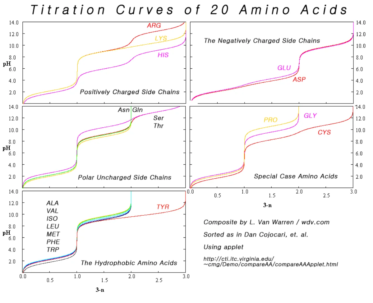
Titration Curve
A plot of pH vs. equivalents of base added shows three regions:
Below pKa₁ (~2): COOH is protonated; molecule is NH₃⁺-COOH (net +1 charge).
Between pKa₁ and pKa₂: zwitterion dominates (NH₃⁺-COO⁻, net zero).
Above pKa₂ (~9): NH₂ is deprotonated; molecule is NH₂-COO⁻ (net -1 charge).
Each pKa corresponds to an inflection point on the titration curve, and the midpoint between them is the pI.
Titration curves for all 20 amino acids grouped by side chain type. The flat "buffering" plateaus correspond to pKa values; steep vertical segments are the equivalence points. Amino acids with ionizable side chains show a third plateau/buffer region around their side chain pKa. Credit: Wikimedia Commons, CC BY-SA
Strecker Synthesis of Amino Acids
Starting materials: an aldehyde, NH₃, and HCN.
Aldehyde + NH₃ → imine (R-CH=NH).
Imine + HCN → aminonitrile (R-CH(NH₂)-CN).
Acid hydrolysis of nitrile → carboxylic acid: R-CH(NH₂)-COOH.
Named after the 19th-century chemist Adolph Strecker. On the AAMC content outline.
Strecker synthesis: aldehyde + NH₃ → imine; imine + HCN → alpha-aminonitrile; acid hydrolysis of the nitrile → alpha-amino acid. A classical route to racemic amino acids from simple starting materials. Credit: Wikimedia Commons, CC BY-SA
Gabriel Synthesis of Primary Amines (and by extension, amino acids)
Problem with direct alkylation of ammonia: over-alkylation (product mixture of 1°, 2°, 3° amines). Gabriel synthesis solves this using potassium phthalimide.
Potassium phthalimide (a “masked” ammonia where the nitrogen is flanked by two carbonyls, preventing further alkylation) + R-X → N-alkyl phthalimide.
Hydrolysis with hydrazine (N₂H₄) or strong acid/base → primary amine + phthalhydrazide (or phthalic acid).
For amino acid synthesis: use an alkyl halide with a pre-installed -COOH (like diethyl alpha-bromomalonate). Phthalimide displaces Br, giving an N-alkyl phthalimide with the ester. Hydrolysis gives the amino acid with clean 1° amine.
D and L Amino Acids
Naturally occurring amino acids are almost exclusively L-amino acids (S configuration at alpha-C for 19 of 20; R for cysteine). D-amino acids appear in bacterial cell walls (peptidoglycan) and a few natural products.
The strict L-selectivity of biology is enforced by aminoacyl-tRNA synthetases, which only accept L-amino acids for protein synthesis. Racemized (D) amino acids cannot be incorporated into standard proteins.
At pH 1, pH 7, and pH 12, what are the ionization states and net charges of glycine (alpha-COOH pKa 2.35, alpha-NH₃⁺ pKa 9.78)?
Click to reveal answer
At pH 1: NH₃⁺-CH₂-COOH (COOH protonated below its pKa, NH₃⁺ protonated far below its pKa). Net charge: +1. At pH 7: NH₃⁺-CH₂-COO⁻ (zwitterion - COOH deprotonated, NH₃⁺ still protonated). Net charge: 0. At pH 12: NH₂-CH₂-COO⁻ (above both pKas - NH₃⁺ deprotonated to NH₂, COO⁻ is still deprotonated). Net charge: -1.
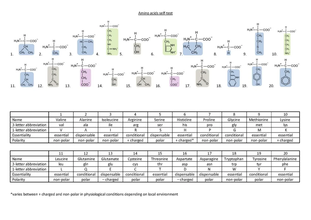
The 20 standard amino acids are grouped by side chain character. The R group determines the amino acid’s role in proteins: hydrophobic side chains pack the protein interior, polar side chains face the surface, and ionizable side chains participate in H-bonding, salt bridges, and catalysis.
The 20 standard amino acids with structures, three-letter and one-letter codes. Grouped by side chain polarity and charge at physiological pH. Memorize the 1-letter codes and side chain classifications - they recur constantly in biochemistry passages. Credit: Wikimedia Commons, CC BY-SA
Nonpolar (Hydrophobic) Side Chains
These have hydrocarbon side chains - no heteroatoms or charges. They cluster together in protein cores to avoid water.
Amino acid
3-letter
1-letter
Side chain
Glycine
Gly
G
-H
Alanine
Ala
A
-CH₃
Valine
Val
V
-CH(CH₃)₂
Leucine
Leu
L
-CH₂CH(CH₃)₂
Isoleucine
Ile
I
-CH(CH₃)CH₂CH₃
Methionine
Met
M
-CH₂CH₂SCH₃
Proline
Pro
P
(cyclic)
Phenylalanine
Phe
F
-CH₂C₆H₅
Tryptophan
Trp
W
indole ring
Polar Uncharged Side Chains
These have heteroatoms (O, N, S) but no net charge at physiological pH. They H-bond with water and are found at protein surfaces.
Amino acid
3-letter
1-letter
Side chain
Serine
Ser
S
-CH₂OH
Threonine
Thr
T
-CH(OH)CH₃
Cysteine
Cys
C
-CH₂SH (sulfhydryl)
Asparagine
Asn
N
-CH₂CONH₂ (amide)
Glutamine
Gln
Q
-CH₂CH₂CONH₂
Tyrosine
Tyr
Y
-CH₂-C₆H₄-OH (phenolic)
Acidic (Negatively Charged at pH 7)
Side chain carboxylic acid with pKa below 5. At pH 7, it is deprotonated (COO⁻).
Amino acid
3-letter
1-letter
Side chain pKa
Aspartate
Asp
D
-CH₂COOH (pKa ~3.9)
Glutamate
Glu
E
-CH₂CH₂COOH (pKa ~4.1)
Basic (Positively Charged at pH 7)
Side chain amine or guanidinium with pKa above 7. At pH 7, it is protonated (NH₃⁺ or guanidinium+).
Amino acid
3-letter
1-letter
Side chain pKa
Lysine
Lys
K
-(CH₂)₄NH₃⁺ (pKa ~10.8)
Arginine
Arg
R
-(CH₂)₃-NH-C(NH)NH₂ (pKa ~12.5)
Histidine
His
H
-CH₂-imidazole (pKa ~6.0)
Note: histidine’s pKa of ~6 is close to physiological pH, which is why it acts as both acid and base in enzyme active sites. At pH 7, histidine is mostly deprotonated but has a significant fraction protonated. This responsiveness to small pH changes is why histidine is so common in enzyme mechanisms.
Calculating pI with Ionizable Side Chains
For amino acids with ionizable side chains, pI is the average of the two pKa values BRACKETING the zwitterionic form (net zero charge).
Aspartate (3 pKa values: 2.0, 3.9, 9.8). Zwitterion is bracketed by pKa₁ (alpha-COOH, 2.0) and pKa₂ (side chain COOH, 3.9). pI = (2.0 + 3.9)/2 = 2.95.
Lysine (3 pKa values: 2.2, 9.0, 10.8). Zwitterion is bracketed by pKa₂ (alpha-NH₃⁺, 9.0) and pKa₃ (side chain NH₃⁺, 10.8). pI = (9.0 + 10.8)/2 = 9.9.
Histidine (3 pKa values: 1.8, 6.0, 9.2). Zwitterion is bracketed by pKa₂ (side chain imidazolium, 6.0) and pKa₃ (alpha-NH₃⁺, 9.2). pI = (6.0 + 9.2)/2 = 7.6.
Special Amino Acids
Glycine (Gly, G): smallest, most flexible. Found in tight turns of proteins (Ramachandran plot shows allowed regions unique to glycine).
Proline (Pro, P): side chain cyclizes back to the alpha-N, creating a rigid ring. Does not fit alpha-helix well - found at helix breaks and in turns.
Cysteine (Cys, C): thiol side chain can form disulfide bonds (S-S bridges) between two cysteines, stabilizing protein tertiary structure.
Tryptophan (Trp, W): the largest amino acid; has an indole ring and intense UV absorbance (used to quantify protein concentration).
Calculate the pI of aspartate, given pKa1 (alpha-COOH) = 2.0, pKa2 (side chain COOH) = 3.9, and pKa3 (alpha-NH₃⁺) = 9.8.
Click to reveal answer
pI = (pKa1 + pKa2) / 2 = (2.0 + 3.9) / 2 = 2.95. Reasoning: the zwitterion of aspartate has both carboxyl groups deprotonated (net -2 from two COO⁻) and the amine protonated (+1 from NH₃⁺), giving net -1. Wait - this means the "zero net charge" form has the side chain COOH protonated AND the alpha-COOH deprotonated. The pKa values bracketing the zero-charge state are the alpha-COOH (2.0) and the side-chain COOH (3.9). pI = average = 2.95. Below pH 2.95 aspartate is net +, above it is net -. At pH 2.95 the molecule is a neutral zwitterion.
A peptide bond is the amide bond that links two amino acids. The -COOH of one amino acid and the -NH₂ of the next condense with loss of water, forming the C-N amide. Cells synthesize peptide bonds at the ribosome using activated aminoacyl-tRNAs as reactive intermediates.
In the lab, this reaction does not work spontaneously because amine + carboxylic acid at neutral pH gives a stable zwitterion salt that does not react. To form peptide bonds in vitro, organic chemists activate the carboxylic acid first (SOCl₂ to give acyl chloride, or DCC to give an O-acylisourea), then add the amine.
Formation (Biological)
Cells activate amino acids by attaching them to tRNA via an ester bond:
Amino acid + ATP + tRNA → aminoacyl-tRNA + AMP + PPi (catalyzed by aminoacyl-tRNA synthetase).
Aminoacyl-tRNA’s carboxyl is activated as an ester (better electrophile than free COOH).
At the ribosome’s peptidyl transferase center, the next aminoacyl-tRNA’s alpha-amine attacks the ester carbonyl of the previous amino acid.
Peptide bond forms; the now-empty tRNA dissociates.
The ribosome is actually a ribozyme - the catalysis is done by rRNA, not protein. Still, the organic mechanism is nucleophilic acyl substitution.
Planarity
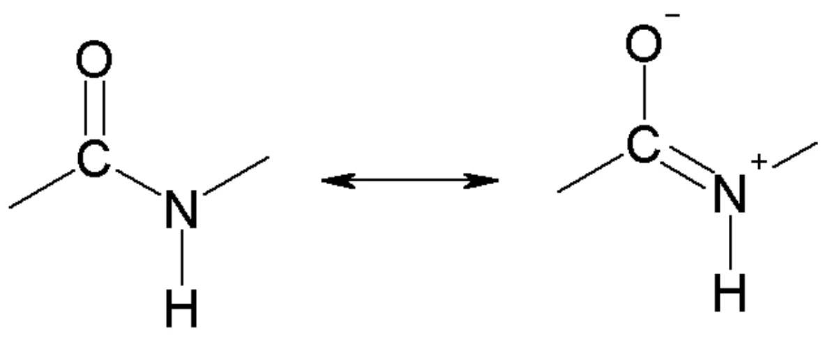
The peptide bond has ~40% double-bond character due to nitrogen lone pair resonance into the carbonyl. All six atoms of the peptide group (Cα, C=O, N, H, adjacent Cα) are coplanar. Rotation around C-N is restricted (barrier ~75 kJ/mol).
Peptide bond resonance: the nitrogen lone pair delocalizes into the C=O pi system. The dipolar contributor has ~40% weight in the hybrid, giving the C-N bond substantial double-bond character and restricting its rotation. Credit: Wikimedia Commons, CC BY-SA
Only the Cα-N bond (phi) and Cα-C bond (psi) allow rotation. The Ramachandran plot shows allowed combinations of phi and psi - the zones that give alpha-helix or beta-sheet secondary structures.
Hydrolysis
Peptide bonds are amide bonds and share amide stability:
Uncatalyzed at pH 7, 37°C: half-life of hundreds of years.
Acid hydrolysis: 6 M HCl, reflux, 24 hours. Classic method for breaking proteins into constituent amino acids. Destroys tryptophan, which must be quantified separately.
Base hydrolysis: concentrated NaOH, reflux. Racemizes chiral alpha-centers, so avoided for analytical protein sequencing.
Enzymatic hydrolysis: proteases (trypsin, chymotrypsin, pepsin, thrombin, etc.) cleave peptide bonds with high specificity at physiological conditions.
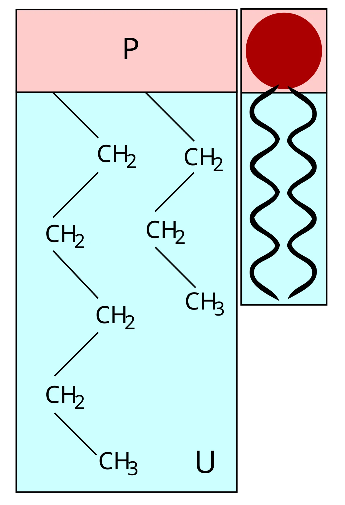
Example of a phospholipid - the base for cell membranes. Structure discussed further in Section 10.11. Credit: Wikimedia Commons, CC BY-SA
Peptide Nomenclature
Peptides are named from the N-terminus (free alpha-NH₃⁺ end) to the C-terminus (free alpha-COO⁻ end). Example: Gly-Ala-Ser is the tripeptide with glycine at the N-terminus, alanine in the middle, serine at the C-terminus.
Dipeptide: 2 amino acids, 1 peptide bond.
Tripeptide: 3 amino acids, 2 peptide bonds.
Polypeptide: many amino acids, many peptide bonds.
Protein: a polypeptide with defined structure and function (often >50 residues).
Specific Proteases
Different proteases cleave at different positions:
Trypsin: cleaves after basic residues (Lys, Arg).
Chymotrypsin: cleaves after aromatic residues (Phe, Trp, Tyr).
Pepsin: cleaves after aromatic and large hydrophobic residues.
Carboxypeptidase: cleaves one residue at a time from the C-terminus.
Aminopeptidase: cleaves one residue at a time from the N-terminus.
These specificities are used to sequence proteins in the Edman degradation method (pre-mass spectrometry era).
Why does biology activate amino acids as aminoacyl-tRNAs (esters with tRNA) before peptide bond formation, rather than just using the free amino acids?
Click to reveal answer
Free amino acids at physiological pH are zwitterions (NH₃⁺-CHR-COO⁻). The COO⁻ is a poor electrophile (negatively charged, not attackable by another amine), and the NH₃⁺ is not nucleophilic (protonated). They cannot react with each other under physiological conditions. Attaching the amino acid to tRNA via an ester bond activates the carboxyl as a reactive electrophile (ester is 10³-10⁴ more reactive than free COOH). The next amino acid’s free amine can then attack the aminoacyl-tRNA, forming the peptide bond and releasing the empty tRNA as the leaving group.
Nitrogen heterocycles are aromatic rings containing one or more nitrogens. They appear everywhere in biology - DNA/RNA bases, amino acid side chains (histidine’s imidazole, tryptophan’s indole), and alkaloid natural products. Understanding aromaticity in these systems requires knowing where each nitrogen’s lone pair lives.
Pyrrole (5-Membered Ring, 1 N)
Structure: 5-membered aromatic ring with one nitrogen (N-H at the ring position).
Key fact: the nitrogen’s lone pair is IN the aromatic pi system. It contributes 2 of the 6 pi electrons needed for Huckel’s 4n+2 rule (n=1).
Consequences:
Pyrrole IS aromatic.
The N-H proton is relatively acidic (pKa ~17) - hydrogen bond donor.
Pyrrole nitrogen is NOT basic (protonating the nitrogen would remove the lone pair from the pi system, breaking aromaticity).
Pyrrole is weakly basic at C2 (electrophilic attack on the ring protonates a ring carbon, which is better than disrupting aromaticity).
Pyrrole units appear in heme (hemoglobin’s oxygen-binding cofactor), chlorophyll, and vitamin B12.
Pyridine (6-Membered Ring, 1 N)
Structure: 6-membered aromatic ring with one nitrogen. No N-H.
Key fact: the nitrogen’s lone pair is NOT in the pi system. It sits in an sp² hybrid orbital in the plane of the ring.
Consequences:
Pyridine IS aromatic (6 pi electrons come from the 3 C=C of the ring, plus the N contributes one p electron to the ring).
The sp² lone pair on N is available for protonation.
Pyridine is a modest base (conjugate acid pKa ~5.2).
Commonly used as a base in organic synthesis (to soak up HCl in acylation reactions).
Imidazole (5-Membered Ring, 2 N)
Structure: 5-membered aromatic ring with 2 nitrogens. One has an H (pyrrole-like), the other does not (pyridine-like).
Pyrrole-like N-H: lone pair is in the pi system (aromatic). N-H is acidic (pKa ~14.5).
Imidazole’s pKa ~7.0 makes it the only basic nitrogen heterocycle in this set with pKa near physiological pH. Histidine’s side chain has an imidazole ring, which is why histidine can serve as both acid and base in enzyme active sites (at pH 7, ~50% protonated, ~50% deprotonated - instantly responsive to pH changes).
Purines and Pyrimidines (DNA/RNA Bases)
DNA and RNA bases are nitrogen heterocycles:
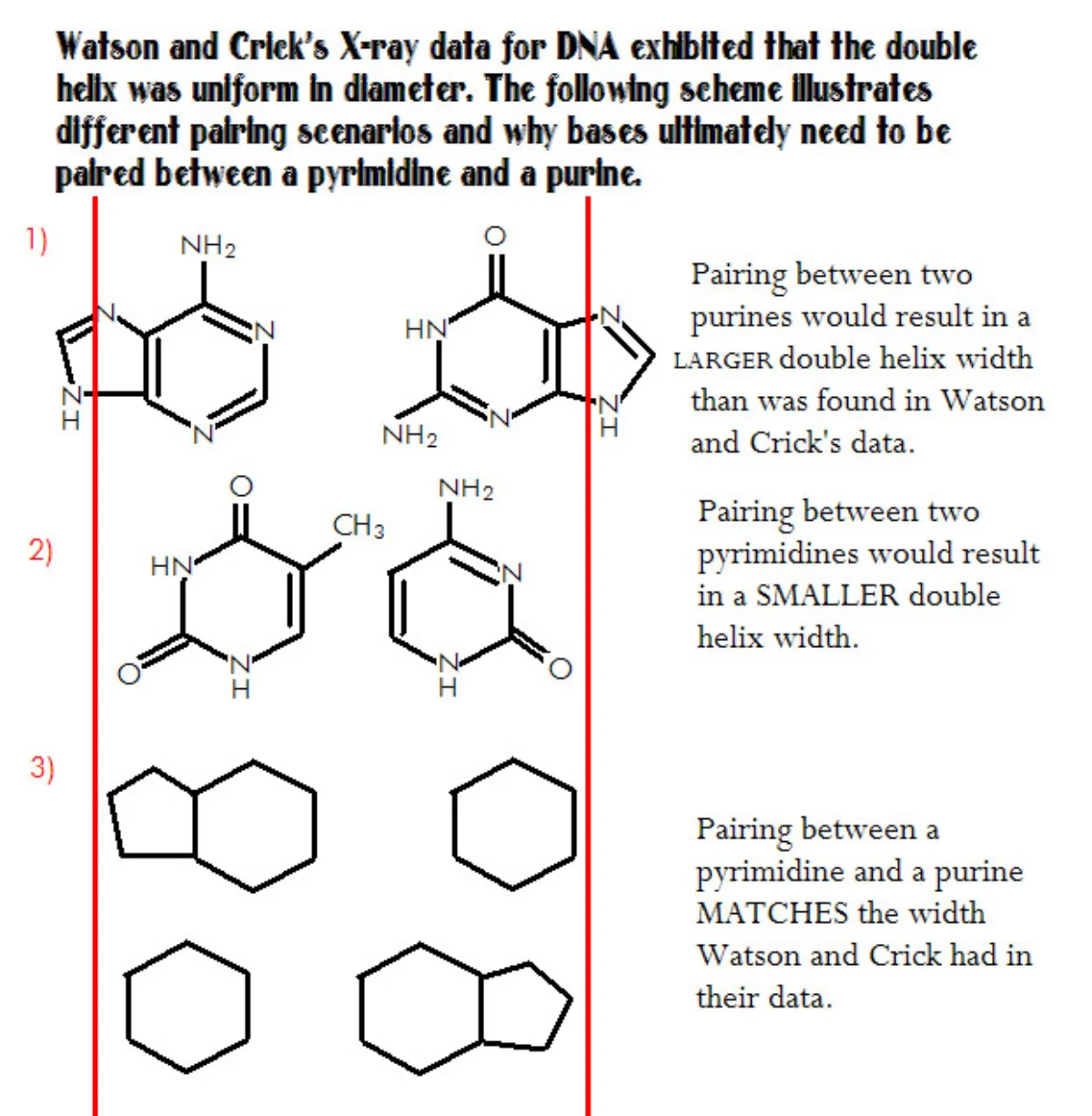
Pyrimidines (6-membered, 2 N): cytosine (C), thymine (T, in DNA only), uracil (U, in RNA only).
The five DNA/RNA nitrogen bases. Purines (adenine, guanine): fused 5+6-ring. Pyrimidines (cytosine, thymine, uracil): single 6-ring with 2 nitrogens. Each has specific H-bond donors and acceptors that enable the Watson-Crick base-pair geometry. Credit: Wikimedia Commons, CC BY-SA
These bases do hydrogen bonding: A pairs with T (or U in RNA) via 2 H-bonds; G pairs with C via 3 H-bonds. The specificity of base pairing is the chemical basis of the genetic code.
The bases are connected to sugar (ribose or deoxyribose) via a glycosidic bond at the N1 of pyrimidines or N9 of purines, producing nucleosides. Adding phosphate gives nucleotides (ATP, GTP, dNTPs, etc.).
Indole and Tryptophan
Indole is a bicyclic aromatic system (benzene fused with pyrrole). It appears as the side chain of tryptophan, the largest amino acid. Indole absorbs UV strongly at 280 nm, which is the basis of protein quantification by UV absorbance (tryptophan dominates the 280 nm protein absorbance).
Indole nitrogen, like pyrrole N, has its lone pair in the pi system - not basic.
Imidazolium in Histidine Catalysis
In the serine protease active site (trypsin, chymotrypsin), histidine’s imidazole acts as a general base to deprotonate serine’s -OH, making it a better nucleophile for attacking the substrate’s peptide bond. The imidazolium then donates the proton back at a different point in the mechanism, regenerating neutral imidazole. This “proton shuttle” is one of the most elegant acid-base roles in enzyme catalysis.
Why is pyridine a modest base (conjugate acid pKa ~5.2) while pyrrole is essentially not basic at nitrogen?
Click to reveal answer
Pyridine's nitrogen lone pair is in an sp² hybrid orbital in the ring plane - NOT part of the aromatic pi system. It is freely available for protonation without disrupting aromaticity. Pyrrole's nitrogen lone pair IS part of the aromatic pi system - it contributes 2 of the 6 pi electrons needed for Huckel's 4n+2 rule. Protonating it would remove electrons from the pi system and destroy aromaticity, an energetically very costly change. So pyrrole's lone pair is "used up" by aromaticity and is not available for basic behavior.
Phosphoric acid (H₃PO₄) is a triprotic acid that forms the backbone of DNA, the high-energy bonds of ATP, and the head groups of phospholipids. Its three acidic protons ionize at different pKa values, giving rise to pH-dependent behavior central to biochemistry.
Phosphoric Acid Structure
Phosphoric acid has a central phosphorus atom bonded to four oxygens: three as -OH groups (ionizable) and one as a P=O double bond. The structure is tetrahedral around P (sp³).
Three pKa values:
Ionization
pKa
H₃PO₄ ⇌ H₂PO₄⁻
2.1
H₂PO₄⁻ ⇌ HPO₄²⁻
7.2
HPO₄²⁻ ⇌ PO₄³⁻
12.7
At physiological pH 7.4, phosphoric acid is between pKa₂ (7.2) and pKa₃ (12.7). The dominant species are roughly 50-50 H₂PO₄⁻ and HPO₄²⁻, with a small amount of each at the far ends. The ~1:1 mix is a key biological buffer (phosphate buffer).
Phosphate Esters
A phosphate ester replaces one or more of phosphoric acid’s -OH groups with -OR. Three types:
Monoester (R-O-PO₃²⁻): one OR, two ionizable -OH. Example: glucose-6-phosphate.
Diester ((RO)₂-PO₂⁻): two OR groups, one ionizable -OH. Example: DNA phosphodiester backbone, phospholipids.
Triester ((RO)₃-PO): three OR groups, no ionizable -OH. Example: organophosphate pesticides (mostly synthetic, rare in biology).
Phosphodiesters are essential in biology: DNA’s phosphate backbone connects one sugar’s 3’-OH to the next sugar’s 5’-OH via a phosphodiester. Phospholipids have one phosphodiester connecting glycerol’s 3-position to a polar head group (choline, serine, etc.).
Hydrolysis
Phosphate esters hydrolyze to the parent alcohol + phosphoric acid (or phosphate anion). Uncatalyzed hydrolysis is slow (half-life of years to millennia at neutral pH). Biology uses phosphatases to accelerate hydrolysis billions of times.
Phosphodiesters (like DNA) are especially slow to hydrolyze uncatalyzed - this is why DNA can preserve genetic information for millions of years in fossil samples. RNA hydrolyzes faster than DNA because the 2’-OH of ribose can attack the phosphate internally (enzymatic or spontaneous).
Phosphate as a Leaving Group
Phosphate can be a good leaving group in biological reactions. Examples:
In glycolysis, a phosphate group leaves when sugar-phosphate becomes pyruvate.
In ATP hydrolysis, the terminal phosphate leaves as Pi.
In protein phosphorylation/dephosphorylation, kinases and phosphatases transfer phosphate between ATP and serine/threonine/tyrosine residues.
The phosphate’s leaving ability is enhanced by its resonance stabilization over four oxygens (when it leaves as HPO₄²⁻ or PO₄³⁻, the charge is delocalized).
Phosphate Transfer Reactions
Many enzymes transfer a phosphate from one molecule to another via a nucleophilic substitution at phosphorus. The mechanism can go through a trigonal bipyramidal intermediate (SN2-like at P) or through a metaphosphate-like intermediate (SN1-like).
For MCAT purposes, recognize that “phosphate transfer” is a type of nucleophilic acyl substitution at phosphorus, analogous to carbonyl NAS but with P instead of C as the central atom.
Phosphate in Buffers
The HPO₄²⁻/H₂PO₄⁻ pair has pKa 7.2, very close to physiological pH. This makes phosphate an excellent biological buffer - small changes in [H⁺] are absorbed by shifting the equilibrium between the two forms without dramatic pH changes.
Inside cells, phosphate buffer maintains cytoplasmic pH near 7.2. In blood, the main buffer is actually bicarbonate (HCO₃⁻/CO₂), but phosphate is a backup.
At physiological pH 7.4, what is the predominant ionization state of phosphoric acid (pKa₁ 2.1, pKa₂ 7.2, pKa₃ 12.7)?
Click to reveal answer
The dominant species at pH 7.4 is HPO₄²⁻ (monohydrogen phosphate, about 60%) and H₂PO₄⁻ (dihydrogen phosphate, about 40%). The ratio is close to 1:1 because pH 7.4 is very close to pKa₂ (7.2). Above pKa₂ (at pH 7.4), the "HPO₄²⁻" form is slightly favored. The ~1:1 mix of HPO₄²⁻ / H₂PO₄⁻ makes phosphate a powerful biological buffer near physiological pH.
Adenosine triphosphate (ATP) is the universal energy currency of the cell. Its hydrolysis to ADP + Pi releases about 7.3 kcal/mol under standard conditions - enough to drive almost every energy-requiring reaction in the cell when coupled properly.
Click Hydrolyze below to cleave the γ-phosphate: the terminal phosphate separates, the energy burst fires, and you are left with ADP + Pᵢ.
ATP hydrolysis
Interactive
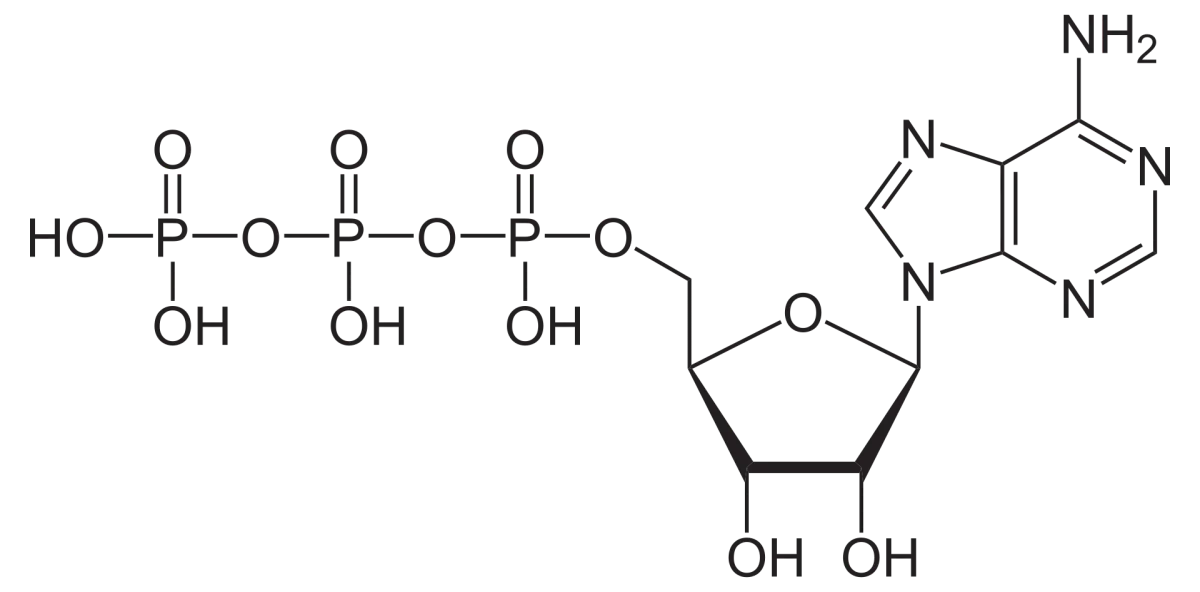
ATP (adenosine triphosphate): adenine base + ribose sugar + three phosphate groups. The ribose-phosphate linkage is a phosphoester bond (low energy). The phosphate-phosphate linkages are phosphoanhydride bonds (high energy). Credit: Wikimedia Commons, CC BY-SA
Structural Components
ATP has three components:
Adenine: the purine base (nitrogen heterocycle with a fused 6+5 ring system).
Ribose: a 5-carbon sugar with hydroxyls at 2’, 3’, and 5’ positions.
Three phosphates: connected in a row to the 5’ position of ribose.
Adenosine = adenine + ribose (a nucleoside). Adding one, two, or three phosphates gives AMP, ADP, or ATP (nucleotides).
The Two Types of Bonds
Phosphoester bond: ribose-phosphate. One P-O-C bond. Low-energy (ΔG hydrolysis ~3 kcal/mol). This is the bond connecting the ribose’s 5’-oxygen to the first phosphate (alpha-phosphate).
Phosphoanhydride bond: phosphate-phosphate. Two phosphates sharing an oxygen via P-O-P. HIGH-energy (ΔG hydrolysis ~7.3 kcal/mol). ATP has TWO phosphoanhydride bonds: alpha-beta and beta-gamma.
Why Phosphoanhydride Bonds Are High-Energy
Three reasons ATP hydrolysis (breaking a phosphoanhydride) is exergonic:
Electrostatic repulsion. The three phosphate groups of ATP each carry negative charges at physiological pH (ATP has about -4 net charge). These charges repel each other. Hydrolysis releases them from proximity, relieving electrostatic strain.
Resonance stabilization of products. After hydrolysis, the released Pi (HPO₄²⁻ or H₂PO₄⁻) is more resonance-stabilized (charge on 4 O) than it was in the ATP (where it was constrained to one P-O-P linkage). The products are more stable than the reactant.
Hydration stabilization. Pi is easier to solvate in water than it was when locked in ATP.
Note: “high-energy bond” does not mean the bond itself is strong - it is actually moderately weak. What makes the bond high-energy is the thermodynamic difference between ATP and ADP+Pi products.
ATP Hydrolysis Reactions
Three main reactions:
ATP → ADP + Pi: terminal (gamma) phosphoanhydride hydrolysis. ~7.3 kcal/mol released. Most common use.
ATP → AMP + PPi (pyrophosphate): alpha-beta phosphoanhydride hydrolysis. The released PPi is often further hydrolyzed to 2 Pi (another 7.3 kcal/mol), making the overall ATP → AMP + 2 Pi reaction very exergonic (~19 kcal/mol combined).
ATP → AMP + Pi + Pi: both phosphoanhydride bonds broken.
Reaction 2 is used for highly unfavorable syntheses (amino acid activation for tRNA loading, fatty acid activation for CoA thioester synthesis). Breaking pyrophosphate provides the extra thermodynamic push.
ATP Synthesis
ATP is synthesized from ADP + Pi by:
Oxidative phosphorylation: ATP synthase, driven by the electrochemical gradient across the inner mitochondrial membrane.
Substrate-level phosphorylation: direct phosphate transfer from a high-energy substrate (like phosphoenolpyruvate or 1,3-bisphosphoglycerate in glycolysis).
The cell continuously cycles between ATP and ADP, with steady-state ATP concentrations around 3 mM.
Other Nucleotide Triphosphates
Other NTPs have similar energy profiles:
GTP: used in protein synthesis (ribosome) and signal transduction (G-proteins).
UTP: activated sugars for polysaccharide synthesis.
All have similar high-energy phosphoanhydride bonds.
In ATP, which bond(s) are phosphoanhydride bonds (high energy), and which bond(s) are phosphoester bonds (low energy)?
Click to reveal answer
Phosphoanhydride bonds (high energy): the two P-O-P linkages between alpha-beta phosphates and between beta-gamma phosphates. These are broken in ATP hydrolysis to release ~7.3 kcal/mol each. Phosphoester bond (low energy): the C-O-P linkage connecting ribose’s 5’-O to the alpha-phosphate. This bond is much more stable. Students sometimes confuse these; the MCAT explicitly tests the distinction.
Phospholipids are amphipathic molecules with a polar phosphate-containing head and hydrophobic fatty acid tails. They spontaneously assemble into bilayers in aqueous solution, forming the basic framework of all cell membranes.
Structure
A typical phospholipid has:
Glycerol backbone: a 3-carbon chain with three hydroxyls.
Two fatty acid tails: esterified to glycerol’s C1 and C2 hydroxyls.
Phosphate group: esterified to glycerol’s C3 hydroxyl.
Polar head group: connected to the phosphate by a second ester. Common head groups: choline (phosphatidylcholine, PC), ethanolamine (PE), serine (PS), inositol (PI).
The two fatty acid tails are hydrophobic (long hydrocarbon chains). The phosphate + head group is hydrophilic (charged, polar). This amphipathic structure is the chemical basis of membrane formation.
Charge at Physiological pH
The phosphate group carries a negative charge at pH 7. Head groups vary:
Choline (+H₃N-CH₂CH₂-O-P-): positive quaternary ammonium, net phosphatidylcholine is zwitterion (net zero).
Ethanolamine (H₃N⁺-CH₂CH₂-O-P-): similar zwitterion.
Serine (HOOC-CH(NH₃⁺)-CH₂-O-P-): net -1 charge (COO⁻ at pH 7, NH₃⁺, P-).
Inositol: neutral cyclohexanol-like ring with hydroxyls; net -1 from the phosphate.
Phosphatidylserine and phosphatidylinositol are the main anionic phospholipids in cell membranes.
Bilayer Assembly
In water, phospholipids assemble into structures that minimize contact between hydrophobic tails and water:
Micelles: small spherical aggregates with tails pointing inward. Only for molecules with one hydrophobic tail (like soap, lysophospholipids).
Bilayers: two-layer sheets with tails inward and heads facing water on both sides. The biological membrane. Favored for molecules with TWO hydrophobic tails (like standard phospholipids).
Liposomes: bilayers curled into spherical vesicles.
The bilayer is ~5 nm thick. The tails are fluid (lateral diffusion is fast) but flip-flop (transfer from one leaflet to another) is very slow without enzyme assistance (flippases).
Cholesterol: stiffens the membrane at high temperature (fills gaps, reduces fluidity) but prevents crystalline packing at low temperature (acts as a buffer).
Temperature: higher T = more fluid.
Membrane Proteins
Membranes are not just lipid - proteins embedded in the bilayer act as transporters, receptors, and enzymes. About 25-50% of a typical cell membrane’s mass is protein. Protein topology (which side of the membrane each segment faces) is controlled during translation.
Biological Significance
Phospholipids enable:
Cell compartmentalization. Membranes separate cytoplasm from extracellular space and create organelles.
Selective permeability. The hydrophobic core is a barrier to polar molecules; channels and transporters provide controlled passage.
Signaling. Phosphoinositides (PIP₂, PIP₃) are key second messengers.
Energy generation. The inner mitochondrial membrane hosts the electron transport chain and ATP synthase.
Lipid Rafts
Specialized membrane microdomains enriched in cholesterol, sphingolipids, and certain proteins. Known as lipid rafts. They serve as platforms for signaling and protein sorting. An active area of biomedical research.
Why do phospholipids form bilayers in water but single-tailed amphipaths (like soap, sodium laurate) form micelles?
Click to reveal answer
The geometry depends on the "cone angle" of the molecule - the relative cross-sectional area of the polar head vs. the hydrophobic tails. Single-tailed soaps have a narrow tail and broad head, giving a conical shape that packs into spherical micelles (narrow ends toward the center). Phospholipids have two tails side by side and a single polar head, giving a more cylindrical shape that packs best into flat bilayers (heads on both sides, tails stacked in the middle). Both are thermodynamically favored arrangements that minimize hydrophobic-water contact.
This synthesis section ties together the organic chemistry of amines, amides, amino acids, and phosphates with their biological roles. Every biomolecule in this chapter has a chemical story rooted in Chs 5-9 mechanism chemistry.
Neurotransmitters
Many neurotransmitters are simple amines:
Acetylcholine = choline (quaternary ammonium) acetylated at the hydroxyl. An ester linking an alcohol to acetate.
Serotonin = 5-hydroxytryptamine, derived from tryptophan by decarboxylation + hydroxylation.
Dopamine, norepinephrine, epinephrine = catecholamines, all aromatic with amine and hydroxyl groups.
Histamine = histidine minus its carboxyl group (decarboxylation).
GABA (gamma-aminobutyric acid) = glutamate minus its alpha-carboxyl (decarboxylation).
Biosynthesis of these involves PLP-dependent decarboxylases and aromatic hydroxylases.
Alkaloid Natural Products
Plant alkaloids are nitrogen-containing natural products with often potent biological effects:
Cocaine (coca leaves): tropane alkaloid, also contains esters.
These are drug templates and targets - many are Schedule I or Schedule II controlled substances because of their psychoactive or toxic effects.
Protein Phosphorylation
Kinases transfer phosphate from ATP to serine, threonine, or tyrosine residues in proteins:
Protein-Ser-OH + ATP → Protein-Ser-OPO₃²⁻ + ADP.
Phosphorylation is one of the main post-translational modifications used for cellular signaling. It alters protein conformation, activity, stability, and localization. Phosphatases remove phosphate, reversing the modification.
About 30% of proteins are regulated by phosphorylation. Major pathways: MAPK, PI3K/AKT, cAMP/PKA, insulin signaling, cell cycle (CDKs). Drug discovery often targets kinases (e.g., tyrosine kinase inhibitors for cancer).
DNA and RNA Backbones
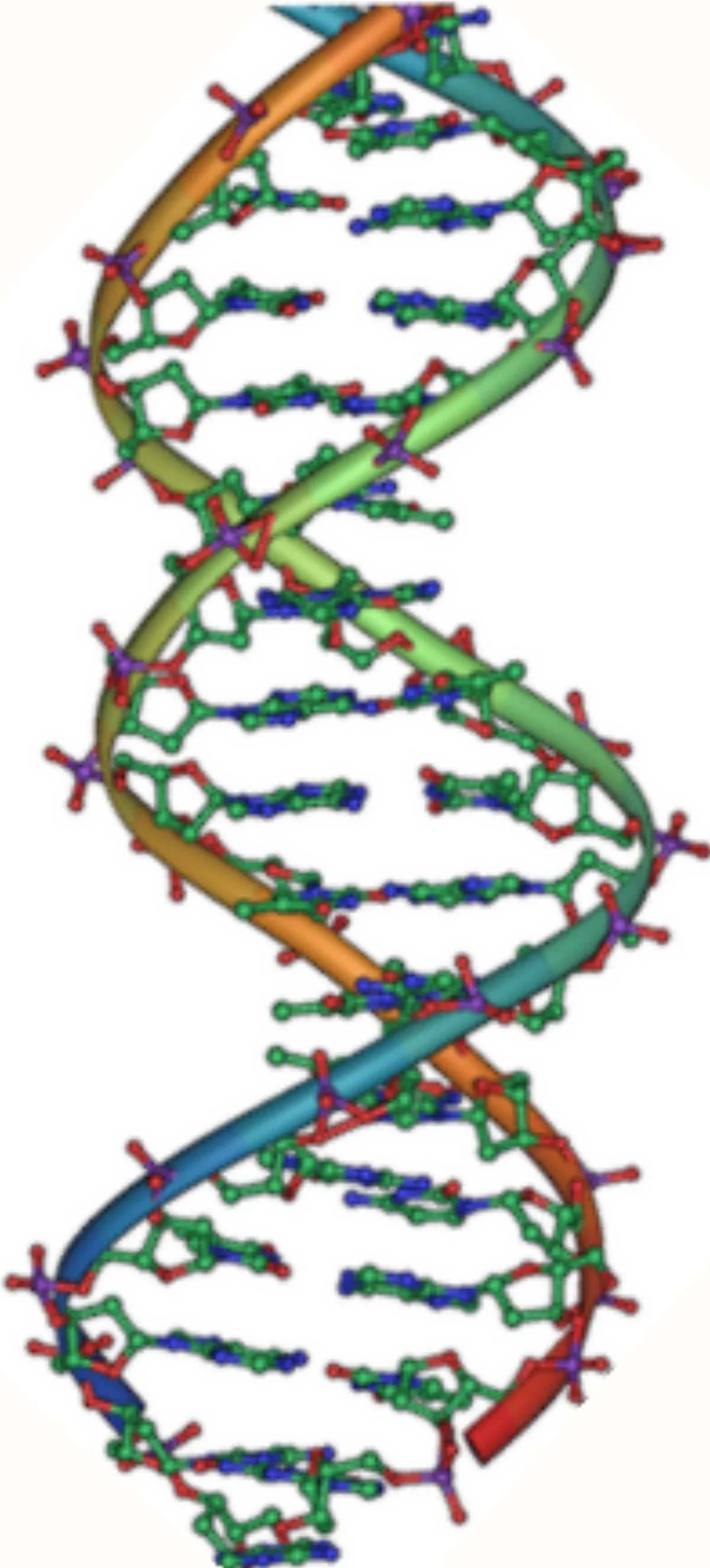
DNA is a polymer of deoxyribonucleotides connected by phosphodiester bonds: 3’-OH of one deoxyribose + 5’-phosphate of the next deoxyribose. The bases (A, T, G, C) hang off the backbone and do the H-bonding that encodes genetic information.
DNA double helix: two antiparallel sugar-phosphate backbones wrap around each other with complementary base pairs H-bonded in the middle (A-T: 2 H-bonds; G-C: 3 H-bonds). The phosphodiester bonds along each backbone give DNA its chemical stability; the H-bonded base pairs encode genetic information. Credit: Wikimedia Commons, CC BY-SA
RNA has the same backbone but with ribose (2’-OH) instead of deoxyribose. The 2’-OH makes RNA more susceptible to hydrolysis (it can attack the phosphate internally). This is why DNA is more stable and is the genetic storage medium, while RNA is more transient.
Hydrolysis of DNA or RNA backbones requires nuclease enzymes. Without them, DNA’s half-life is millions of years; RNA’s is on the order of hours to days depending on conditions.
Second Messengers
Several phosphate-containing molecules serve as signaling second messengers:
cAMP (cyclic AMP): a cyclic phosphodiester, the classic second messenger downstream of Gs-coupled receptors.
cGMP: similar to cAMP, downstream of vision and nitric oxide signaling.
IP3 (inositol trisphosphate): a heavily phosphorylated inositol ring that triggers Ca²⁺ release from the ER.
DAG (diacylglycerol): not a phosphate but paired with IP3 from PIP2 cleavage.
All of these are produced enzymatically from phospholipid or ATP substrates.
Urea Cycle and Nitrogen Disposal
Nitrogen (from protein breakdown) is toxic as NH₃/NH₄⁺. Mammals convert it to urea (H₂N-CO-NH₂, a diamide of carbonic acid) via the urea cycle. Urea is excreted in urine; it is chemically stable and non-toxic.
Urea’s diamide structure explains its stability (both amide bonds are unreactive under physiological conditions) and its water solubility (two amide groups are strong H-bond donors and acceptors).
The Big Picture
Nitrogen and phosphorus are unique among elements in their biological versatility:
Nitrogen: four bonds to C and H give amines (basic), amides (neutral, planar), heterocycles (aromatic), nitrates (leaving groups). Spans multiple oxidation states (-3 to +5).
Phosphorus: spanning oxidation states (-3 to +5), five-coordinate transition states in enzyme reactions, phosphoester and phosphoanhydride bonds for energy.
Together, they form the chemical framework for proteins (amides), nucleic acids (phosphodiesters), membranes (phospholipids), and the universal energy currency (ATP).
Acetylcholine (ACh) consists of choline + acetate. What kind of bond connects them, and how is ACh degraded in the synaptic cleft?
Click to reveal answer
Acetylcholine has an ester bond: choline's -OH is esterified with acetic acid, giving CH₃-CO-O-CH₂CH₂-N⁺(CH₃)₃. Acetylcholinesterase (AChE) enzymatically hydrolyzes this ester bond to release acetate + choline, terminating synaptic transmission. Nerve agents and some pesticides (organophosphates) inhibit AChE, causing prolonged muscle contraction and toxicity. Reversal of nerve agent poisoning uses pralidoxime (2-PAM), which displaces the organophosphate from AChE.