
Peptide Bonds
A peptide bond is the amide bond that links two amino acids. The -COOH of one amino acid and the -NH₂ of the next condense with loss of water, forming the C-N amide. Cells synthesize peptide bonds at the ribosome using activated aminoacyl-tRNAs as reactive intermediates.
Formation (In Vitro)
Condensation: H₂N-CHR₁-COOH + H₂N-CHR₂-COOH → H₂N-CHR₁-CO-NH-CHR₂-COOH + H₂O.
In the lab, this reaction does not work spontaneously because amine + carboxylic acid at neutral pH gives a stable zwitterion salt that does not react. To form peptide bonds in vitro, organic chemists activate the carboxylic acid first (SOCl₂ to give acyl chloride, or DCC to give an O-acylisourea), then add the amine.
Formation (Biological)
Cells activate amino acids by attaching them to tRNA via an ester bond:
- Amino acid + ATP + tRNA → aminoacyl-tRNA + AMP + PPi (catalyzed by aminoacyl-tRNA synthetase).
- Aminoacyl-tRNA’s carboxyl is activated as an ester (better electrophile than free COOH).
- At the ribosome’s peptidyl transferase center, the next aminoacyl-tRNA’s alpha-amine attacks the ester carbonyl of the previous amino acid.
- Peptide bond forms; the now-empty tRNA dissociates.
The ribosome is actually a ribozyme - the catalysis is done by rRNA, not protein. Still, the organic mechanism is nucleophilic acyl substitution.
Planarity
The peptide bond has ~40% double-bond character due to nitrogen lone pair resonance into the carbonyl. All six atoms of the peptide group (Cα, C=O, N, H, adjacent Cα) are coplanar. Rotation around C-N is restricted (barrier ~75 kJ/mol).
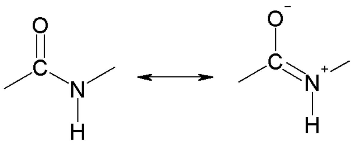
Only the Cα-N bond (phi) and Cα-C bond (psi) allow rotation. The Ramachandran plot shows allowed combinations of phi and psi - the zones that give alpha-helix or beta-sheet secondary structures.
Hydrolysis
Peptide bonds are amide bonds and share amide stability:
- Uncatalyzed at pH 7, 37°C: half-life of hundreds of years.
- Acid hydrolysis: 6 M HCl, reflux, 24 hours. Classic method for breaking proteins into constituent amino acids. Destroys tryptophan, which must be quantified separately.
- Base hydrolysis: concentrated NaOH, reflux. Racemizes chiral alpha-centers, so avoided for analytical protein sequencing.
- Enzymatic hydrolysis: proteases (trypsin, chymotrypsin, pepsin, thrombin, etc.) cleave peptide bonds with high specificity at physiological conditions.
Peptide Nomenclature
Peptides are named from the N-terminus (free alpha-NH₃⁺ end) to the C-terminus (free alpha-COO⁻ end). Example: Gly-Ala-Ser is the tripeptide with glycine at the N-terminus, alanine in the middle, serine at the C-terminus.
- Dipeptide: 2 amino acids, 1 peptide bond.
- Tripeptide: 3 amino acids, 2 peptide bonds.
- Polypeptide: many amino acids, many peptide bonds.
- Protein: a polypeptide with defined structure and function (often >50 residues).
Specific Proteases
Different proteases cleave at different positions:
- Trypsin: cleaves after basic residues (Lys, Arg).
- Chymotrypsin: cleaves after aromatic residues (Phe, Trp, Tyr).
- Pepsin: cleaves after aromatic and large hydrophobic residues.
- Carboxypeptidase: cleaves one residue at a time from the C-terminus.
- Aminopeptidase: cleaves one residue at a time from the N-terminus.
These specificities are used to sequence proteins in the Edman degradation method (pre-mass spectrometry era).