Before diving into specific disorders, we need a framework for classifying them. The MCAT wants you to know the dominant diagnostic manual and the main theoretical lenses used to understand mental illness.
The DSM-5
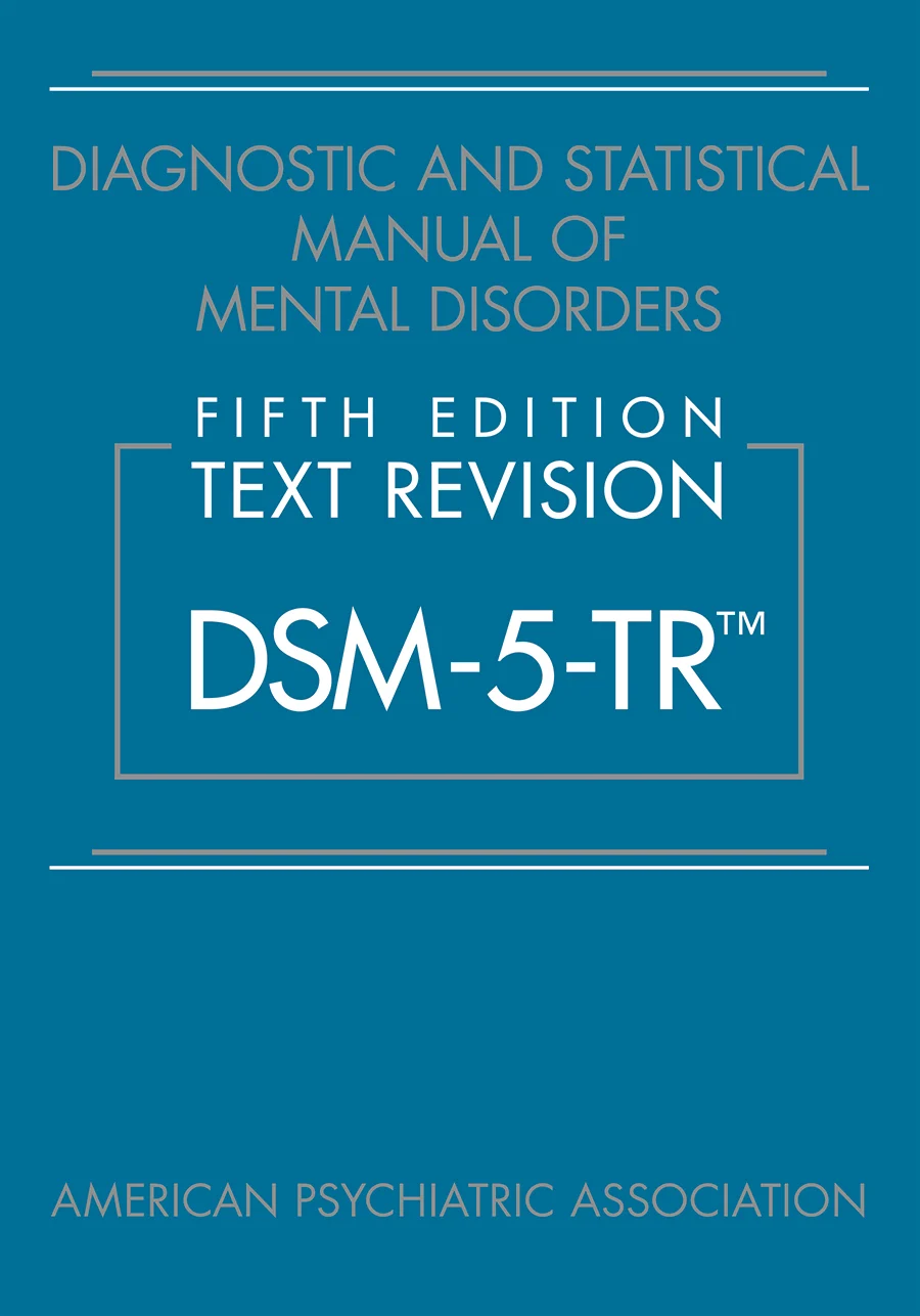
The Diagnostic and Statistical Manual of Mental Disorders, 5th Edition (DSM-5), published by the American Psychiatric Association, is the standard reference for diagnosing mental disorders in the United States. It lists hundreds of disorders, each with specific diagnostic criteria.
The DSM-5 (here in its 2022 Text Revision) is the gold-standard diagnostic reference in U.S. psychiatry. MCAT passages routinely quote DSM-5 criteria verbatim, so know the framework: defined symptom clusters plus significant distress or impairment plus a minimum duration. Credit: American Psychiatric Association via Wikimedia Commons (Public Domain).
A diagnosis requires:
Presence of specified symptoms.
Symptoms cause significant distress or impairment in daily functioning.
Symptoms are not better explained by another condition (e.g., medical illness, substance use).
For many disorders, symptoms must persist for a specified duration.
The DSM-5 replaced the multi-axial system of DSM-IV with a single-axis approach, integrating psychiatric and medical conditions together.
The Biopsychosocial Model
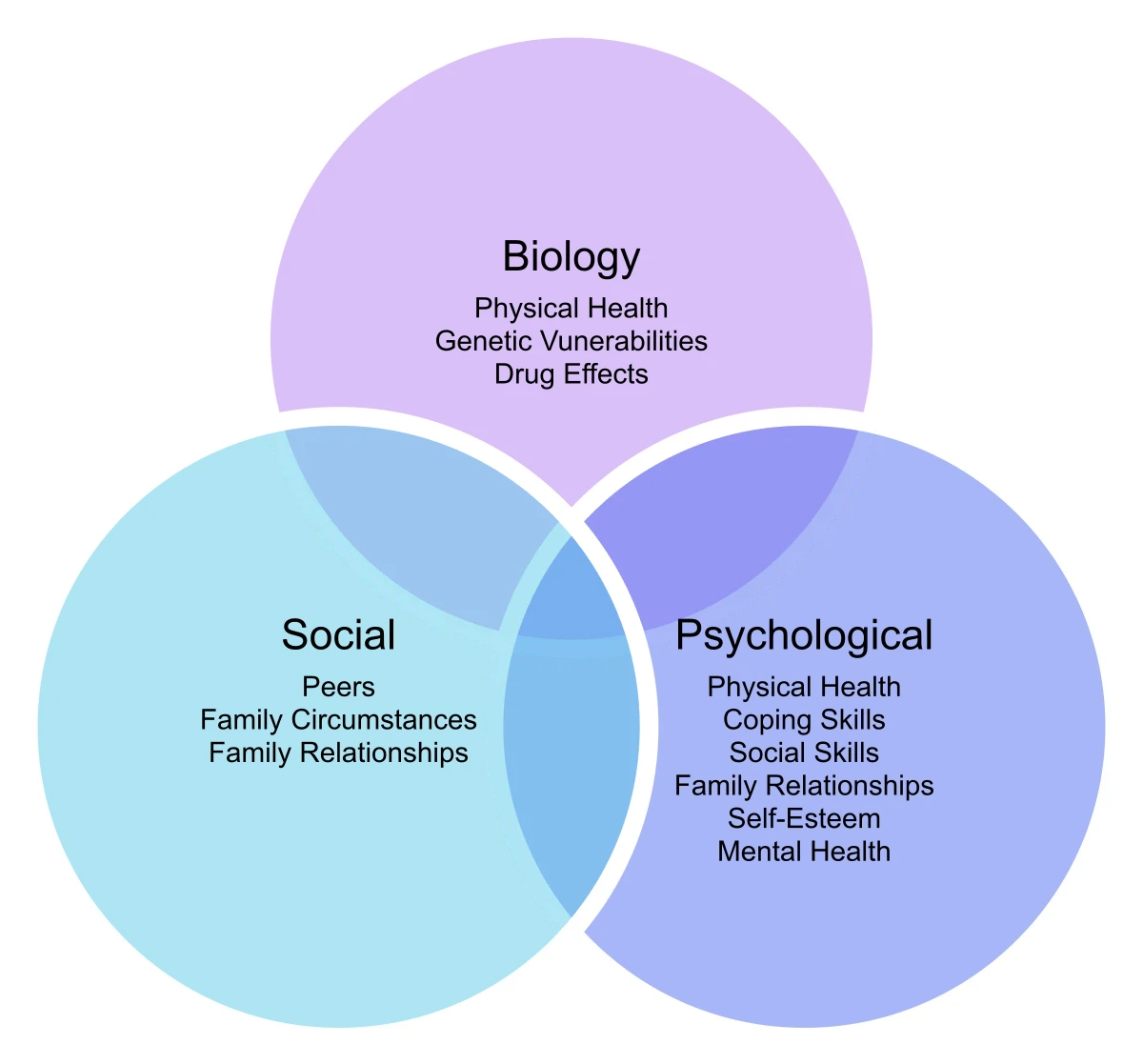
Mental disorders emerge from interacting factors at three levels:
Sociocultural. Family dynamics, social support, stressors, poverty, discrimination, cultural norms.
No single level tells the whole story. Depression can be triggered by a serotonin system vulnerability (bio), by rumination and hopelessness (psych), or by job loss and isolation (social) - usually all three together.
The biopsychosocial model: mental disorders emerge from the interaction of biological, psychological, and sociocultural factors. The MCAT loves this framework as a contrast to the older, narrower biomedical model. Credit: Seth Falco via Wikimedia Commons (CC BY-SA 4.0).
What Counts as “Abnormal”?
The DSM leans on several overlapping criteria:
Statistical deviance. The behavior is far from the norm (rare).
Maladaptive behavior. It interferes with daily functioning.
Personal distress. The person feels significant suffering.
Violation of social norms. The behavior deviates from what the culture considers acceptable.
None alone is sufficient. Genius is statistically deviant but not disordered. Someone in a dangerous job has normal distress. Cultural norms vary. Modern diagnosis requires a cluster of these with documented impairment.
Stigma and the Biomedical Model
Stigma is a major barrier to treatment. People with mental illness face discrimination, self-stigma (internalized shame), and courtesy stigma (affecting family and caregivers). Disclosure risks employment, relationships, and insurance.
The biomedical model frames mental disorders as brain diseases, like diabetes or cancer. This framing can reduce stigma (“it’s not your fault, your brain is sick”) but can also risk reductionism - ignoring the psychological and social dimensions. Modern psychiatry tries to integrate biomedical and psychological perspectives through the biopsychosocial framework.
Comorbidity
Mental disorders often co-occur. A person with major depression may also have anxiety, substance use disorder, or a personality disorder. This comorbidity complicates diagnosis and treatment - the clinician must sort out what’s primary, what’s secondary, and what pattern the patient actually lives with.
What is the DSM-5, and what does a diagnosis require?
Click to reveal answer
The Diagnostic and Statistical Manual of Mental Disorders, 5th Edition. A diagnosis requires specified symptoms, significant distress or impairment, and exclusion of other explanations. Many disorders require symptoms to persist for a minimum duration.
Name the three levels of the biopsychosocial model.
Click to reveal answer
Biological (genes, neurotransmitters, brain), Psychological (cognitions, coping, personality), Sociocultural (family, support, stressors, culture). Good clinical care addresses all three levels.
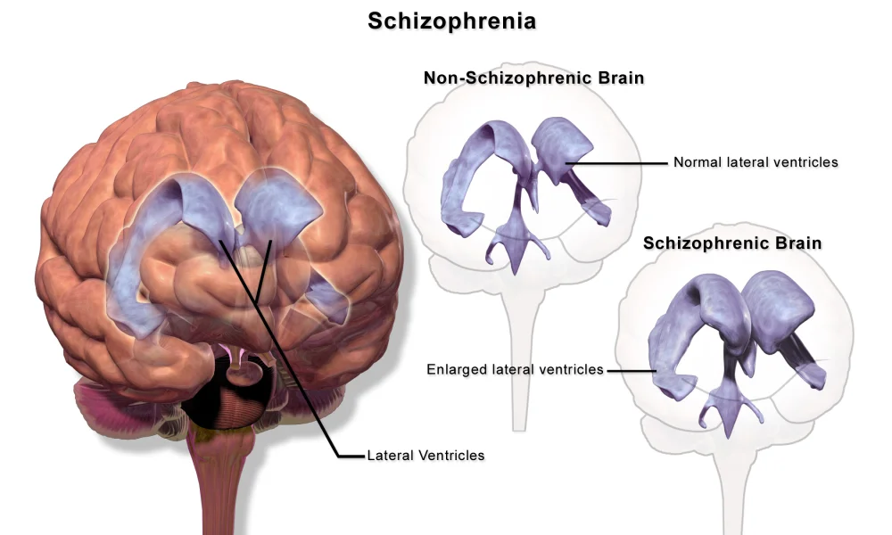
Schizophrenia is a severe psychotic disorder characterized by distorted thinking, perception, and emotion. It affects about 1% of the population worldwide, usually emerging in late adolescence or early adulthood (earlier in men, later in women). Despite stereotypes, schizophrenia is not “split personality” - that’s dissociative identity disorder.
One of the most reproducible structural findings in schizophrenia: enlarged lateral ventricles, which reflect underlying brain tissue loss. Credit: BruceBlaus via Wikimedia Commons (CC BY-SA 4.0).
Symptoms
DSM-5 requires at least two of the following for a significant portion of a month, with at least one being hallucinations, delusions, or disorganized speech:
Positive Symptoms (Excess of Normal Function)
Things that are “added” compared to healthy function.
Hallucinations. Sensory experiences without external stimuli. Auditory (voices) are most common; visual, tactile, olfactory also occur. Auditory hallucinations often comment on the person’s behavior or issue commands.
Delusions. Fixed false beliefs held despite contrary evidence. Categories:
Persecutory (being followed, harassed, poisoned).
Grandeur (being a famous person, having special powers).
Reference (innocuous events refer specifically to oneself - the TV anchor is sending me messages).
Control (an external force is controlling one’s thoughts or behavior).
Disorganized speech. Loose associations, word salad, neologisms (made-up words), derailment (drifting between topics).
Disorganized or catatonic behavior. Grossly inappropriate behavior, rigid postures, repetitive movements, or lack of movement.
Negative Symptoms (Reduction of Normal Function)
Things that are “taken away” compared to healthy function.
Flat/blunted affect. Reduced emotional expression in face and voice.
Alogia. Poverty of speech; few words.
Avolition. Lack of motivation; difficulty initiating goal-directed activities.
Anhedonia. Inability to experience pleasure.
Social withdrawal. Retreat from relationships.
Negative symptoms are often more disabling over time and are harder to treat with medication.
Cognitive Symptoms
Impairments in attention, working memory, and executive function - less dramatic than positive symptoms but major contributors to long-term disability.
Course
The disorder often progresses through:
Prodromal phase. Social withdrawal, odd beliefs, declining function, lasting months to years before full onset.
Active phase. Full psychotic symptoms emerge - delusions, hallucinations, disorganization.
Residual phase. Positive symptoms attenuate; negative and cognitive symptoms persist. Relapses can send the patient back to active phase.
Early treatment during the prodromal or first-episode phase significantly improves long-term outcomes.
Biological Basis
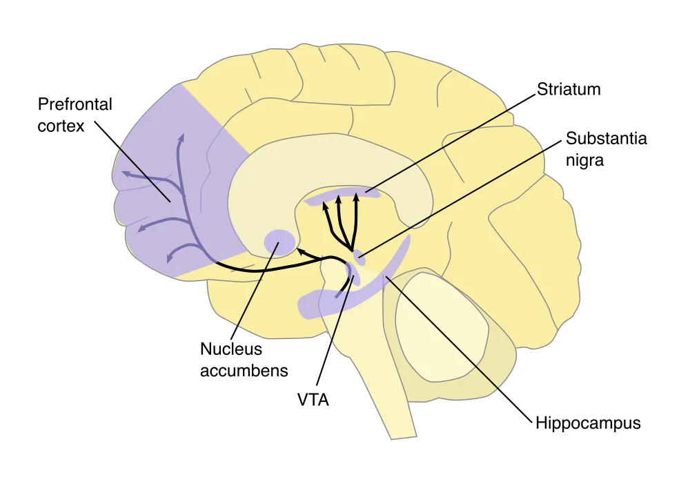
Dopamine Hypothesis
Schizophrenia involves dysregulation of dopamine signaling.
The four major dopamine pathways. The dopamine hypothesis of schizophrenia centers on two of them: a hyperactive mesolimbic pathway (VTA → nucleus accumbens) drives positive symptoms; a hypoactive mesocortical pathway (VTA → prefrontal cortex) drives negative symptoms. The nigrostriatal pathway (substantia nigra → striatum) is the one degenerated in Parkinson's disease - and the one D2 antagonists hit to cause movement side effects. Credit: NIDA / Quasihuman via Wikimedia Commons (Public Domain).
Evidence:
Amphetamines (which boost dopamine) can induce psychosis in healthy people.
First-generation antipsychotics block D2 dopamine receptors and reduce positive symptoms.
Levodopa (dopamine precursor, used for Parkinson’s) can trigger psychotic symptoms.
Glutamate Hypothesis
Evidence for glutamate dysfunction, especially at NMDA receptors. PCP and ketamine (NMDA antagonists) can produce both positive and negative symptoms.
Genetic. Heritability around 80%. Concordance ~50% in monozygotic twins, ~10% in dizygotic twins.
Environmental triggers. Prenatal viral infections, birth complications, cannabis use (especially heavy adolescent use), and severe stress in genetically vulnerable individuals.
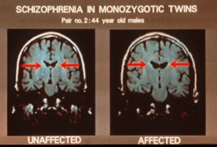
Coronal MRI slices from monozygotic twins discordant for schizophrenia: the affected twin (right) shows enlarged lateral ventricles compared to the genetically identical unaffected twin (left). Because their genomes are essentially identical, the structural difference highlights the role of environmental and developmental factors on top of inherited risk. Credit: Daniel Weinberger, NIMH via Wikimedia Commons (Public Domain).
Treatment
Antipsychotic Medications
First-generation (typical) antipsychotics (haloperidol, chlorpromazine) - block D2 receptors. Effective for positive symptoms, less effective for negative symptoms, and cause movement-related side effects (tardive dyskinesia - involuntary repetitive movements).
Second-generation (atypical) antipsychotics (risperidone, olanzapine, clozapine) - also block D2 but act on serotonin receptors. Better for negative symptoms, fewer movement side effects, but carry metabolic risks (weight gain, diabetes). Clozapine is the most effective but requires blood monitoring due to risk of agranulocytosis.
Psychosocial Treatment
Family therapy - reducing high expressed emotion (harsh criticism, over-involvement) in family reduces relapse.
Cognitive behavioral therapy - helping patients challenge delusional beliefs and cope with hallucinations.
Supported employment, social skills training - helping patients maintain function.
Distinguish positive from negative symptoms of schizophrenia, with examples.
Click to reveal answer
Positive symptoms are ADDITIONS to normal functioning (hallucinations, delusions, disorganized speech). Negative symptoms are SUBTRACTIONS from normal functioning (flat affect, avolition, alogia, anhedonia).
According to the dopamine hypothesis, which pathway is hyperactive in schizophrenia's positive symptoms?
Click to reveal answer
The mesolimbic pathway (VTA → nucleus accumbens). Hypoactivity in the mesocortical pathway (VTA → prefrontal cortex) is associated with negative symptoms. First-generation antipsychotics reduce positive symptoms by blocking D2 receptors.
What is the typical course of schizophrenia in terms of phases?
Click to reveal answer
Prodromal (subtle decline, social withdrawal, odd beliefs) → Active (full psychotic symptoms) → Residual (positive symptoms attenuated, negative/cognitive persist). Early treatment during prodromal or first-episode phase improves outcomes.
Mood disorders span a continuum from depression at one end to mania at the other. The DSM-5 separates depressive disorders from bipolar disorders, recognizing they have different courses, genetics, and treatments.
Major Depressive Disorder (MDD)
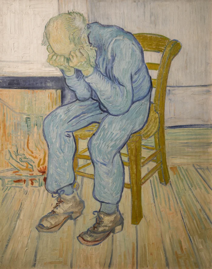
Vincent van Gogh's At Eternity's Gate (1890) - one of the most enduring portraits of clinical depression. The posture (hands clutching head, slumped collapse) captures the psychomotor retardation, hopelessness, and exhaustion that define a major depressive episode. Van Gogh painted it while himself institutionalized for what we would now classify as a mood disorder. Credit: GoldenArtists via Wikimedia Commons (CC BY-SA 4.0).
Major depressive disorder requires a major depressive episode: at least five symptoms present for at least 2 weeks, including either depressed mood or anhedonia (loss of interest/pleasure). Other symptoms include:
Significant weight change or appetite change.
Insomnia or hypersomnia.
Psychomotor agitation or retardation.
Fatigue or low energy.
Feelings of worthlessness or excessive guilt.
Impaired concentration or indecisiveness.
Recurrent thoughts of death or suicide.
Mnemonic: SIG E CAPS (Sleep, Interest, Guilt, Energy, Concentration, Appetite, Psychomotor, Suicide) plus mood.
Women are diagnosed with MDD about twice as often as men. Peak onset is in the 20s-30s. Lifetime prevalence is 15-20%.
Persistent Depressive Disorder (Dysthymia)
Persistent depressive disorder is a chronic, milder form of depression lasting at least 2 years (1 year in children). Symptoms overlap with MDD but are less severe, and there are no symptom-free periods longer than 2 months. Sometimes called “low-grade depression” - the person functions, but poorly and unhappily.
Bipolar Disorders
Bipolar disorders involve alternating episodes of depression and mania.
Manic Episode
At least 1 week of elevated, expansive, or irritable mood and increased energy, plus at least 3 additional symptoms (4 if mood is only irritable):
Inflated self-esteem or grandiosity.
Decreased need for sleep.
More talkative than usual; pressure to keep talking.
Mnemonic: DIG FAST (Distractibility, Irritability, Grandiosity, Flight of ideas, Agitation/Activity, Sleep decreased, Talkativeness).
Mania can severely impair judgment and often requires hospitalization.
Bipolar I
At least one manic episode. Most people with bipolar I also have depressive episodes, but depression is not required for diagnosis.
Bipolar II
At least one major depressive episode and at least one hypomanic episode (a less severe form of mania that lasts at least 4 days and doesn’t cause marked impairment). Never a full manic episode - the presence of one upgrades the diagnosis to bipolar I.
Cyclothymic Disorder
A chronic but milder form: alternating periods of hypomanic and depressive symptoms for at least 2 years, without meeting criteria for a full episode.
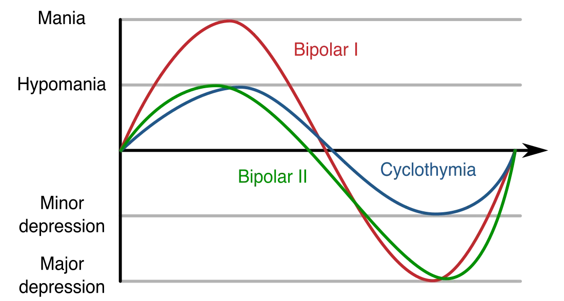
The bipolar spectrum visualized as mood swings over time. Bipolar I reaches full mania and full depression. Bipolar II tops out at hypomania but still drops into major depression. Cyclothymia oscillates within a milder band. The MCAT loves to test these distinctions - especially the bipolar I vs. II line: a single full manic episode upgrades a diagnosis to bipolar I. Credit: Blacktc via Wikimedia Commons (CC BY 4.0).
Biological Basis
Monoamine Hypothesis of Depression
Depression involves deficiency in monoamine neurotransmitters, particularly serotonin, norepinephrine, and dopamine. Evidence:
SSRIs (selective serotonin reuptake inhibitors) and other antidepressants that boost these monoamines effectively treat depression in many patients.
Reserpine (old antihypertensive that depleted monoamines) caused depression.
Brain imaging often shows altered activity in prefrontal cortex, amygdala, hippocampus.
The hypothesis is incomplete - antidepressants boost monoamines within hours but take weeks to produce clinical benefit, suggesting downstream neuroplastic changes matter more than the neurotransmitter level itself.
Other Findings
Anterior cingulate cortex shows reduced serotonin responsiveness in depression.
Chronic stress elevates cortisol and damages the hippocampus; people with depression often have smaller hippocampi.
Learned helplessness (Seligman) is an animal model of depression.
Genetic. Heritability ~40% for MDD, ~70% for bipolar.
Treatment
Antidepressants
SSRIs (fluoxetine, sertraline, escitalopram) - block serotonin reuptake. First-line for depression. Minor side effects, low overdose risk.
SNRIs (venlafaxine, duloxetine) - block serotonin and norepinephrine reuptake.
Tricyclic antidepressants (amitriptyline, nortriptyline) - older, more side effects, higher overdose risk.
MAOIs - block monoamine oxidase, preventing breakdown of monoamines. Effective but require dietary restrictions (tyramine crisis) and have many drug interactions.
All antidepressants take 4-6 weeks for full effect.
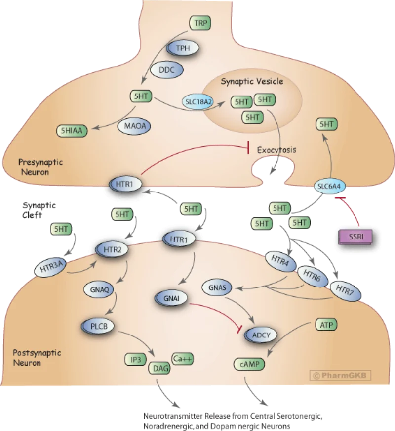
How an SSRI works. Selective serotonin reuptake inhibitors block the SLC6A4 serotonin transporter on the presynaptic neuron, preventing reabsorption of released serotonin. More serotonin lingers in the synaptic cleft, prolonging postsynaptic receptor activation. The clinical benefit takes weeks - longer than the receptor-level change - which suggests downstream neuroplastic adaptations matter as much as the immediate transmitter effect. Credit: PharmGKB via Wikimedia Commons (CC BY-SA 4.0).Fluoxetine (Prozac), one of the first SSRIs and still one of the most widely prescribed antidepressants in the world. SSRIs displaced the older tricyclics and MAOIs as first-line therapy because of their cleaner side-effect profile and much lower overdose lethality. Credit: Tom Varco via Wikimedia Commons (CC BY-SA 3.0).
Mood Stabilizers for Bipolar
Lithium - the classic mood stabilizer. Reduces mania and prevents relapse. Narrow therapeutic window - blood levels must be monitored. Mechanism incompletely understood.
Anticonvulsants (valproate, lamotrigine, carbamazepine) - also effective as mood stabilizers.
Antidepressants given alone to bipolar patients can trigger a manic episode - so mood stabilizers are often co-prescribed.
Lithium - the prototypical mood stabilizer. Despite being the simplest drug in the psychiatric pharmacopeia (just an alkali metal), it remains one of the most effective treatments for bipolar mania and a strong reducer of suicide risk. Its narrow therapeutic window means patients need regular blood-level monitoring to avoid toxicity. Credit: P3829 via Wikimedia Commons (CC0).
Interpersonal therapy - focuses on relationship patterns.
Electroconvulsive therapy (ECT) - for severe, treatment-resistant depression. Induces brief seizures under anesthesia. Highly effective but carries memory side effects.
Transcranial magnetic stimulation (TMS) - non-invasive alternative to ECT, approved for treatment-resistant depression.
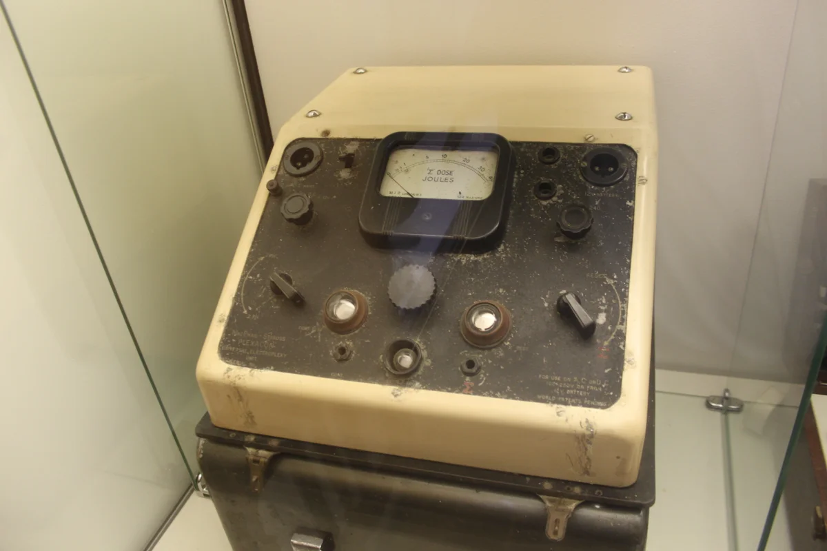
An older-generation Siemens Konvulsator III ECT device. Modern ECT, performed under general anesthesia and muscle relaxation, remains the most effective treatment for severe, medication-resistant depression and severe catatonia. Despite its grim reputation in pop culture, it has high response rates - the principal side effect is short-term memory loss around the treatment period. Credit: Nasko via Wikimedia Commons (CC0).
A diagnosis of major depressive disorder requires at least how many symptoms for how long?
Click to reveal answer
At least 5 symptoms present for at least 2 weeks, including either depressed mood or anhedonia as one of them. Mnemonic: SIG E CAPS (Sleep, Interest, Guilt, Energy, Concentration, Appetite, Psychomotor, Suicide) plus mood.
Distinguish bipolar I from bipolar II disorder.
Click to reveal answer
Bipolar I requires at least one full MANIC episode. Bipolar II requires at least one hypomanic episode plus at least one major depressive episode, but NO full manic episode. A full manic episode upgrades bipolar II to bipolar I.
Why might an antidepressant taken ALONE trigger a manic episode in a bipolar patient?
Click to reveal answer
Antidepressants can 'switch' bipolar patients from depression into mania. This is why mood stabilizers (lithium, anticonvulsants) are typically co-prescribed, or why antidepressant monotherapy is avoided in bipolar disorder.
Anxiety is a normal response to threat. Anxiety disorders involve anxiety that is excessive, persistent, and disruptive, far out of proportion to actual danger. This group and the closely related OCD and PTSD make up the most common psychiatric conditions - the lifetime prevalence of any anxiety disorder is around 30%.
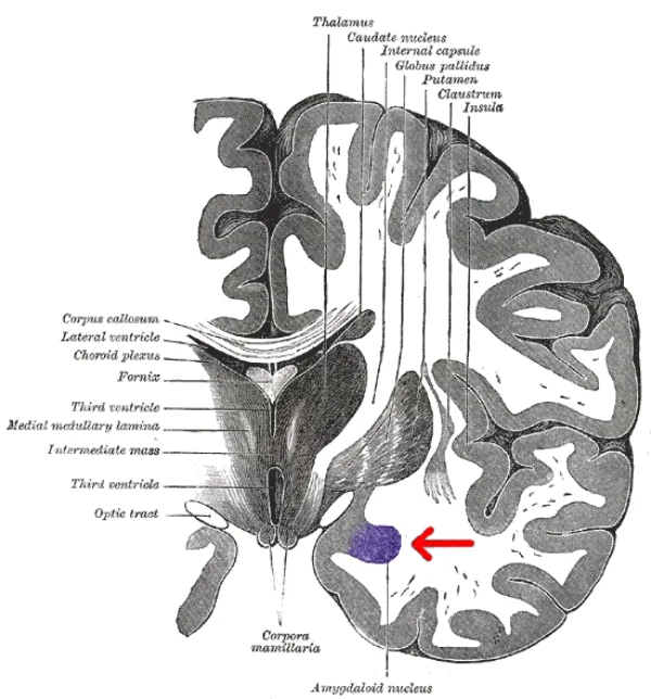
The amygdala (highlighted in purple), the brain's threat-detection hub. It is hyperactive in anxiety disorders, panic disorder, and PTSD - and treatments that work (SSRIs, exposure-based CBT) tend to dampen its reactivity over time. Credit: Henry Vandyke Carter (Gray's Anatomy, 1918) via Wikimedia Commons (Public Domain).
Generalized Anxiety Disorder (GAD)
Persistent, excessive worry about a range of life situations, lasting at least 6 months, with at least three of:
Restlessness, feeling “on edge.”
Easily fatigued.
Difficulty concentrating.
Irritability.
Muscle tension.
Sleep disturbance.
The worry is hard to control and is about everyday things (money, family, work) rather than specific triggers.
Panic Disorder
Recurrent, unexpected panic attacks plus persistent worry about future attacks or maladaptive behavior changes to avoid them.
A panic attack is a discrete episode of intense fear that peaks within minutes, with at least 4 of:
Pounding heart, palpitations.
Sweating, shaking, trembling.
Shortness of breath.
Choking sensation.
Chest pain.
Nausea.
Dizziness or faintness.
Chills or hot flashes.
Numbness or tingling.
Derealization (feeling unreal) or depersonalization (detachment from self).
Fear of losing control or “going crazy.”
Fear of dying.
The physical symptoms are so intense that first panic attacks are often mistaken for heart attacks. The fear of having another attack - and avoiding situations where one might happen - drives the disorder’s course.
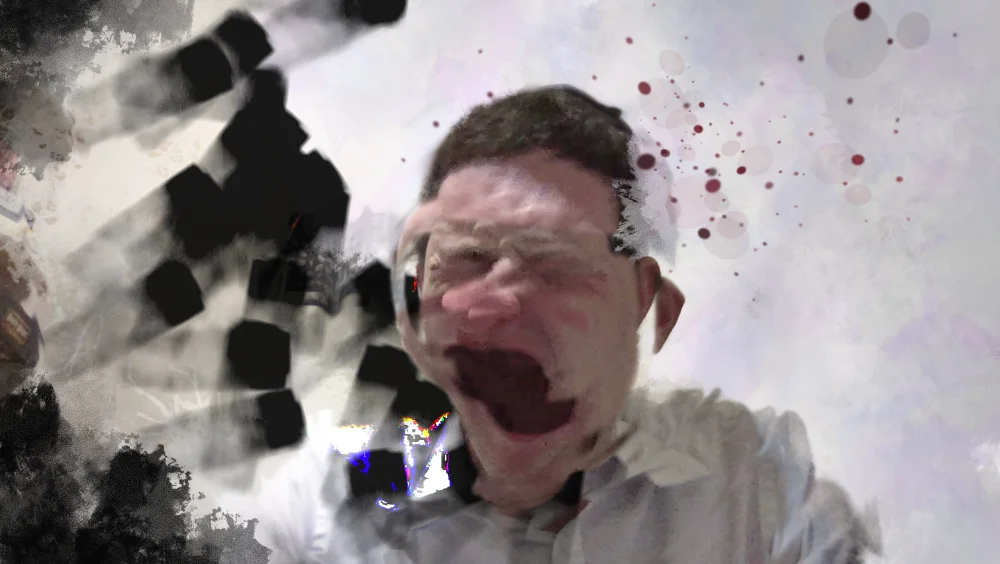
An artist's rendering of the subjective experience of a panic attack: derealization, sensory distortion, and the overwhelming "I'm dying" terror that defines the moment. The first attack is so visceral that patients often present to the ER convinced they are having a heart attack. Credit: Yitzilitt via Wikimedia Commons (CC BY-SA 4.0).
Agoraphobia
Intense fear of being in places or situations from which escape would be difficult or embarrassing if panic or incapacitation struck. Classic examples: crowded markets, open spaces, enclosed spaces, public transportation, being far from home alone.
Often co-occurs with panic disorder - once someone has had a panic attack in a supermarket, they may avoid supermarkets.
Specific Phobias
Marked, persistent fear of a specific object or situation that is disproportionate to actual danger. Exposure almost always provokes anxiety. Common phobias include:
Recall from Chapter 3 that preparedness theory explains why ancestrally dangerous stimuli (snakes, heights) produce phobias more readily than modern dangers (electrical outlets, cars).
Spiders are one of the most common targets of specific phobia (arachnophobia) - and a textbook example of preparedness theory: humans acquire phobias of ancestrally dangerous stimuli (spiders, snakes, heights) much more readily than of modern dangers like cars or electrical outlets, even though the modern dangers actually kill far more people. Credit: Karin Schmitt via Wikimedia Commons (Public Domain).
Social Anxiety Disorder
Intense fear of social situations in which one might be scrutinized or judged. Public speaking is the most common trigger, but broader social anxiety disorder extends to most social interactions. Causes significant avoidance of social, occupational, and academic opportunities.
Obsessive-Compulsive Disorder (OCD)
OCD was reclassified out of the anxiety disorder group in DSM-5 but is still conceptually related.
Obsessions. Unwanted, intrusive, recurrent thoughts that cause anxiety (fear of contamination, fear of harming someone, unwanted taboo thoughts, need for symmetry).
Compulsions. Repetitive behaviors or mental acts the person feels driven to perform in response to obsessions (washing, checking, counting, arranging, silent prayers).
The compulsions temporarily relieve the anxiety caused by the obsessions but reinforce the cycle. Compulsions become time-consuming (at least 1 hour/day for diagnosis) and often severely impair functioning.
Compulsive handwashing in response to contamination obsessions is one of the most recognizable OCD presentations. The act briefly reduces anxiety, which negatively reinforces the behavior - creating the loop that keeps the disorder going. Exposure and response prevention (ERP) breaks the loop by exposing the patient to the obsession trigger while preventing the compulsion. Credit: Lars Klintwall Malmqvist via Wikimedia Commons (Public Domain).
Biology:
Abnormalities in the cortico-striatal-thalamic loop.
Genetic component - about 25% of first-degree relatives have OCD or related conditions.
Serotonin dysfunction - SSRIs at high doses are first-line medical treatment.
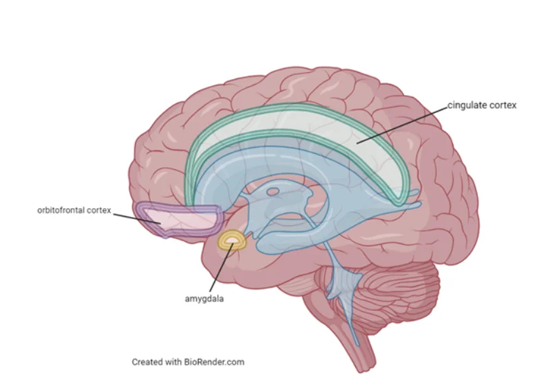
Brain regions implicated in OCD - the orbitofrontal cortex, anterior cingulate cortex, and amygdala - which together form part of the cortico-striato-thalamo-cortical (CSTC) loop. Hyperactivity in this circuit is hypothesized to drive the intrusive thoughts and the urge to neutralize them. Credit: Created with BioRender via Wikimedia Commons (Public Domain).
Cognitive-behavioral therapy with exposure and response prevention (ERP) - deliberately exposing the patient to triggers while preventing the compulsive response - is the most effective psychological treatment.
Post-Traumatic Stress Disorder (PTSD)
Also reclassified in DSM-5 into “trauma- and stressor-related disorders.”
Requires:
Exposure to actual or threatened death, serious injury, or sexual violence (direct, witnessed, or learned about in a close other).
Avoidance - of memories, thoughts, feelings, or external reminders of the trauma.
Negative alterations in cognition/mood - inability to remember aspects of the trauma, persistent negative beliefs, persistent negative emotions, detachment from others.
Partial PTSD is common - not everyone exposed to trauma develops PTSD, and prior trauma plus inadequate social support raise risk.
Treatments include trauma-focused CBT, prolonged exposure therapy, eye movement desensitization and reprocessing (EMDR), and SSRIs.
Treatment of Anxiety Disorders
Medications
SSRIs - first-line for most anxiety disorders, OCD, and PTSD. Also treat depression (common comorbidity).
SNRIs - also effective.
Benzodiazepines - rapid anxiety relief but risk of dependence. Used short-term or acutely (pre-flight, during a panic attack). Not first-line for chronic treatment.
Beta blockers - reduce physical symptoms of anxiety (racing heart, trembling). Used for performance anxiety.
Psychotherapy
Cognitive-behavioral therapy (CBT) - gold standard. Challenges catastrophic thoughts and gradually exposes patients to feared situations.
Systematic desensitization (Wolpe) - relaxation paired with a graded fear hierarchy.
Flooding/implosive therapy - full-intensity exposure in a safe context.
What distinguishes obsessions from compulsions in OCD?
Click to reveal answer
Obsessions are unwanted intrusive THOUGHTS that cause anxiety (fear of contamination, need for symmetry). Compulsions are repetitive BEHAVIORS or mental acts performed to reduce the anxiety (washing, checking, counting). Compulsions temporarily relieve but reinforce the cycle.
What is the gold-standard psychological treatment for OCD, and how does it work?
Click to reveal answer
Exposure and response prevention (ERP), a form of CBT. The patient is exposed to obsession-triggering stimuli but prevented from performing compulsions. Over time, the anxiety extinguishes without reinforcement, and the OCD loop breaks.
A patient experiences recurrent nightmares, intrusive memories, and hyperarousal 3 months after a car accident. Which diagnosis?
Click to reveal answer
Post-traumatic stress disorder (PTSD). Requires exposure to trauma, plus intrusion symptoms (nightmares, flashbacks), avoidance, negative cognitions/mood changes, and arousal/reactivity alterations, lasting at least 1 month.
Three final disorder groups round out the chapter. They’re less common on the MCAT than schizophrenia or depression, but each appears in passages, and each has a distinct clinical picture.
Dissociative Disorders
Dissociation is a disconnect between consciousness, memory, identity, and perception. It can be a normal experience (daydreaming, highway hypnosis) but becomes a disorder when it’s severe or impairing.
An artistic rendering of dissociation - a felt sense of fragmentation between thought, memory, identity, and perception. Mild dissociation is normal (daydreaming, highway hypnosis); severe or chronic dissociation defines disorders such as DID, dissociative amnesia, and depersonalization/derealization. Credit: Dyversions via Wikimedia Commons (CC0).
Dissociative Identity Disorder (DID)
Formerly called multiple personality disorder. Two or more distinct personality states (alters) that recurrently take control of behavior, with gaps in memory between states. Each alter may have its own name, voice, mannerisms, and even handwriting.
Strongly associated with severe childhood trauma (especially repeated abuse). The idea: the mind fragments as a defense, splitting unbearable experience into separate identities.
DID is rare and controversial in psychiatry - debated in terms of prevalence and the role of suggestion. The MCAT does test the basic concept.
Dissociative Amnesia
Inability to recall important autobiographical information, usually traumatic, not explained by ordinary forgetting. May be localized (specific event), selective (specific aspects), or generalized (entire life history). Can include dissociative fugue - purposeful travel with amnesia for past identity, sometimes adopting a new one.
Depersonalization/Derealization Disorder
Persistent or recurrent experiences of:
Depersonalization - feeling detached from your own thoughts, feelings, or body, as if watching yourself from outside.
Derealization - feeling the external world is unreal, dreamlike, visually distorted.
Reality testing remains intact (the person knows this isn’t real), distinguishing it from psychotic disorders.
Somatic Symptom and Related Disorders
The common thread: physical symptoms without adequate medical explanation, or disproportionate to any underlying medical condition.
Somatic Symptom Disorder
One or more distressing somatic symptoms, plus excessive thoughts, feelings, and behaviors regarding those symptoms. Persists for at least 6 months. The symptoms are real to the patient - often medically unexplained or only partially explained by findings.
Neurological symptoms (blindness, paralysis, seizures) that aren’t explained by any neurological disease. Historically called “hysterical conversion” - Freud thought psychic conflict was “converted” into physical symptoms.
Preoccupation with having or acquiring a serious illness. Physical symptoms are minimal or absent. The person may perform excessive health-related checking, research symptoms obsessively, or avoid medical appointments out of fear of bad news.
Factitious Disorder
Deliberately producing or feigning symptoms for the purpose of assuming the sick role (no external reward). Distinct from malingering, where symptoms are faked for external gain (disability, drugs, avoiding work). Factitious disorder imposed on another (formerly Munchausen by proxy) involves inducing symptoms in someone else, typically a child.
Personality Disorders
Personality disorders are enduring patterns of inner experience and behavior that deviate markedly from cultural expectations, are pervasive and inflexible, begin by early adulthood, and cause distress or impairment. Ten specific disorders are organized into three clusters.
Cluster A: Odd or Eccentric
Paranoid. Distrustful, suspicious. Sees benign remarks as threats. Not psychotic.
Schizoid. Detached from social relationships. Limited emotional expression. “A loner” by preference.
Schizotypal. Eccentric thinking, speech, and behavior; magical thinking or unusual beliefs. Does not meet criteria for schizophrenia but shares some features.
Mnemonic: “weird.”
Cluster B: Dramatic, Emotional, or Erratic
Antisocial. Disregard for others’ rights; deception, impulsivity, aggression, lack of remorse. Was called psychopathy/sociopathy. Requires evidence of conduct disorder before age 15.
Borderline. Unstable relationships, self-image, and emotions. Intense fear of abandonment. Impulsivity (spending, sex, substance use, self-harm). Chronic feelings of emptiness. Suicidal/self-injurious behavior common.
Narcissistic. Grandiosity, need for admiration, lack of empathy. Exploits others, expects special treatment.
Mnemonic: “wild.”
Cluster C: Anxious or Fearful
Avoidant. Social inhibition, feelings of inadequacy, hypersensitivity to criticism. Wants relationships but is too afraid of rejection.
Dependent. Excessive need to be cared for. Submissive, clinging. Fears separation and inability to function alone.
Obsessive-compulsive personality disorder (OCPD). Preoccupation with order, perfectionism, and control. DIFFERENT from OCD - OCPD involves personality traits that feel ego-syntonic (the person thinks their rigidity is correct); OCD involves ego-dystonic obsessions/compulsions that distress the person.
Mnemonic: “worried.”
Treatment
Personality disorders are notoriously difficult to treat. Pharmacotherapy targets specific symptoms (mood, anxiety, psychosis) but doesn’t treat the underlying pattern. Dialectical behavior therapy (DBT), developed by Marsha Linehan, is the evidence-based psychotherapy for borderline personality disorder. Long-term psychotherapy can gradually shift rigid patterns but progress is slow.
What is the key distinction between DID and schizophrenia?
Click to reveal answer
DID (dissociative identity disorder) involves multiple distinct personality states with memory gaps between them - a dissociative disorder, often linked to childhood trauma. Schizophrenia involves psychotic symptoms (hallucinations, delusions, disorganized thought) and one identity. Popular culture often confuses the two, but they are unrelated.
Somatic symptom disorder: real distressing symptoms + excessive concern. Conversion disorder: neurological-like symptoms with no medical basis. Illness anxiety disorder: preoccupation with having/getting an illness, minimal symptoms. Factitious disorder: deliberately producing/faking symptoms to assume sick role (malingering does so for external gain).
A patient has unstable relationships, intense fear of abandonment, chronic emptiness, and self-harming behavior. Which personality disorder?
Click to reveal answer
Borderline personality disorder (Cluster B). Evidence-based psychotherapy is DBT (dialectical behavior therapy), developed by Marsha Linehan.
What distinguishes OCD from OCPD?
Click to reveal answer
OCD: obsessions and compulsions that are ego-dystonic (the person finds them distressing and irrational). OCPD: pervasive personality pattern of rigidity, perfectionism, and control that is ego-syntonic (the person thinks their approach is correct and doesn't see a problem).
Alzheimer’s and Parkinson’s are the two most common progressive neurodegenerative diseases affecting behavior. Both are biological disorders with psychological and behavioral consequences, so they bridge the “biological bases” chapter with the psychological disorders chapter.
Alzheimer’s Disease
Alzheimer’s disease (AD) is the most common form of dementia - the umbrella term for significant, progressive cognitive decline that interferes with daily functioning. Roughly 60-70% of dementia cases are AD.
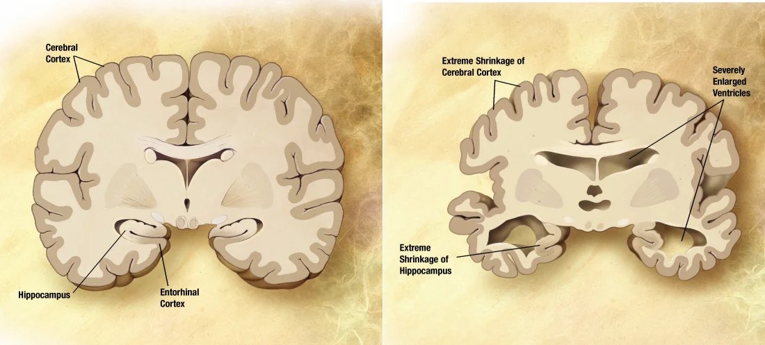
Coronal slice comparison: a healthy brain (left) versus a brain in late-stage Alzheimer's disease (right). Note the cortical atrophy, the dramatic shrinkage of the hippocampus, and the compensatory enlargement of the ventricles. The hippocampal involvement explains why early Alzheimer's so disproportionately wipes out short-term memory. Credit: NIA/ADEAR via Wikimedia Commons (Public Domain).
Clinical Course
AD unfolds in stages over years:
Early. Memory loss (particularly short-term - forgetting recent conversations, appointments). Word-finding difficulty. Procedural and remote memory are relatively preserved.
Middle. Worsening memory, language problems, disorientation (time and place), difficulties with self-care. Personality changes - apathy, irritability.
Late. Severe memory loss (may fail to recognize family). Loss of motor skills, incontinence, mutism. Bedridden. Death often from pneumonia or infection.
Average life expectancy after diagnosis is 4-8 years, though the range varies widely.
Pathology
Two hallmark findings at autopsy:
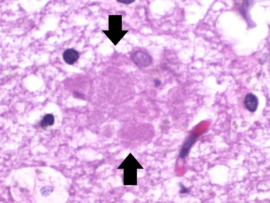
Amyloid plaques (senile plaques). Extracellular clumps of beta-amyloid protein. Thought to damage neurons and trigger inflammation.
Neurofibrillary tangles. Intracellular clumps of hyperphosphorylated tau protein. Disrupt normal neuronal function.
An amyloid (senile) plaque seen on H&E-stained brain histology. The pale, fluffy extracellular deposit is composed of aggregated beta-amyloid protein and is one of the two diagnostic hallmarks of Alzheimer's disease at autopsy. Credit: Mikael Häggström, M.D. via Wikimedia Commons (CC0).A neurofibrillary tangle inside a neuron - the second diagnostic hallmark of Alzheimer's. The dense intracellular fibers are made of hyperphosphorylated tau protein, which normally stabilizes microtubules but in AD aggregates and disrupts cellular transport. Credit: Mikael Häggström, M.D. via Wikimedia Commons (CC0).
Neuronal loss is widespread but particularly severe in:
Hippocampus - explaining early memory loss.
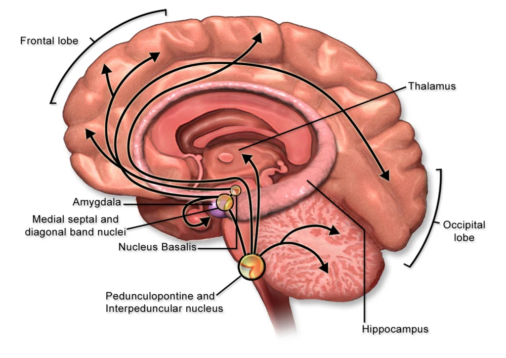
Basal forebrain cholinergic neurons - explaining widespread cognitive decline. This is the target of cholinesterase inhibitors (donepezil, rivastigmine), which boost acetylcholine levels and modestly slow symptom progression.
Cerebral cortex - explaining language and reasoning problems.
The cholinergic projection system. Cholinergic neurons in the basal forebrain (especially the nucleus basalis of Meynert) project diffusely to the cortex and hippocampus. These neurons preferentially die in Alzheimer's disease - the rationale for cholinesterase inhibitors (donepezil, rivastigmine), which slow the breakdown of remaining acetylcholine. Credit: BruceBlaus via Wikimedia Commons (CC BY-SA 4.0).
The brain literally shrinks over time; ventricles enlarge to fill the space.
Risk Factors
Age - primary risk factor. Prevalence roughly doubles every 5 years after 65.
Genetics - APOE ε4 allele significantly increases risk; rare familial forms with autosomal dominant inheritance cause early-onset AD.
Cardiovascular risk factors - hypertension, diabetes, high cholesterol.
Head trauma. Lower educational attainment and lack of mental engagement (possibly through reduced “cognitive reserve”).
Treatment
No cure. Cholinesterase inhibitors and memantine (NMDA receptor modulator) offer modest symptomatic benefit. Newer antibody therapies targeting amyloid (aducanumab, lecanemab) show small effects in trials but remain controversial. Most care is supportive - managing symptoms, planning for progressive decline, supporting caregivers.
Parkinson’s Disease
Parkinson’s disease (PD) is a progressive motor disorder caused by loss of dopamine-producing neurons in the substantia nigra of the midbrain. Those neurons normally project to the striatum (caudate and putamen), enabling smooth motor control. As they die, motor function deteriorates.
The classic Parkinsonian stance, drawn by neurologist Sir William Gowers in 1886: stooped posture, flexed elbows and knees, mask-like face. Despite a century-and-a-half of progress in pathology, the bedside clinical picture has barely changed. Credit: Sir William Richard Gowers (1886) via Wikimedia Commons (Public Domain).
Clinical Features
Classic cardinal signs:
Tremor. Resting tremor (most noticeable when the limb is at rest), classically “pill-rolling” in the hands. Disappears with voluntary movement.
Bradykinesia. Slowness of voluntary movement. Difficulty initiating movement. Reduced arm swing while walking.
Postural instability. Poor balance. Falls common.
Other features:
Masked facies. Reduced facial expression - the person looks emotionally flat even when not.
Shuffling gait.
Micrographia - very small handwriting.
Non-motor symptoms. Depression, sleep disturbance (especially REM sleep behavior disorder as an early sign), anosmia, constipation, and eventually dementia in about 30% of advanced cases.
Pathology
At autopsy:
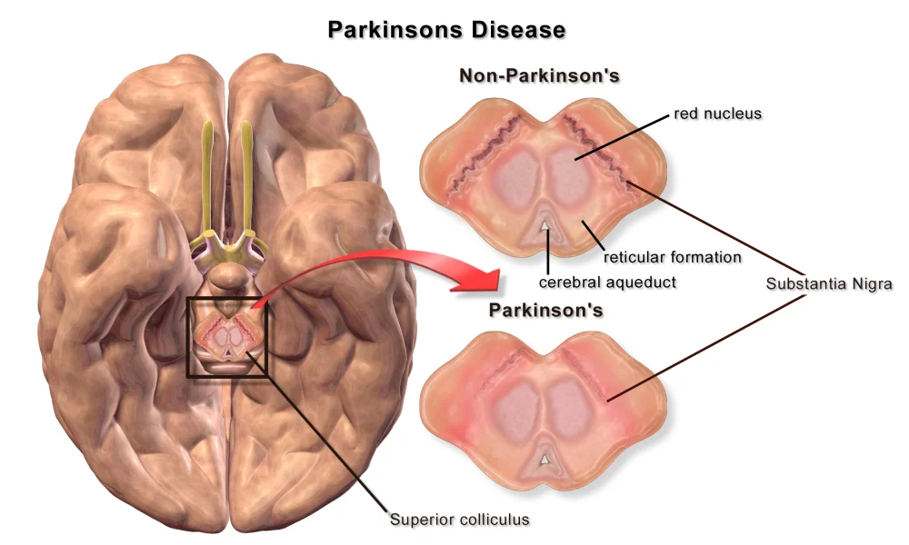
Loss of pigmented (dopaminergic) neurons in the substantia nigra.
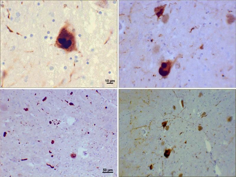
Lewy bodies - intracellular inclusions composed mostly of alpha-synuclein protein - in surviving neurons.
The defining lesion of Parkinson's disease: loss of pigmented dopaminergic neurons in the substantia nigra of the midbrain. Compare the dark, intact substantia nigra in the non-Parkinson's section to the faded, pigment-poor band in the Parkinson's section. These neurons normally project to the striatum via the nigrostriatal pathway; their loss is what causes the cardinal motor signs (tremor, rigidity, bradykinesia, postural instability). Credit: BruceBlaus / Blausen Medical via Wikimedia Commons (CC BY 3.0).Lewy bodies (top panels) and thread-like Lewy neurites (bottom panels) seen on immunohistochemistry for alpha-synuclein. These intracellular protein inclusions are the second pathological hallmark of Parkinson's disease. When Lewy bodies extend into the cortex, they cause dementia with Lewy bodies, with prominent visual hallucinations and fluctuating cognition. Credit: Suraj Rajan via Wikimedia Commons (CC BY-SA 3.0).
Dementia with Lewy bodies is a related disorder in which Lewy bodies appear throughout the cortex, producing dementia with fluctuating cognition, visual hallucinations, and Parkinsonian motor features.
Treatment
No cure. Management focuses on replacing the lost dopamine.
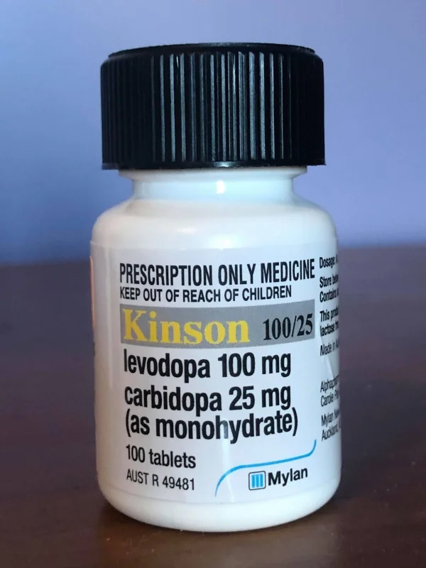
L-DOPA (levodopa). Dopamine precursor that crosses the blood-brain barrier (dopamine itself cannot). Usually given with carbidopa, which inhibits peripheral breakdown and allows more L-DOPA to reach the brain. Effective but becomes less so over time, and can cause dyskinesias.
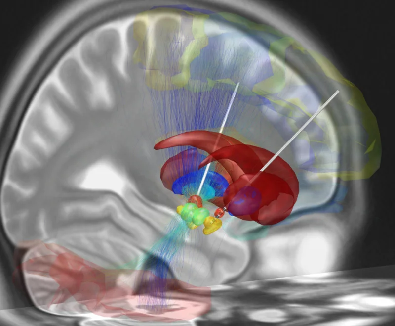
Deep brain stimulation (DBS) - surgical implantation of electrodes into the subthalamic nucleus. Effective in carefully selected patients.
A combination levodopa/carbidopa preparation - the workhorse Parkinson's medication. Levodopa, the dopamine precursor, crosses the blood-brain barrier (dopamine itself cannot) and is converted to dopamine in the brain. Carbidopa blocks peripheral breakdown of levodopa so a higher fraction reaches the CNS, reducing nausea and dyskinesia from peripheral dopamine. Credit: Revion101 via Wikimedia Commons (CC BY-SA 4.0).Reconstruction of deep brain stimulation (DBS) electrode placement targeting the subthalamic nucleus - a surgical option for Parkinson's patients whose motor symptoms can no longer be controlled with medication alone. Continuous high-frequency stimulation effectively suppresses tremor, rigidity, and bradykinesia. Credit: Andreashorn via Wikimedia Commons (CC BY-SA 4.0).
What are the two hallmark pathological findings in Alzheimer's disease?
Click to reveal answer
Amyloid plaques (extracellular beta-amyloid aggregates) and neurofibrillary tangles (intracellular hyperphosphorylated tau protein). Along with widespread neuronal loss, especially in the hippocampus and basal forebrain.
Which neurotransmitter system is most severely affected in Alzheimer's, and how does that inform treatment?
Click to reveal answer
Cholinergic system - basal forebrain neurons that project acetylcholine to the cortex die. Cholinesterase inhibitors (donepezil, rivastigmine) slow the breakdown of remaining acetylcholine, producing modest symptomatic improvement.
List the four cardinal motor signs of Parkinson's disease.
Click to reveal answer
Resting tremor (classically 'pill-rolling'), rigidity (cogwheel), bradykinesia (slow movement), and postural instability. Caused by dopamine loss in the substantia nigra → striatum (nigrostriatal) pathway.
Why is L-DOPA given rather than dopamine directly for Parkinson's disease?
Click to reveal answer
Dopamine cannot cross the blood-brain barrier. L-DOPA (levodopa) is the precursor to dopamine and crosses the BBB; once inside the brain, it is converted to dopamine. Usually given with carbidopa, which blocks peripheral L-DOPA breakdown so more reaches the brain.