Alzheimer’s and Parkinson’s are the two most common progressive neurodegenerative diseases affecting behavior. Both are biological disorders with psychological and behavioral consequences, so they bridge the “biological bases” chapter with the psychological disorders chapter.
Alzheimer’s Disease
Alzheimer’s disease (AD) is the most common form of dementia - the umbrella term for significant, progressive cognitive decline that interferes with daily functioning. Roughly 60-70% of dementia cases are AD.
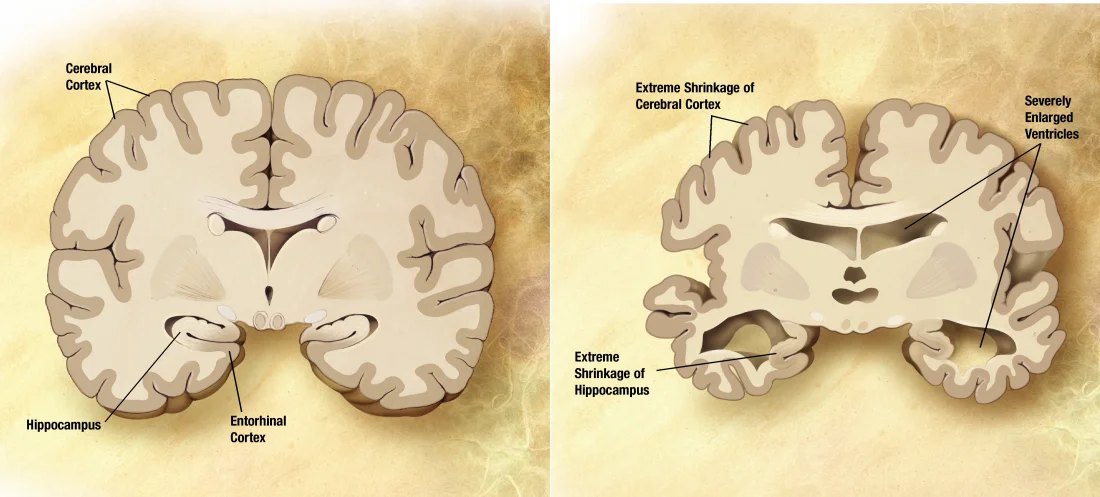
Coronal slice comparison: a healthy brain (left) versus a brain in late-stage Alzheimer's disease (right). Note the cortical atrophy, the dramatic shrinkage of the hippocampus, and the compensatory enlargement of the ventricles. The hippocampal involvement explains why early Alzheimer's so disproportionately wipes out short-term memory. Credit: NIA/ADEAR via Wikimedia Commons (Public Domain).
Clinical Course
AD unfolds in stages over years:
Early. Memory loss (particularly short-term - forgetting recent conversations, appointments). Word-finding difficulty. Procedural and remote memory are relatively preserved.
Middle. Worsening memory, language problems, disorientation (time and place), difficulties with self-care. Personality changes - apathy, irritability.
Late. Severe memory loss (may fail to recognize family). Loss of motor skills, incontinence, mutism. Bedridden. Death often from pneumonia or infection.
Average life expectancy after diagnosis is 4-8 years, though the range varies widely.
Pathology
Two hallmark findings at autopsy:
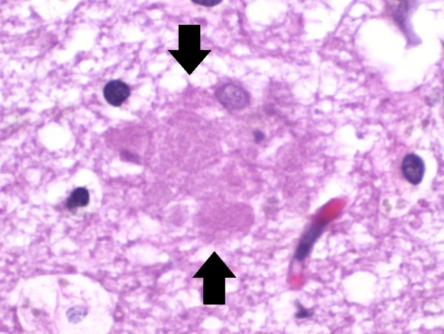
Amyloid plaques (senile plaques). Extracellular clumps of beta-amyloid protein. Thought to damage neurons and trigger inflammation.
Neurofibrillary tangles. Intracellular clumps of hyperphosphorylated tau protein. Disrupt normal neuronal function.
An amyloid (senile) plaque seen on H&E-stained brain histology. The pale, fluffy extracellular deposit is composed of aggregated beta-amyloid protein and is one of the two diagnostic hallmarks of Alzheimer's disease at autopsy. Credit: Mikael Häggström, M.D. via Wikimedia Commons (CC0).A neurofibrillary tangle inside a neuron - the second diagnostic hallmark of Alzheimer's. The dense intracellular fibers are made of hyperphosphorylated tau protein, which normally stabilizes microtubules but in AD aggregates and disrupts cellular transport. Credit: Mikael Häggström, M.D. via Wikimedia Commons (CC0).
Neuronal loss is widespread but particularly severe in:
Hippocampus - explaining early memory loss.
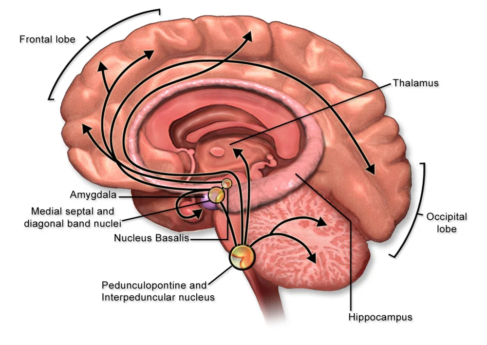
Basal forebrain cholinergic neurons - explaining widespread cognitive decline. This is the target of cholinesterase inhibitors (donepezil, rivastigmine), which boost acetylcholine levels and modestly slow symptom progression.
Cerebral cortex - explaining language and reasoning problems.
The cholinergic projection system. Cholinergic neurons in the basal forebrain (especially the nucleus basalis of Meynert) project diffusely to the cortex and hippocampus. These neurons preferentially die in Alzheimer's disease - the rationale for cholinesterase inhibitors (donepezil, rivastigmine), which slow the breakdown of remaining acetylcholine. Credit: BruceBlaus via Wikimedia Commons (CC BY-SA 4.0).
The brain literally shrinks over time; ventricles enlarge to fill the space.
Risk Factors
Age - primary risk factor. Prevalence roughly doubles every 5 years after 65.
Genetics - APOE ε4 allele significantly increases risk; rare familial forms with autosomal dominant inheritance cause early-onset AD.
Cardiovascular risk factors - hypertension, diabetes, high cholesterol.
Head trauma. Lower educational attainment and lack of mental engagement (possibly through reduced “cognitive reserve”).
Treatment
No cure. Cholinesterase inhibitors and memantine (NMDA receptor modulator) offer modest symptomatic benefit. Newer antibody therapies targeting amyloid (aducanumab, lecanemab) show small effects in trials but remain controversial. Most care is supportive - managing symptoms, planning for progressive decline, supporting caregivers.
Parkinson’s Disease
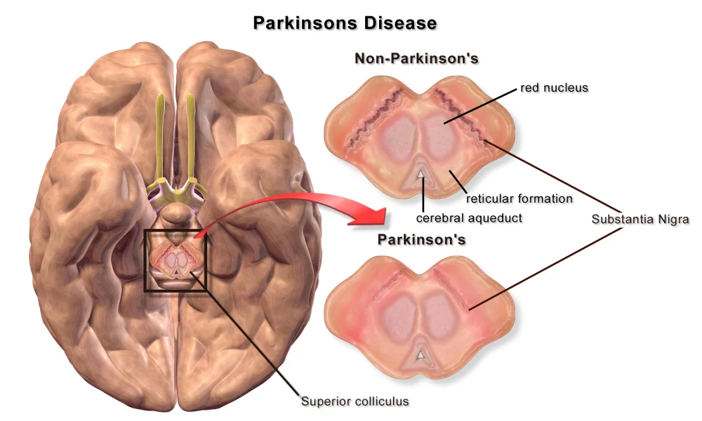
Parkinson’s disease (PD) is a progressive motor disorder caused by loss of dopamine-producing neurons in the substantia nigra of the midbrain. Those neurons normally project to the striatum (caudate and putamen), enabling smooth motor control. As they die, motor function deteriorates.
The classic Parkinsonian stance, drawn by neurologist Sir William Gowers in 1886: stooped posture, flexed elbows and knees, mask-like face. Despite a century-and-a-half of progress in pathology, the bedside clinical picture has barely changed. Credit: Sir William Richard Gowers (1886) via Wikimedia Commons (Public Domain).
Clinical Features
Classic cardinal signs:
Tremor. Resting tremor (most noticeable when the limb is at rest), classically “pill-rolling” in the hands. Disappears with voluntary movement.
Bradykinesia. Slowness of voluntary movement. Difficulty initiating movement. Reduced arm swing while walking.
Postural instability. Poor balance. Falls common.
Other features:
Masked facies. Reduced facial expression - the person looks emotionally flat even when not.
Shuffling gait.
Micrographia - very small handwriting.
Non-motor symptoms. Depression, sleep disturbance (especially REM sleep behavior disorder as an early sign), anosmia, constipation, and eventually dementia in about 30% of advanced cases.
Pathology
At autopsy:
Loss of pigmented (dopaminergic) neurons in the substantia nigra.
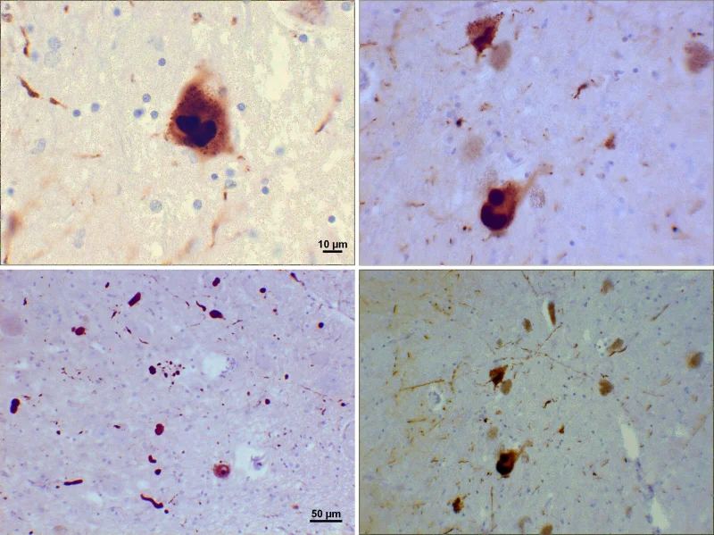
Lewy bodies - intracellular inclusions composed mostly of alpha-synuclein protein - in surviving neurons.
The defining lesion of Parkinson's disease: loss of pigmented dopaminergic neurons in the substantia nigra of the midbrain. Compare the dark, intact substantia nigra in the non-Parkinson's section to the faded, pigment-poor band in the Parkinson's section. These neurons normally project to the striatum via the nigrostriatal pathway; their loss is what causes the cardinal motor signs (tremor, rigidity, bradykinesia, postural instability). Credit: BruceBlaus / Blausen Medical via Wikimedia Commons (CC BY 3.0).Lewy bodies (top panels) and thread-like Lewy neurites (bottom panels) seen on immunohistochemistry for alpha-synuclein. These intracellular protein inclusions are the second pathological hallmark of Parkinson's disease. When Lewy bodies extend into the cortex, they cause dementia with Lewy bodies, with prominent visual hallucinations and fluctuating cognition. Credit: Suraj Rajan via Wikimedia Commons (CC BY-SA 3.0).
Dementia with Lewy bodies is a related disorder in which Lewy bodies appear throughout the cortex, producing dementia with fluctuating cognition, visual hallucinations, and Parkinsonian motor features.
Treatment
No cure. Management focuses on replacing the lost dopamine.
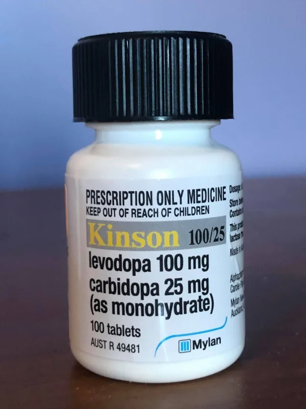
L-DOPA (levodopa). Dopamine precursor that crosses the blood-brain barrier (dopamine itself cannot). Usually given with carbidopa, which inhibits peripheral breakdown and allows more L-DOPA to reach the brain. Effective but becomes less so over time, and can cause dyskinesias.
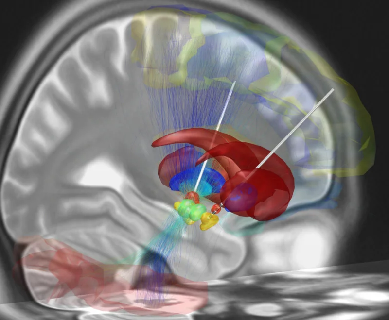
Deep brain stimulation (DBS) - surgical implantation of electrodes into the subthalamic nucleus. Effective in carefully selected patients.
A combination levodopa/carbidopa preparation - the workhorse Parkinson's medication. Levodopa, the dopamine precursor, crosses the blood-brain barrier (dopamine itself cannot) and is converted to dopamine in the brain. Carbidopa blocks peripheral breakdown of levodopa so a higher fraction reaches the CNS, reducing nausea and dyskinesia from peripheral dopamine. Credit: Revion101 via Wikimedia Commons (CC BY-SA 4.0).Reconstruction of deep brain stimulation (DBS) electrode placement targeting the subthalamic nucleus - a surgical option for Parkinson's patients whose motor symptoms can no longer be controlled with medication alone. Continuous high-frequency stimulation effectively suppresses tremor, rigidity, and bradykinesia. Credit: Andreashorn via Wikimedia Commons (CC BY-SA 4.0).
What are the two hallmark pathological findings in Alzheimer's disease?
Click to reveal answer
Amyloid plaques (extracellular beta-amyloid aggregates) and neurofibrillary tangles (intracellular hyperphosphorylated tau protein). Along with widespread neuronal loss, especially in the hippocampus and basal forebrain.
Which neurotransmitter system is most severely affected in Alzheimer's, and how does that inform treatment?
Click to reveal answer
Cholinergic system - basal forebrain neurons that project acetylcholine to the cortex die. Cholinesterase inhibitors (donepezil, rivastigmine) slow the breakdown of remaining acetylcholine, producing modest symptomatic improvement.
List the four cardinal motor signs of Parkinson's disease.
Click to reveal answer
Resting tremor (classically 'pill-rolling'), rigidity (cogwheel), bradykinesia (slow movement), and postural instability. Caused by dopamine loss in the substantia nigra → striatum (nigrostriatal) pathway.
Why is L-DOPA given rather than dopamine directly for Parkinson's disease?
Click to reveal answer
Dopamine cannot cross the blood-brain barrier. L-DOPA (levodopa) is the precursor to dopamine and crosses the BBB; once inside the brain, it is converted to dopamine. Usually given with carbidopa, which blocks peripheral L-DOPA breakdown so more reaches the brain.