The primary structure is the simplest thing in biology and the most important. It is just the order of amino acids in the chain, written from N-terminus to C-terminus. Nothing more.
Yet that one-dimensional string of letters determines everything: how the protein folds, what it binds, how fast it works, how long it lasts. Change one letter and you can destroy the whole protein - or, occasionally, build a new one.
How the Primary Structure is Specified
The gene dictates the primary sequence through the genetic code. Each codon (3 bases of mRNA) corresponds to one amino acid. The ribosome reads codons left to right and adds amino acids to the growing chain N-terminus first. Once translation finishes, the entire primary structure is set - nothing can change it without breaking a peptide bond.
Why Primary Structure is Hierarchically the Most Important
All other levels of structure are consequences of primary structure. The sequence “causes” the folding. Change the sequence and you change (potentially) everything downstream.
This is why biochemists say “the primary structure determines the tertiary structure.” Christian Anfinsen’s famous 1961 experiment showed this directly: ribonuclease was denatured (unfolded), then allowed to refold on its own in water. It regained its full enzymatic activity without any cellular help. The sequence carried all the information needed to fold.
Single Amino Acid Substitutions: Sickle Cell
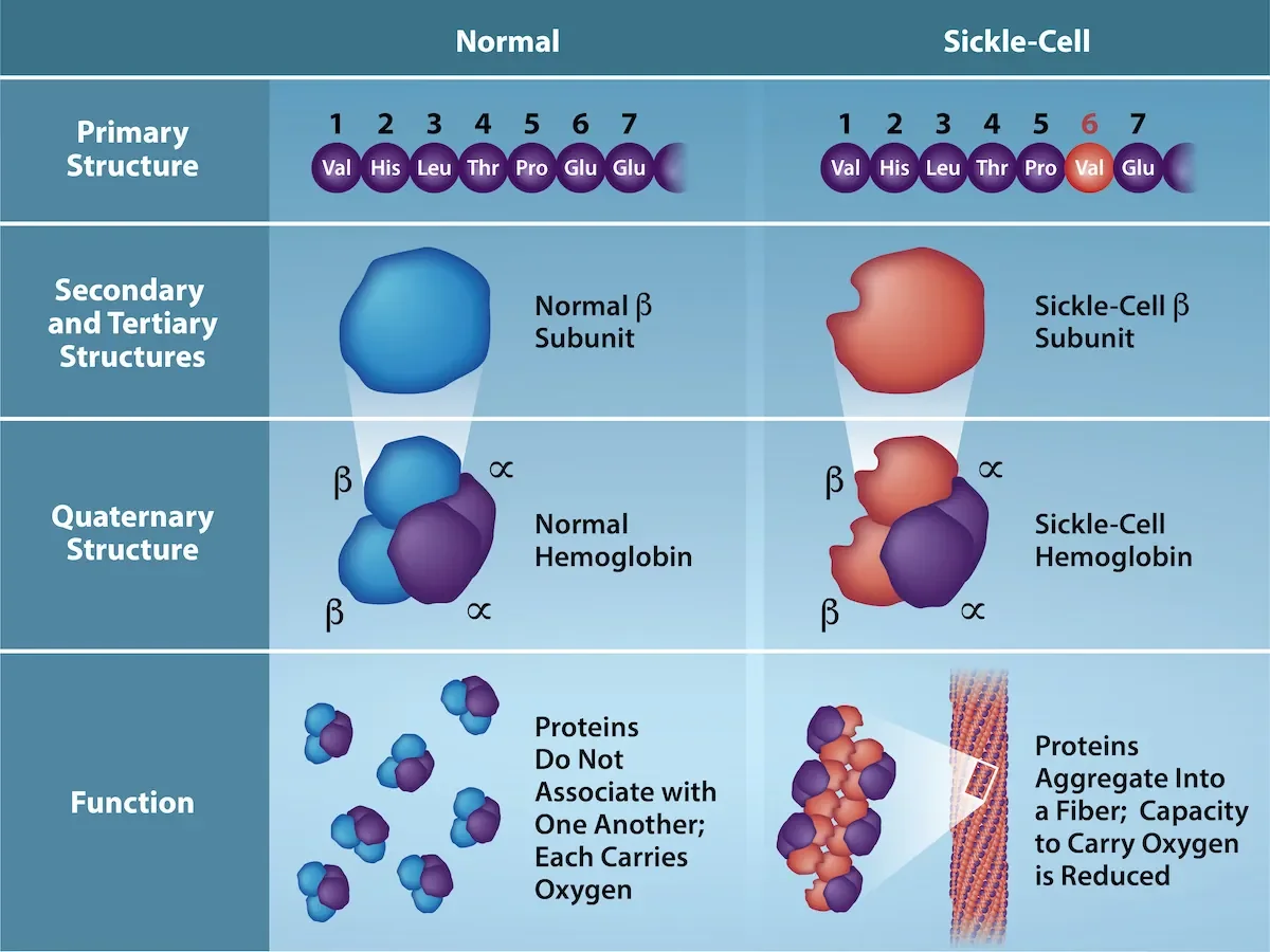
The beta chain of adult hemoglobin is 146 amino acids long. In sickle cell disease, position 6 is changed from glutamate (E, negatively charged, hydrophilic) to valine (V, nonpolar, hydrophobic). One letter in 146. The rest of the sequence is identical.
Consequences of that single swap:
Valine on the surface creates a hydrophobic sticky patch.
At low oxygen, these hydrophobic patches on neighboring hemoglobin molecules stick together.
The hemoglobin polymerizes into long rigid fibers.
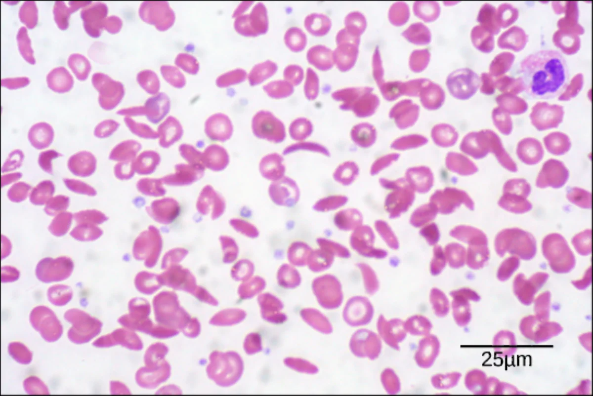
The fibers warp red blood cells into the characteristic sickle shape.
Sickled cells clog capillaries and cause ischemic pain, organ damage, and anemia.
A single amino acid substitution in the hemoglobin beta chain (glutamate to valine at position 6) propagates through secondary, tertiary, and quaternary structure to distort the entire red blood cell. Primary structure controls everything downstream. Credit: OpenStax Biology 2e, CC BY 4.0Real scanning electron micrograph of red blood cells. Normal cells are round and flexible; sickled cells are rigid crescents. All from one amino acid change in the hemoglobin primary sequence. Credit: Lumen Learning / OpenStax Biology 2e, CC BY 4.0
Determining Primary Structure in the Lab
Historically proteins were sequenced by Edman degradation: the N-terminal residue is cleaved off one at a time, identified by chromatography, and the process repeats. This is slow.
Modern methods use mass spectrometry. A protease (like trypsin) cuts the protein at specific residues (trypsin cuts after K or R), the fragments are analyzed by mass spec, and the sequence is reconstructed. A whole proteome can be identified this way.
What defines the primary structure of a protein?
Click to reveal answer
The linear sequence of amino acids from the N-terminus to the C-terminus, linked by covalent peptide bonds. The primary structure is encoded by the gene (via the genetic code) and contains all the information needed to determine how the protein will fold.
Why does the single Glu-to-Val mutation in hemoglobin beta cause sickle cell disease?
Click to reveal answer
Glutamate (E) is negatively charged and hydrophilic; valine (V) is nonpolar and hydrophobic. Placing a hydrophobic residue on the protein surface creates a sticky patch. At low oxygen, these patches on neighboring hemoglobins associate, polymerizing hemoglobin into rigid fibers that distort red blood cells into sickle shapes.
What does "primary structure determines tertiary structure" mean, and whose experiment demonstrated it?
Click to reveal answer
It means the amino acid sequence alone contains enough information to specify how a protein folds into its 3D shape. Christian Anfinsen showed this by denaturing ribonuclease and letting it refold in water without any cellular machinery - the enzyme regained full activity, proving the sequence alone encoded the fold.