Some proteins are lone wolves - one polypeptide chain folds, does its job, and that is the whole story (myoglobin, for example). Other proteins are teams - two or more polypeptide chains assemble into a functional complex. That team assembly is the quaternary structure.
Only proteins with more than one polypeptide chain have a quaternary structure. A single-chain protein has primary, secondary, and tertiary structure. Add a second chain and you have quaternary.
The Vocabulary
Monomer: single polypeptide chain.
Dimer: two subunits. A homodimer has two identical subunits; a heterodimer has two different ones.
Trimer: three subunits (e.g., collagen is a triple helix).
Tetramer: four subunits (e.g., hemoglobin).
Oligomer: general term for any multisubunit complex.
Hemoglobin: the Classic Example
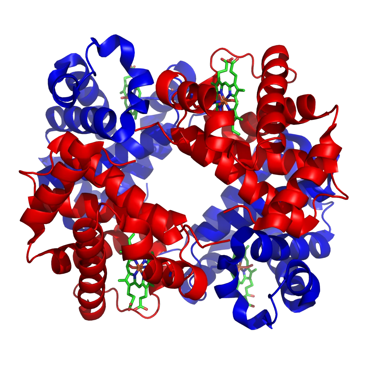
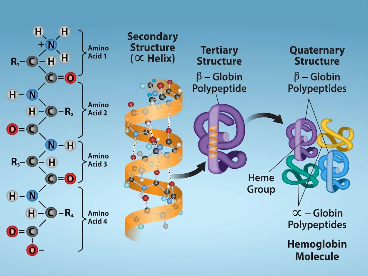
Adult hemoglobin is a heterotetramer of two alpha chains and two beta chains (α2β2). Each subunit has its own tertiary structure and binds one heme group. The four subunits fit together in a compact roughly tetrahedral arrangement.
Hemoglobin, the classic quaternary-structure protein. Two alpha and two beta subunits (α2β2) assemble around four heme groups to create the oxygen carrier. Credit: Wikimedia Commons / Richard Wheeler (Zephyris), CC BY-SA 3.0 (from PDB 1GZX)The four levels of protein structure, using hemoglobin as the example. Primary (sequence) builds secondary (helix), which folds into tertiary (one globin chain), which assembles into quaternary (full hemoglobin tetramer). Credit: OpenStax Biology 2e, CC BY 4.0
Why Quaternary Structure Matters
Two big reasons quaternary structure shows up all over biology:
Cooperativity. When one subunit binds a ligand, it can change its shape and transmit that change to the others, making them more (or less) eager to bind. Hemoglobin does this with oxygen: binding O2 at one subunit pulls the other three into their high-affinity state. This is why the hemoglobin-oxygen curve is sigmoidal while single-subunit myoglobin’s curve is hyperbolic.
Allosteric regulation. A molecule can bind at one site and affect activity at a distant site on another subunit. This is how many enzymes are controlled. Multisubunit proteins have more surface area and more interfaces for regulatory molecules to latch onto.
Subunit Interfaces Use the Same Forces as Tertiary Structure
Quaternary assembly uses the same bond types that build tertiary structure: hydrophobic contacts at the subunit interface, ionic interactions, hydrogen bonds, and sometimes disulfide bonds that bridge two chains (like the inter-chain disulfides in insulin and immunoglobulins). There is no new chemistry at the quaternary level - just a bigger playing field.
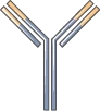
IgG antibody schematic. Two heavy chains and two light chains assemble into a Y-shape held together by disulfide bonds - a textbook example of quaternary structure. Credit: Servier Medical Art, CC BY 4.0
What defines quaternary structure, and which proteins have it?
Click to reveal answer
Quaternary structure is the assembly of two or more polypeptide chains (subunits) into a functional complex. Only multisubunit proteins have quaternary structure. Examples: hemoglobin (α2β2 tetramer), antibodies (2 heavy + 2 light chains), collagen (triple helix). Single-chain proteins like myoglobin do not have quaternary structure.
Why does hemoglobin show a sigmoidal O2 binding curve while myoglobin shows a hyperbolic one?
Click to reveal answer
Hemoglobin has four cooperating subunits; myoglobin has only one. When one hemoglobin subunit binds O2, it nudges the other subunits into a higher-affinity state (positive cooperativity), giving the S-shaped sigmoidal curve. Myoglobin has no subunits to cooperate with, so its binding follows a simple hyperbolic curve.
A protein runs as one band on native gel but as two bands of different sizes on SDS-PAGE with a reducing agent. What does this tell you?
Click to reveal answer
The protein has quaternary structure with two different subunits held together by disulfide bonds. Native gel keeps the complex intact (one band). SDS-PAGE with a reducing agent (like DTT or β-mercaptoethanol) denatures the protein and breaks disulfide bonds, separating the complex into its individual subunits. The fact that two bands appear shows two non-identical chains.