Primary structure is a flat sequence. Secondary structure is local folding (helices and sheets). Tertiary structure is the whole polypeptide chain folded into its final 3D shape, with the helices and sheets packed against each other and a stable core in the middle.
Tertiary structure is where the protein becomes functional. An enzyme’s active site is a tertiary feature. A binding pocket on a hormone receptor is a tertiary feature. If tertiary structure is wrong, the protein does not work.
The Five Forces That Stabilize Tertiary Structure
Force
What it is
Strength
R groups involved
Hydrophobic interactions
Nonpolar R groups cluster away from water
Strongest cumulative driver of folding
Val, Leu, Ile, Phe, Trp, Met, Ala
Disulfide bonds
Covalent -S-S- between two cysteines
Strongest per-bond (covalent)
Cys only
Salt bridges (ionic)
Attraction between oppositely charged side chains
Medium
Asp/Glu with Lys/Arg/His
Hydrogen bonds
Between polar side chains (or backbone)
Medium-weak
Ser, Thr, Tyr, Asn, Gln, His
Van der Waals forces
Weak, cumulative close-range attractions
Individually weak, collectively important
All residues
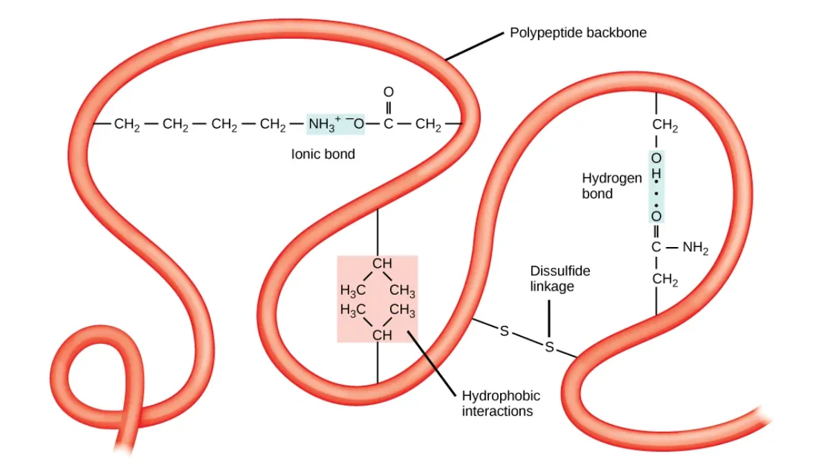
The main forces that stabilize tertiary structure. Hydrophobic clustering (at center) drives the overall fold, while disulfide bonds, ionic salt bridges, and hydrogen bonds lock the final shape in place. Credit: OpenStax Biology 2e, CC BY 4.0
Hydrophobic Interactions: the Main Engine
Nonpolar R groups cannot hydrogen bond with water. When a protein folds, exposed nonpolar residues disrupt the water’s hydrogen bond network, forcing water molecules to organize into “cages” around them (entropy penalty). Burying those nonpolar residues in a protein interior releases those ordered water molecules back into the bulk. That entropy gain is the driving force of folding.
Disulfide Bonds
Two cysteines can be oxidized to form a covalent -S-S- bridge. This is the strongest single bond stabilizing tertiary structure - a true covalent bond, not a weak interaction.
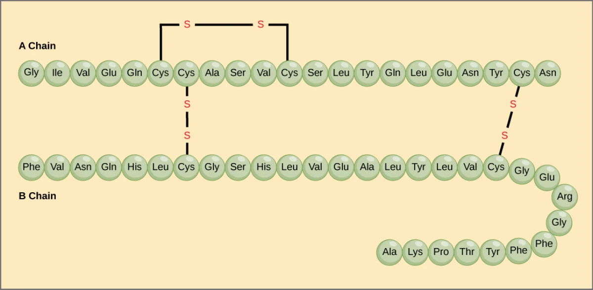
Disulfide bonds require an oxidizing environment. Inside the cytoplasm (reducing environment), they rarely form. In the endoplasmic reticulum and in extracellular space (oxidizing environments), they are common. That is why secreted proteins (insulin, antibodies, digestive enzymes) tend to be rich in disulfide bonds while cytosolic proteins are not.
Insulin has two short polypeptide chains held together by three disulfide bonds (two between chains, one within the A chain). Disulfide bonds are common in secreted proteins. Credit: Lumen Learning / OpenStax Biology 2e, CC BY 4.0
Salt Bridges (Ionic Interactions)
Salt bridges form between a negatively charged side chain (Asp or Glu) and a positively charged one (Lys, Arg, His). They are weaker than covalent bonds but can have a significant effect on stability when buried inside the protein where no water can interfere.
Salt bridges are pH-sensitive. Lowering pH (protonating Asp/Glu) or raising pH (deprotonating Lys/Arg) removes the charge and breaks the bridge - one reason proteins denature at extreme pH.
Hydrogen Bonds and Van der Waals
Polar side chains (Ser, Thr, Tyr, Asn, Gln, His) can hydrogen bond with each other or with the backbone. Van der Waals forces (instantaneous dipoles) act between all atoms when they are close enough. Individually weak, but thousands of van der Waals contacts in a tightly packed protein interior add up.
What is the MOST important force driving protein folding, and why?
Click to reveal answer
The hydrophobic effect. Burying nonpolar side chains in the protein interior releases ordered water molecules that were trapped around exposed nonpolar surfaces. That increase in water's entropy drives the overall fold. Disulfide bonds are stronger per bond but are rare; hydrophobic interactions are everywhere in every protein and are the overall driver.
Why are disulfide bonds common in secreted proteins like insulin and antibodies but rare in cytoplasmic proteins?
Click to reveal answer
Disulfide bond formation requires an oxidizing environment. The cytoplasm is reducing (high glutathione), so -SH groups stay reduced. The endoplasmic reticulum and extracellular space are oxidizing, so cysteines can form -S-S- bridges. Secreted proteins traverse the ER and end up outside the cell, where disulfide bonds help stabilize them.
Between which amino acids would a salt bridge most likely form?
Click to reveal answer
Between a negatively charged side chain (aspartate or glutamate, D/E) and a positively charged one (lysine, arginine, or histidine; K/R/H). At physiological pH, these opposite charges attract. The interaction is called a salt bridge (ionic interaction) and it is pH-sensitive - protonation or deprotonation of either partner breaks it.