
Secondary Structure
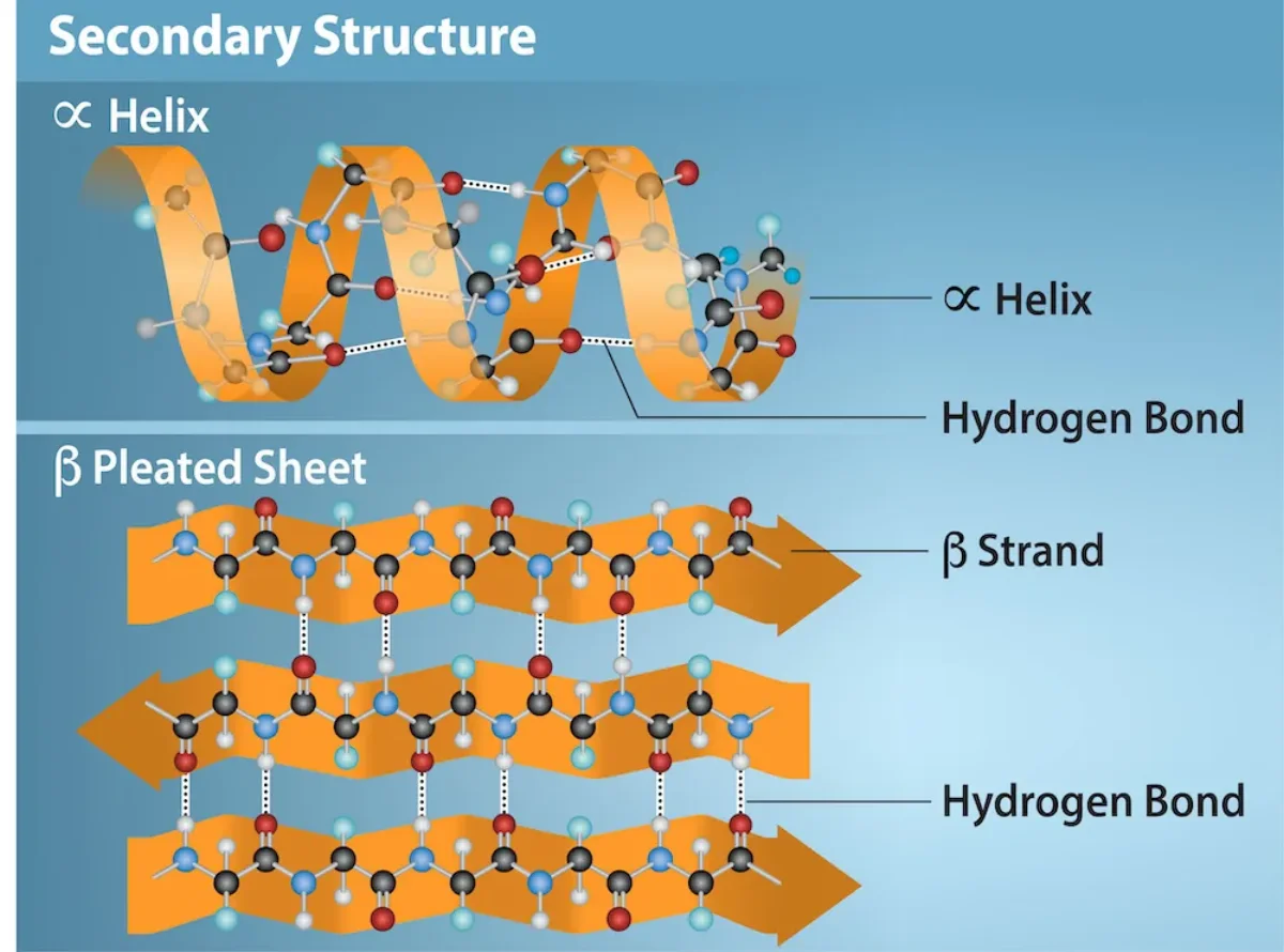
After translation, the polypeptide chain begins to fold as it leaves the ribosome. The first folds are local - not the whole protein collapsing, just small regions snapping into repeating patterns. These local patterns are secondary structure, and they come in two flavors: alpha helices and beta pleated sheets.
Both are held together by the same force: hydrogen bonds between backbone amide groups. Not R groups. The backbone N-H of one residue hydrogen bonds to the backbone C=O of another residue a few positions away. R groups do not participate - they stick out to the side.
The Alpha Helix
The alpha helix is a right-handed corkscrew. The backbone spirals around an imaginary central axis. Every residue’s carbonyl oxygen (C=O) hydrogen bonds to the amide N-H exactly four residues ahead in the chain. Side chains project outward, away from the helix core.
Key numbers to know:
- 3.6 residues per turn
- 5.4 Å (0.54 nm) per turn - so 1.5 Å rise per residue
- Hydrogen bond is from residue n to residue n+4
- Most alpha helices are right-handed
Helix Breakers
Two amino acids disrupt alpha helices:
- Proline has a ring that locks its Phi angle and removes the amide N-H needed for hydrogen bonding. Proline almost never appears inside a helix (only at the N-terminal “cap” position).
- Glycine is too flexible. Its side chain is just hydrogen, so it samples too many conformations. It prefers turns and loops over the regular helix geometry.
The Beta Pleated Sheet
Beta sheets form between beta strands - segments of backbone running in an extended zigzag. Two or more strands line up next to each other and form hydrogen bonds sideways between adjacent strands. The result is a pleated sheet that looks like an accordion.
Beta sheets come in two flavors:
- Parallel: both strands run in the same N-to-C direction. Hydrogen bonds are slightly skewed. Less stable than antiparallel.
- Antiparallel: strands run in opposite N-to-C directions. Hydrogen bonds are linear (straight across). More stable.
Beta strands are separated in sequence but close in space. Between two adjacent strands in a sheet, there must be a connecting loop or “beta turn.”
Loops and Turns
Not every residue participates in a helix or sheet. In between, the backbone makes turns (short reversals of direction, often 3-4 residues) and loops (longer irregular segments). These regions often sit on the protein surface and contain active sites, binding loops, or antibody recognition loops.
Glycine and proline are over-represented in loops and turns. Glycine provides flexibility; proline enforces a sharp kink.
Why Secondary Structure Exists
Hydrogen bonding the backbone amides is energetically cheap and there are a lot of them in a polypeptide. If the protein did NOT form secondary structure, those H-bond donors (N-H) and acceptors (C=O) would need to find other partners - usually water. Burying the backbone in the protein interior requires first satisfying all those H-bonds internally. That is exactly what alpha helices and beta sheets do.