
Double Helix
DNA is a right-handed double helix of two antiparallel polynucleotide strands. The backbones run on the outside; the bases pair in the center.
What a nucleotide is, and what holds two strands together
Scroll sideways to see the whole map.
Antiparallel, and what followsOne strand runs 5' to 3' and its partner runs 3' to 5'. Because every polymerase can only build toward a 3' end, that single geometric fact forces the leading and lagging strands in replication, sets the direction of transcription, and defines what a template strand is.
Reading a sequenceBy convention a sequence is written 5' to 3' unless stated otherwise. The complement of 5'-ATGC-3' is 3'-TACG-5', which written conventionally is 5'-GCAT-3'. Half the mistakes on this topic are from forgetting to flip.
Purines and pyrimidinesPURe As Gold: purines are Adenine and Guanine, and they have two rings. CUT the PY: pyrimidines are Cytosine, Uracil, and Thymine, with one ring. Every pair is one of each, which is what keeps the helix a constant width.
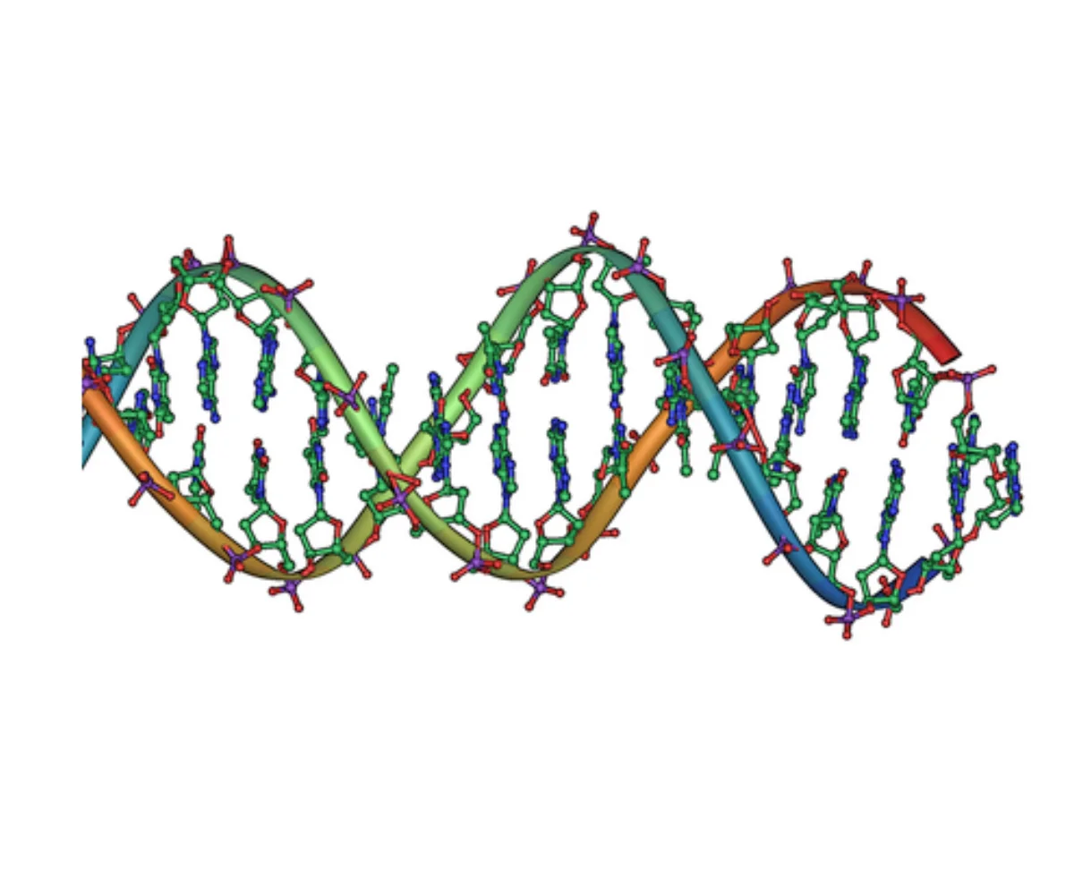
Base Pairing
The two strands are held together by hydrogen bonds between paired bases. Watson-Crick base pairing follows strict rules:
- A pairs with T via 2 hydrogen bonds.
- G pairs with C via 3 hydrogen bonds.
A purine always pairs with a pyrimidine, keeping the helix diameter constant.
Antiparallel Strands
The two strands run in opposite directions. One strand is 5’ → 3’ top to bottom; the other is 5’ → 3’ bottom to top. Each base on one strand pairs with the complementary base directly across from it on the other strand.
This antiparallel arrangement has a major consequence: since DNA polymerases only synthesize 5’ → 3’, the two strands at a replication fork must be copied differently. One is made continuously (leading strand), one is made in fragments (lagging strand).
Grooves
The helix has two grooves where proteins can read the base sequence without unwinding the helix:
- Major groove: wider; exposes more of the base edges. Most transcription factors recognize DNA sequences here.
- Minor groove: narrower; less information. Some specialized proteins read sequences here.
Chargaff’s Rules
Before Watson and Crick, Erwin Chargaff showed that for any double-stranded DNA sample:
- The amount of A equals the amount of T, and the amount of G equals the amount of C.
- The percent of purines (A + G) equals the percent of pyrimidines (T + C), which is always 50%.
These relationships make sense only if A pairs with T and G pairs with C. Chargaff’s data was a critical clue in Watson and Crick’s 1953 model.
DNA Denaturation, Reannealing, and Hybridization
The hydrogen bonds between paired bases are much weaker than the covalent backbone, so you can pull the two strands apart without breaking the sequence. This is denaturation (sometimes called “melting”). Heat is the usual cause in the lab; extreme pH or chaotropes (urea, formamide) also work.
- Melting temperature (Tm): the temperature at which half of the DNA is single-stranded and half is double-stranded. Higher GC content = higher Tm (three H-bonds per GC vs. two per AT). Longer DNA and higher salt also raise Tm.
- Reannealing: if cooling is slow after denaturation, the separated strands will find their complements again and reform the double helix. The optimal reannealing temperature is about 20-25°C below Tm.
- Hybridization: single-stranded DNA (or RNA) from one source pairs with complementary single-stranded nucleic acid from another source. This is how probes work in Southern and Northern blots, how primers find their targets in PCR, and how microarrays read gene expression.
Chromatin Structure in the Nucleus
A single human cell contains about 2 meters of DNA packaged into a nucleus only a few microns across. The packaging solution is chromatin - DNA wound around proteins and folded hierarchically.
- Histones: small basic proteins rich in lysine and arginine (positively charged to bind the negative DNA backbone). Five histone types: H2A, H2B, H3, H4, and the linker H1.
- Nucleosome: ~147 bp of DNA wrapped about 1.65 turns around a histone octamer (two copies each of H2A, H2B, H3, H4). Nucleosomes are the fundamental repeating unit of chromatin, often described as “beads on a string.”
- 30-nm fiber: nucleosomes coil into thicker fibers, with H1 stabilizing the coil.
- Higher-order folding produces the condensed metaphase chromosome, which is ~10,000-fold shorter than the naked DNA.
Euchromatin vs. Heterochromatin
- Euchromatin: loosely packed, transcriptionally active. Appears lighter under microscopy.
- Heterochromatin: densely packed, transcriptionally silent. Appears darker. Constitutive heterochromatin (always condensed, like centromeres and telomeres) vs. facultative heterochromatin (tissue- or time-specific silencing, like the inactivated X chromosome).
Telomeres and Centromeres
- Telomeres: repetitive sequences (TTAGGG in humans) at chromosome ends. They cap the ends to prevent fusion and degradation, and they shorten with each round of replication unless telomerase extends them. Shortening is linked to cellular aging; telomerase reactivation is a hallmark of many cancers.
- Centromeres: heterochromatic repetitive regions in the middle of each chromosome that anchor sister chromatids together and serve as the assembly site for the kinetochore during mitosis.
Single-Copy vs. Repetitive DNA
Only about 1-2 percent of the human genome codes for protein. The rest includes regulatory elements, introns, and large stretches of repetitive DNA.
- Single-copy DNA: most protein-coding genes. Present as one (or a few) copies per genome.
- Repetitive DNA: tandem repeats (like centromeric and telomeric repeats) and interspersed repeats (like transposon-derived SINEs and LINEs). Some repetitive regions are structural; others are evolutionary relics.