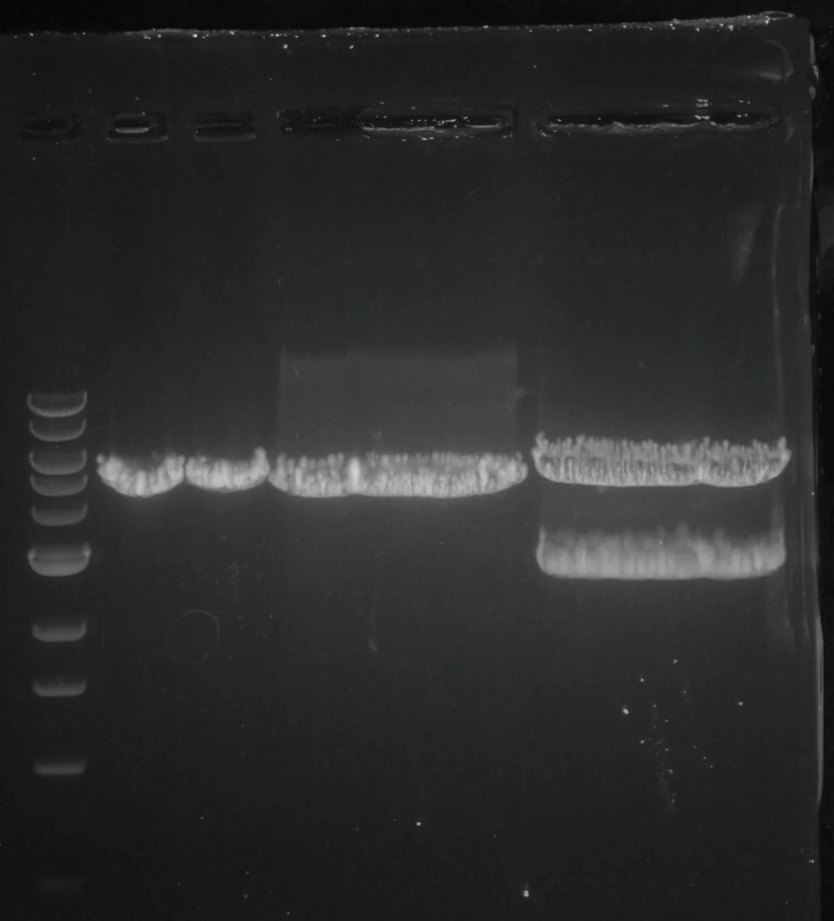
Gel electrophoresis is the standard way to separate DNA fragments by size. The DNA sample is loaded into wells at one end of a porous gel (usually agarose). An electric field drives the DNA through the gel. Small fragments travel quickly; large fragments travel slowly.
An agarose gel stained with ethidium bromide. DNA bands appear white/orange under UV light. The leftmost lane is a DNA ladder of known fragment sizes for reference. Credit: Wikimedia Commons, CC BY-SA
How It Works
DNA has a uniformly negative charge from its phosphate backbone. In an electric field, it moves toward the positive electrode (anode). The gel matrix has tiny pores that slow fragments based on size:
Small fragments: slip through easily → travel far.
Large fragments: get caught in the mesh → travel less distance.
After the run, fragments are visualized by adding a DNA-binding dye:
Ethidium bromide: intercalates between base pairs and fluoresces under UV light.
SYBR Safe and other modern dyes: less toxic alternatives.
The pattern of bands along each lane is the “gel image” - a visual sorting of the fragments by size.
Reading a Gel
A typical gel image has:
A ladder lane: DNA fragments of known sizes (e.g., 100, 200, 300, 500, 1000, 2000 bp). This is the ruler.
Sample lanes: your DNA samples. Compare bands to the ladder to estimate each fragment’s size.
The wells are at the top (or negative end). Bands near the top are large; bands near the bottom are small.
Agarose vs. Polyacrylamide
Two gel materials are common:
Agarose: best for DNA fragments 100 bp to 50+ kb. Large pores. Lower resolution.
Polyacrylamide: best for small DNA (<500 bp) and for proteins. Smaller pores, higher resolution. Proteins on polyacrylamide (SDS-PAGE) were covered in Chapter 3.
The choice depends on the size range you care about. For routine cloning checks (1-5 kb plasmid fragments), agarose is standard.
Why does DNA migrate toward the positive electrode in gel electrophoresis?
Click to reveal answer
The phosphate backbone of DNA carries many negative charges - one per nucleotide. Under an electric field, the negatively charged DNA is attracted to the positive electrode (anode). Proteins, by contrast, have variable net charges depending on their amino acids and pH, which is why SDS is needed to give proteins uniform negative charge.
In agarose gel electrophoresis, where on the gel do the smallest DNA fragments end up?
Click to reveal answer
Farthest from the wells, closest to the bottom (positive electrode). Small DNA fragments travel more easily through the gel pores, so they migrate faster and end up at the leading edge. Large fragments are stuck in the matrix near the wells. This is the opposite of size exclusion chromatography (big elutes first) because gel electrophoresis physically restricts larger molecules in pores.
How is a band's size estimated from a gel?
Click to reveal answer
By comparison to a ladder - a set of DNA fragments with known sizes run in a parallel lane. The sample band's migration distance is compared to the ladder's bands to estimate the sample's size. More precisely, migration distance is approximately proportional to -log(fragment size), so linear interpolation between adjacent ladder bands gives a good estimate.