
Electrophoresis
Electrophoresis separates charged molecules by driving them through a gel under an electric field. Small or highly charged molecules move fast. Large or weakly charged molecules move slowly. In protein biochemistry, electrophoresis is the standard way to check how pure a preparation is and to estimate molecular weights.
SDS-PAGE - Size Only
SDS-PAGE is the gold-standard technique for separating proteins by size. SDS (sodium dodecyl sulfate) is a detergent that does three things:
- Coats every protein with many SDS molecules, giving all proteins a uniformly strongly negative charge proportional to length.
- Denatures (unfolds) the proteins into extended rods.
- Disrupts non-covalent interactions.
Beta-mercaptoethanol (or dithiothreitol) is added to reduce disulfide bonds, so multi-chain proteins are separated into individual subunits.
After SDS treatment, every protein has roughly the same charge-to-mass ratio. In the polyacrylamide gel, separation depends only on size: small proteins move quickly through the gel pores, large proteins move slowly.
Native PAGE - No SDS
In native PAGE, you leave the protein folded and natively charged. Separation depends on a combination of size, shape, and net charge. Native PAGE is useful when you want to preserve biological activity (a protein that is still functional after the gel). It gives less clean size information than SDS-PAGE because shape and charge vary among proteins.
Isoelectric Focusing (IEF) - Charge Only
A gel is cast with a pH gradient. When an electric field is applied, a protein migrates toward the pole opposite its net charge. As it moves, it crosses pH zones that change its protonation state. When it reaches the pH that equals its isoelectric point (pI), the protein has net zero charge and stops moving. Each protein “focuses” at its own pI.
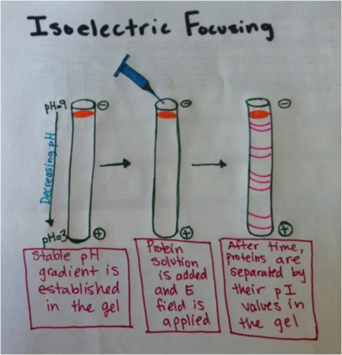
IEF is often used to detect small charge differences between protein variants - for example, distinguishing isoforms that differ by a single amino acid charge or by a post-translational modification.
2D Gel Electrophoresis - Charge Then Size
Combining IEF and SDS-PAGE gives 2D gel electrophoresis, the highest-resolution method for separating complex protein mixtures.
- Run IEF in one dimension (horizontal), separating by pI.
- Rotate the strip 90 degrees, apply SDS, and run SDS-PAGE in the perpendicular dimension (vertical), separating by size.
The result is a 2D “spot pattern” where each protein appears as a single dot at unique coordinates of (pI, molecular weight). Thousands of proteins can be resolved in one gel, and the pattern can be compared between healthy and diseased tissue to find differentially expressed proteins.
Detecting the Bands
After the gel is run, you need to visualize the proteins:
- Coomassie blue or silver stain binds proteins nonspecifically and shows all bands.
- Western blot: transfer gel contents to a membrane, then probe with antibodies to detect one specific protein. This is the standard technique for proving a specific protein is present.