Structural proteins are the beams, cables, and sheets of the body. They are designed to resist force. Most are long, repetitive, and fibrous rather than globular. The MCAT focuses on three: collagen, keratin, and elastin, plus a passing nod to the cytoskeletal proteins (actin, microtubules, intermediate filaments).
Collagen
Collagen is the most abundant protein in the body - about 25-30 percent of total protein. It is the main component of bone, tendons, cartilage, skin, blood vessels, and basement membranes. Its job is to resist stretching.
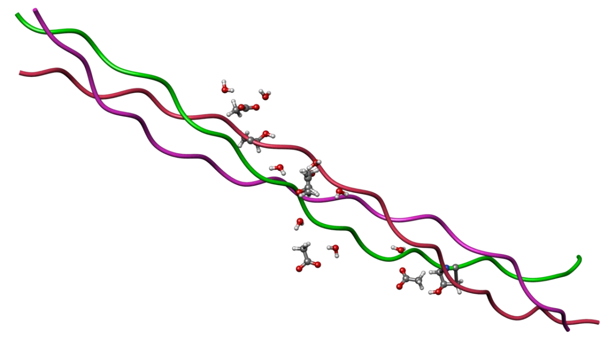
Collagen has a distinctive triple helix structure. Three polypeptide chains (called alpha chains) wrap around each other in a right-handed superhelix. Each chain has the repeating sequence Gly-X-Y, where X is often proline and Y is often hydroxyproline. Glycine’s tiny R group (just -H) is essential because only glycine is small enough to fit in the tight interior of the triple helix. Every third position MUST be glycine.
Collagen triple helix. Three alpha chains, each with a Gly-X-Y repeat, wrap around one another. Every third residue is glycine. Credit: Wikimedia Commons, CC BY-SA
Why Collagen Needs Vitamin C
The hydroxyprolines and hydroxylysines are not encoded in DNA. They are made after translation by enzymes called prolyl hydroxylase and lysyl hydroxylase, which need vitamin C (ascorbate) as a cofactor. Vitamin C keeps the iron in these enzymes in the Fe2+ state, which is required for hydroxylation.
Without hydroxylation, collagen fibers cannot form the hydrogen bonds that stabilize the triple helix. Weak collagen means weak blood vessels, gums, and connective tissue. That is scurvy: bleeding gums, loose teeth, poor wound healing, fragile skin.
Keratin
Keratin is the protein in hair, nails, horns, feathers, and the outer layer of skin. Two types:
Alpha-keratin is rich in alpha helices coiled around each other in coiled coils. Found in hair, wool, nails, and epidermis.
Beta-keratin is rich in beta sheets. Found in feathers, scales, and beaks.
Keratins also contain many cysteines, which form cross-linking disulfide bonds between chains. The more disulfide bonds, the tougher the keratin (nails and claws are harder than hair).
Elastin
Elastin is what allows stretchy tissues (skin, lungs, large arteries) to snap back to their original shape after being deformed. It is a rubber-like protein. Elastin forms a loose, cross-linked network rather than a rigid fiber. When the tissue stretches, the chains pull apart; when force is released, they spring back - entropy drives the recoil.
Elastin is cross-linked by an unusual amino acid called desmosine (formed from 4 lysines). The AAMC outline does not require you to memorize desmosine, but you should know that elastin is extensively cross-linked.
Cell Adhesion Molecules
Proteins at the cell surface decide what sticks to what. The MCAT tests three families - cadherins, integrins, and selectins - and their specific roles in tissue architecture, wound healing, and leukocyte migration.
CAM family
What it binds
Calcium-dependent?
Signature role
Cadherins
Identical cadherins on a neighboring cell (homophilic, cell-cell)
Yes (Ca2+ required)
Hold epithelial tissues together at adherens junctions
Integrins
Extracellular matrix proteins (fibronectin, laminin, collagen) via RGD motifs
Yes (Ca2+/Mg2+)
Anchor cells to the ECM; trigger inside-out and outside-in signaling
Selectins
Carbohydrate ligands on other cells (e.g., sialyl-Lewis X)
Yes
Initial tethering and rolling of leukocytes on endothelium
Cytoskeletal Proteins
Inside cells, three main polymer systems form the cytoskeleton:
Polymer
Monomer
Diameter
Jobs
Actin / microfilaments
G-actin
~7 nm
Cell shape, muscle contraction, cytokinesis
Microtubules
Tubulin dimers (alpha + beta)
~25 nm
Tracks for motor proteins, mitotic spindle
Intermediate filaments
Varies (keratin, lamin, vimentin, neurofilament)
~10 nm
Mechanical strength, nuclear envelope support
Actin and microtubules are dynamic (assembly and disassembly are regulated). Intermediate filaments are mostly stable and provide tensile strength.
Why must every third amino acid in collagen's polypeptide chains be glycine?
Click to reveal answer
Glycine's R group is just a hydrogen. In the tightly wound triple helix, the third position points into the interior where no larger side chain could fit. Any mutation that replaces a glycine disrupts the helix and causes connective tissue disease (e.g., osteogenesis imperfecta).
How does vitamin C deficiency cause scurvy?
Click to reveal answer
Vitamin C is required by prolyl and lysyl hydroxylases that hydroxylate proline and lysine residues in collagen. Without hydroxylation, the collagen triple helix cannot form stable hydrogen bonds, so new collagen is weak. Connective tissues fail - bleeding gums, loose teeth, poor wound healing.
What structural feature of keratin makes it so mechanically tough?
Click to reveal answer
Disulfide cross-links between cysteine residues. Alpha-keratin is based on coiled coils of alpha helices, and beta-keratin on beta sheets, but both rely on abundant cysteine to form covalent S-S bridges that lock the structure. The more cysteines, the harder the keratin (compare soft hair vs. hard nails).
Motor proteins convert chemical energy (ATP hydrolysis) into mechanical work. They literally walk along cellular tracks, carrying cargo or pulling on filaments to cause contraction. Three MCAT-relevant motor proteins, all walking different tracks.
Motor
Walks on
Direction
Typical cargo/job
Myosin
Actin filaments
Plus end (most myosins)
Muscle contraction; vesicle transport
Kinesin
Microtubules
Plus end (toward periphery, anterograde)
Axonal transport outward from cell body
Dynein
Microtubules
Minus end (toward center, retrograde)
Axonal transport inward; cilia and flagella beating
Myosin and Muscle Contraction
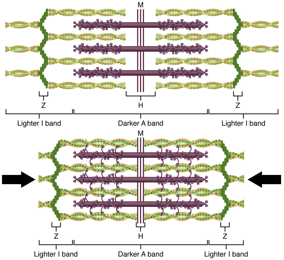
In skeletal muscle, myosin is organized into thick filaments. Actin forms thin filaments. Together they slide past one another to produce contraction. The unit of contraction is the sarcomere, bounded by two Z-discs.
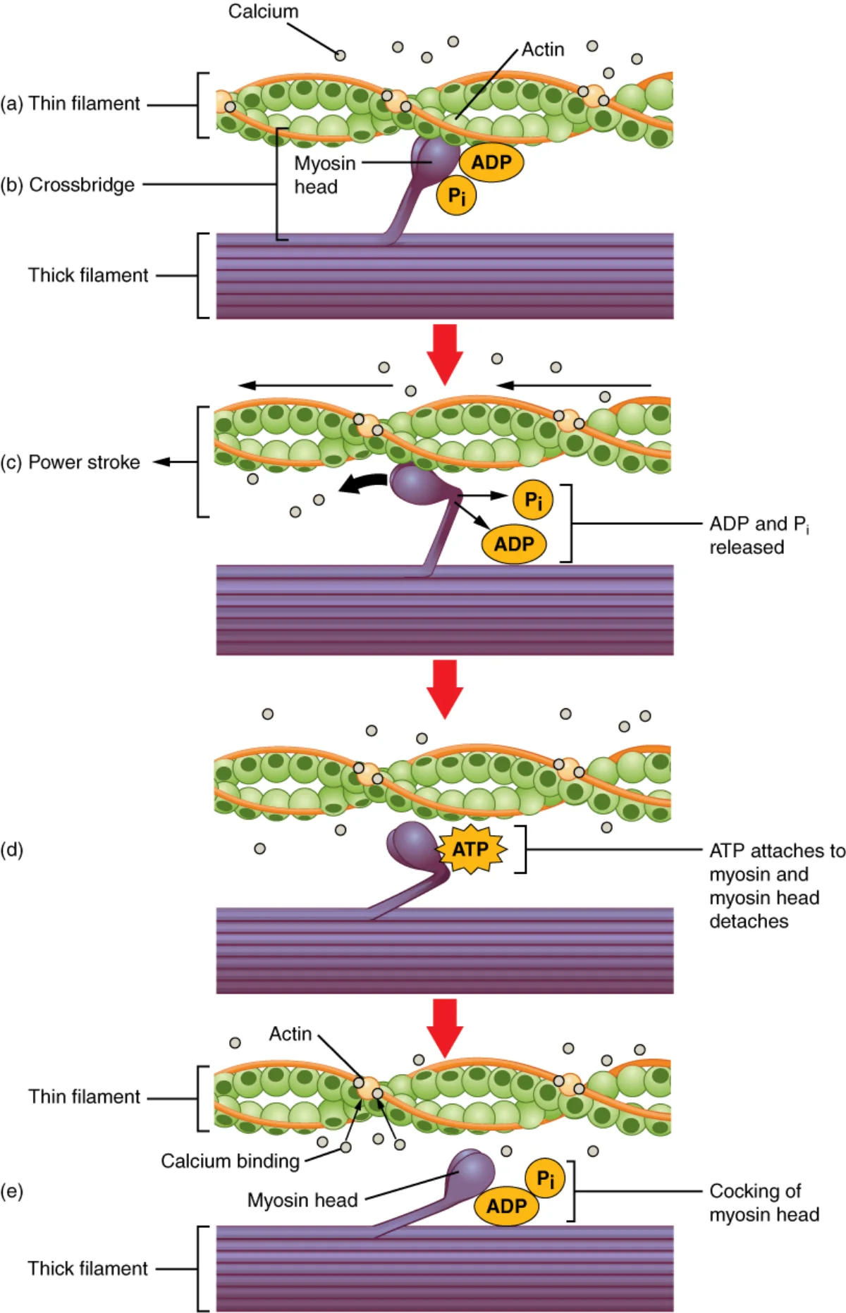
The cross-bridge cycle is the four-step ATP-driven process that makes one myosin head “walk” along actin:
ATP binds the myosin head, releasing it from actin.
ATP hydrolysis cocks the myosin head into a high-energy position (like a spring being loaded).
Myosin binds actin and releases inorganic phosphate.
Power stroke: myosin swings back to its original shape, pulling actin toward the center of the sarcomere. ADP leaves.
The cross-bridge cycle. Each cycle consumes one ATP and produces one "step" of myosin along actin. Dozens of cycles in parallel produce muscle contraction. Credit: OpenStax Anatomy and Physiology 2e, CC BY 4.0
Rigor Mortis
After death, cells run out of ATP. Without ATP, myosin cannot release actin (step 1 of the cycle). Muscles seize up in a contracted state called rigor mortis. A few hours later, proteases begin degrading the myofibrils and the muscle relaxes again.
Sliding filament model. During contraction, actin thin filaments slide past myosin thick filaments toward the sarcomere center, shortening the sarcomere. Credit: OpenStax Anatomy and Physiology 2e, CC BY 4.0
Kinesin
Kinesin is a two-headed motor that walks toward the plus end of microtubules. It has a heavy chain with motor domains, a stalk, and light chains that attach to cargo (vesicles, mitochondria, RNA granules). Each step hydrolyzes one ATP and covers about 8 nm.
The classic example is axonal transport: kinesin carries neurotransmitter vesicles and mitochondria down the axon, from the cell body to the synapse. Without kinesin, a long neuron could not supply its distant axon terminals with fresh proteins.
Dynein
Dynein is the opposite-direction partner. It walks toward the minus end of microtubules (back toward the cell body). It transports endocytosed material, recycles old organelles, and - in a completely different role - powers the beating of cilia and flagella.
In cilia and flagella, dynein molecules between adjacent microtubule pairs pull past one another, producing the wave-like bending that propels cells (sperm flagella, airway cilia). The axoneme structure - nine outer microtubule doublets plus two central singlets, “9+2” - is bound together by dynein arms and nexin links.
On which cytoskeletal filament does each motor protein walk: myosin, kinesin, dynein?
Click to reveal answer
Myosin walks on actin filaments. Kinesin and dynein both walk on microtubules. Kinesin moves toward the plus end (anterograde, outward in axons). Dynein moves toward the minus end (retrograde, inward).
Why does muscle go into rigor mortis after death?
Click to reveal answer
After death, ATP production stops. Myosin needs ATP to release from actin. Without ATP, the cross-bridges cannot detach and the muscle stays contracted. Rigor mortis resolves hours later when lysosomal proteases begin to digest the muscle fibers.
Which motor protein family is responsible for the beating of cilia and flagella?
Click to reveal answer
Dynein. Axonemal dyneins between adjacent microtubule doublets slide past one another while tethered by nexin links, producing the wave-like bending that moves cilia and flagella. The "9+2" arrangement of microtubules in cilia and flagella depends on dynein arms.
Binding proteins grab small molecules or ions and carry them somewhere. They do not transform their cargo chemically. Immunoglobulins are a specialized class of binding proteins that grab foreign antigens to mark them for destruction. Both are essential MCAT topics.
Plasma Binding Proteins
The most abundant protein in blood plasma is albumin, which binds and transports fatty acids, bilirubin, steroid hormones, thyroid hormones, and many drugs. Albumin also contributes most of the plasma oncotic pressure (the osmotic pull that keeps fluid inside blood vessels). Low albumin causes edema.
Other important binding proteins:
Transferrin binds iron (Fe3+) and delivers it to cells via the transferrin receptor.
Ferritin stores iron inside cells.
Sex hormone binding globulin (SHBG) carries testosterone and estradiol.
Hemoglobin is the quintessential binding protein (it carries O2 inside red blood cells). Covered in detail in sections 3.5 and 3.6.
Immunoglobulins (Antibodies)
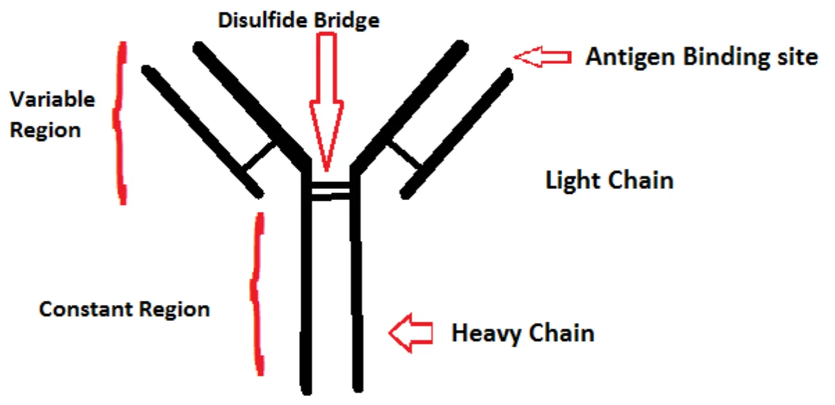
An antibody is a Y-shaped protein that recognizes a specific shape (an epitope on an antigen) and binds it with very high affinity. Each antibody is made of two identical heavy chains and two identical light chains, held together by disulfide bonds.
IgG antibody structure. The two arms (Fab regions) carry the antigen-binding site. The stem (Fc region) interacts with immune cells and complement. Credit: Wikimedia Commons, CC BY-SA
Heavy and Light Chain Architecture
Every IgG has exactly 4 polypeptide chains:
2 heavy (H) chains: ~50 kDa each. Each has one variable domain (VH) at the N-terminal tip, followed by three or four constant domains (CH1, CH2, CH3).
2 light (L) chains: ~25 kDa each. Each has one variable domain (VL) and one constant domain (CL). Light chains come in two flavors: kappa (κ) or lambda (λ).
The chains are held together by disulfide bonds: two interchain disulfides link the two heavy chains to each other in the hinge region, and one disulfide links each light chain to its heavy-chain partner. Intrachain disulfides fold each domain into a compact immunoglobulin fold (a beta sandwich).
Variable vs. Constant Regions
Variable (V) regions are at the N-terminal tips of the Fab arms. They are encoded by V, D, and J gene segments that rearrange during B-cell development, producing a near-infinite library of binding shapes. The three hypervariable loops (complementarity-determining regions, CDRs) from VH and VL together form the antigen-binding site.
Constant (C) regions are identical within an isotype. They determine antibody class (IgG vs. IgA vs. IgM, etc.) and effector function - which immune cells and complement proteins the antibody recruits.
Fab vs. Fc
Fab (fragment antigen-binding) - the two arms of the Y. Each arm is built from one whole light chain + the N-terminal half of one heavy chain (VH + CH1). Antigen binds at the VH/VL tip.
Fc (fragment crystallizable) - the stem of the Y. Made entirely of the two heavy chains’ C-terminal constant domains (CH2 + CH3). This is where Fc receptors on immune cells and complement C1q bind after the antibody has grabbed its antigen.
The Five Immunoglobulin Classes
The class of an antibody is determined by its heavy chain constant region. Five MCAT-relevant classes, each with distinctive biology:
Class
Structure
Where found
Function
IgG
Monomer
Plasma, crosses placenta
Main circulating antibody; long-term immunity
IgA
Dimer (with a J chain)
Mucosal secretions, breast milk, saliva
Mucosal immunity
IgM
Pentamer
Blood
First antibody made in a new infection; activates complement
IgE
Monomer
Bound to mast cells
Allergies; parasite defense
IgD
Monomer
B-cell surface
B-cell receptor for naive B cells
Antibody Specificity
The variable region of an antibody is generated by random recombination of V, D, and J gene segments during B-cell development. The variable region on each arm has three hypervariable loops (also called complementarity-determining regions, CDRs) that physically contact the antigen. Small differences in the CDRs produce the enormous diversity that lets your immune system recognize almost any foreign molecule.
Monoclonal Antibodies
A monoclonal antibody (mAb) is a cloned population of identical antibodies, all recognizing the same epitope. Monoclonals are the fastest-growing class of drugs - examples include trastuzumab (HER2-targeted cancer therapy), adalimumab (anti-TNF for autoimmune disease), and many COVID-19 treatments. You do not need to memorize drug names, but you should know that monoclonals are a major real-world use of antibody specificity.
What are the Fab and Fc regions of an antibody responsible for?
Click to reveal answer
Fab (fragment antigen-binding) contains the variable regions and binds the antigen with high specificity. Fc (fragment crystallizable) is the constant stem that interacts with immune cells (via Fc receptors) and complement proteins, triggering effector responses after antigen binding.
Which immunoglobulin class is a pentamer produced first in a new infection?
Click to reveal answer
IgM. It is the first antibody produced by B cells in a primary immune response, exists as a pentamer joined by a J chain, and is very effective at activating complement. Later in the response, class switching produces IgG and other isotypes with the same specificity.
What is the physiologic role of albumin in plasma?
Click to reveal answer
Albumin is the most abundant plasma protein. It transports fatty acids, bilirubin, steroid and thyroid hormones, and many drugs. It also provides most of the plasma oncotic pressure, which holds fluid inside blood vessels. Low albumin causes edema and altered drug pharmacokinetics.
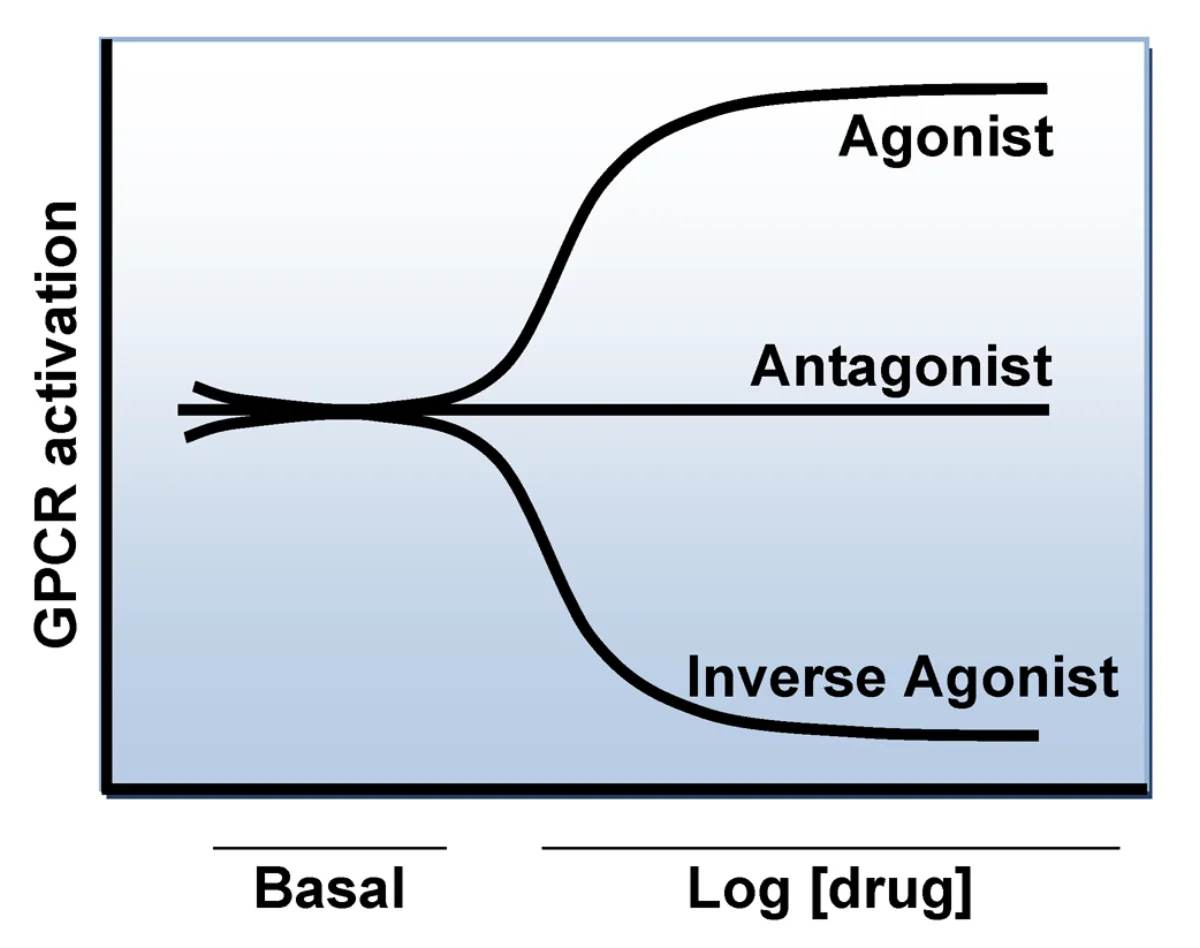
Cells need to sense what is happening outside - hormone levels, neurotransmitters, tastes, smells, light. Receptors on the cell surface grab signaling molecules and transmit the signal inside. The MCAT focuses on two major receptor families (GPCRs and RTKs) and the second messengers they produce.
G-Protein Coupled Receptors (GPCRs)
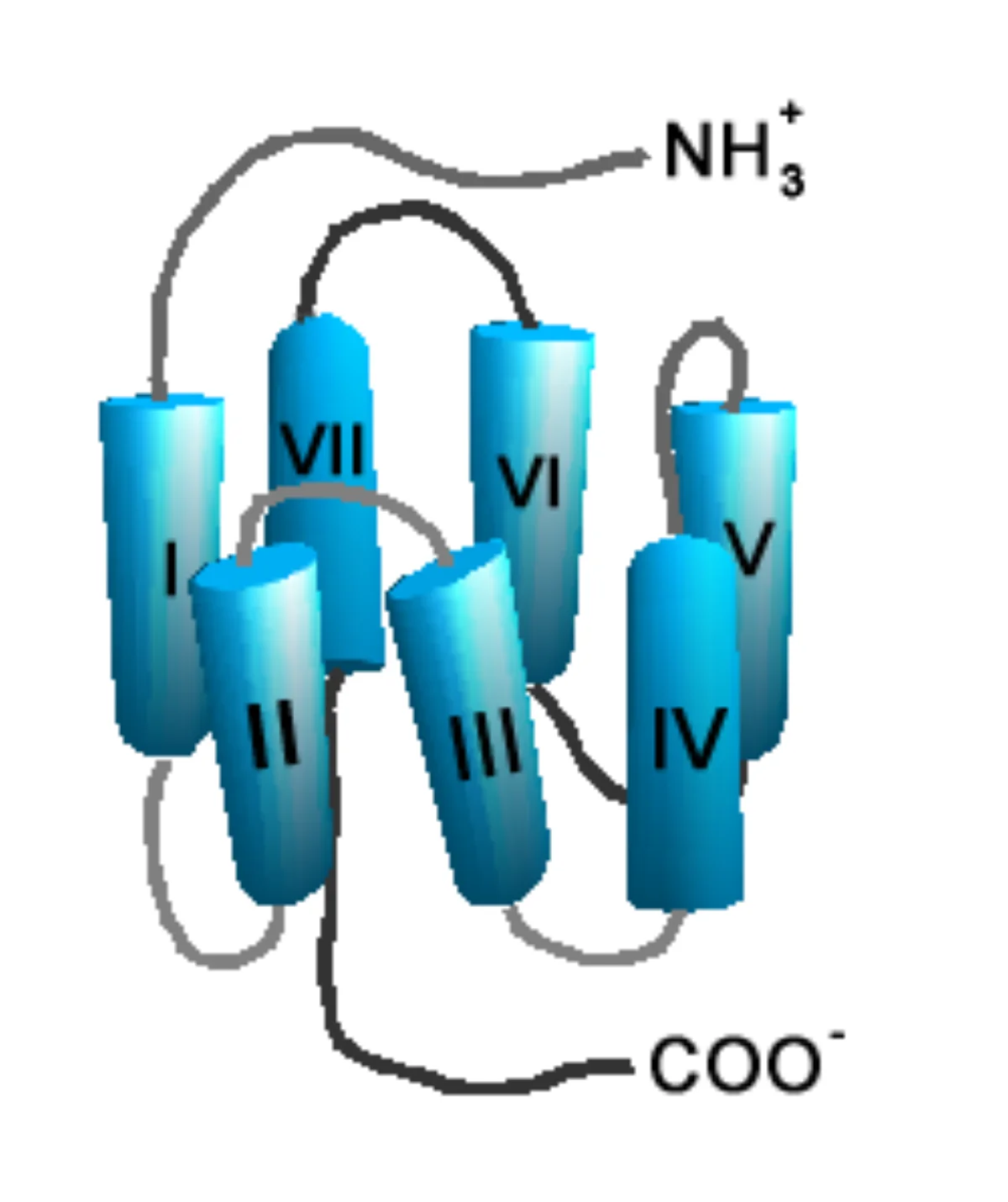
GPCRs are the largest family of cell-surface receptors. They have seven transmembrane alpha helices (so they are called 7TM receptors), an extracellular ligand-binding face, and an intracellular face that couples to a heterotrimeric G protein (alpha, beta, gamma subunits).
GPCR architecture. Seven helices thread through the membrane, creating a ligand-binding pocket on the outside and a G-protein coupling face on the inside. Credit: Wikimedia Commons, CC BY-SA
GPCR Activation Cycle
Ligand binds the extracellular face of the receptor.
The receptor changes shape, which activates the Gα subunit - it swaps GDP for GTP.
Gα separates from the βγ dimer and drifts along the inner membrane to a downstream enzyme (adenylyl cyclase, phospholipase C, etc.).
The downstream enzyme produces a second messenger (cAMP, IP3, DAG, Ca2+) that amplifies the signal inside the cell.
Gα hydrolyzes GTP to GDP (it has slow intrinsic GTPase activity), turns off, and reassociates with βγ.
GPCR signaling cycle. Ligand binding triggers G-protein activation, which activates downstream enzymes until GTP is hydrolyzed back to GDP. Credit: Wikimedia Commons, CC BY-SA
The Main Second Messengers
Second messenger
Made by
Downstream effect
cAMP
Adenylyl cyclase (from ATP)
Activates protein kinase A (PKA)
cGMP
Guanylyl cyclase (from GTP)
Activates protein kinase G
IP3
Phospholipase C (from PIP2)
Opens Ca2+ channels on the ER
DAG
Phospholipase C (from PIP2)
Activates protein kinase C (PKC)
Ca2+
Released from ER by IP3
Binds calmodulin, activates many enzymes
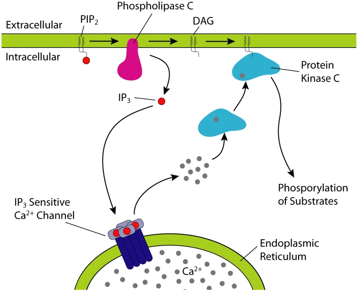
The IP3 / DAG signaling pathway. PLC cleaves membrane PIP2 into IP3 (water-soluble, triggers Ca2+ release from ER) and DAG (membrane-bound, activates PKC). This is the Gq-coupled receptor output. Credit: Wikimedia Commons, CC BY-SA
Different Gα Subtypes
The MCAT expects you to distinguish a few Gα flavors:
Gq: activates phospholipase C, generates IP3 and DAG (and raises cytoplasmic Ca2+). Example: alpha-1 adrenergic receptor.
Receptor Tyrosine Kinases (RTKs)
RTKs are single-pass transmembrane receptors whose intracellular domain is a kinase. When a ligand (usually a growth factor like insulin, EGF, or FGF) binds, two receptor molecules dimerize and cross-phosphorylate each other on tyrosine residues. The phosphorylated tyrosines recruit downstream signaling proteins, typically triggering the Ras/MAP kinase pathway.
RTKs control cell growth, differentiation, and survival. Many cancer-driving mutations affect RTKs or downstream components (EGFR mutations in lung cancer, HER2 amplification in breast cancer, BCR-ABL in chronic myeloid leukemia).
Receptor Ion Channels
Ligand-gated ion channels are receptors that double as ion channels. When neurotransmitter binds, the channel opens and ions flow. The nicotinic acetylcholine receptor (at the neuromuscular junction) and the GABA-A receptor (an inhibitory chloride channel in the brain) are classic examples.
How many transmembrane helices does a GPCR have, and what is the role of its G protein?
Click to reveal answer
Seven transmembrane alpha helices. The intracellular face couples to a heterotrimeric G protein (α, β, γ). When the receptor is activated, the Gα subunit exchanges GDP for GTP, dissociates from βγ, and activates a downstream effector enzyme (adenylyl cyclase, phospholipase C, etc.) that generates second messengers.
Which second messengers does a Gq-coupled receptor produce, and what do they do?
Click to reveal answer
Gq activates phospholipase C, which cleaves PIP2 into IP3 and DAG. IP3 binds receptors on the endoplasmic reticulum, releasing Ca2+ into the cytoplasm. DAG remains in the membrane and activates protein kinase C. The combined result: a calcium signal plus PKC phosphorylation of downstream targets.
How does a receptor tyrosine kinase activate itself when ligand binds?
Click to reveal answer
Ligand binding causes two receptor monomers to dimerize. The intracellular kinase domains then cross-phosphorylate each other on tyrosine residues. Those phosphotyrosines become docking sites for SH2-domain-containing signaling proteins, triggering downstream pathways like Ras/MAPK. Insulin, EGF, and FGF receptors all work this way.
Hemoglobin is the textbook example of a cooperative allosteric protein. It carries oxygen from the lungs to every tissue in your body. The MCAT tests hemoglobin in every exam cycle, so learn this section cold.
Structure
Hemoglobin is a tetramer of four subunits. Adult hemoglobin (HbA) has two alpha chains and two beta chains (α2β2). Each chain wraps around a heme group, and each heme holds one Fe2+ ion that binds one O2 molecule. So each hemoglobin tetramer carries four O2 molecules at full capacity.
The heme group is a planar porphyrin ring with an iron at its center. Only the Fe2+ (ferrous) form binds oxygen. Oxidation to Fe3+ (ferric) gives methemoglobin, which cannot carry O2.
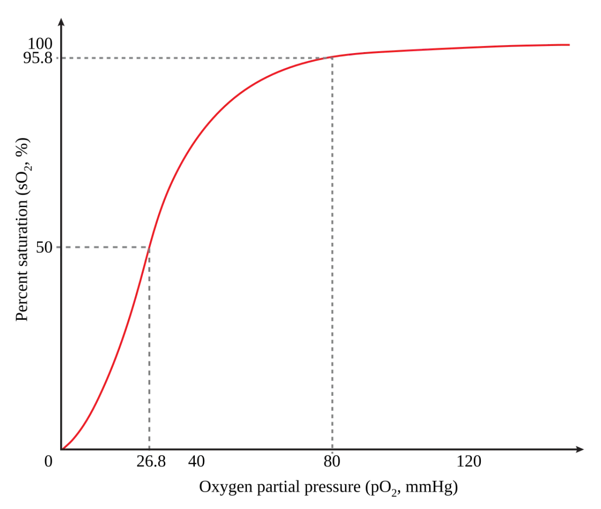
Hemoglobin's oxygen saturation curve is sigmoidal (S-shaped), a signature of cooperative binding. In the lungs (high pO2, right side) hemoglobin loads O2. In tissues (lower pO2, left side) it releases O2. Credit: Wikimedia Commons, public domain
Cooperativity and the Sigmoidal Curve
When the first O2 binds a heme, it pulls its iron slightly into the plane of the porphyrin ring, which tugs on the surrounding protein and shifts the whole tetramer from the T (tense) state to the R (relaxed) state. In the R state, the other three hemes have much higher O2 affinity. Binding becomes progressively easier with each additional O2.
The result: hemoglobin releases most of its O2 over a narrow range of pO2, exactly where cells need it. At the high pO2 of the lungs (~100 mmHg), hemoglobin is almost fully saturated. At the low pO2 of working tissues (~20-40 mmHg), it rapidly dumps most of its O2.
T vs. R State
Property
T (tense) state
R (relaxed) state
Oxygen affinity
Low
High
Stabilized by
Low pO2, high CO2, low pH, high 2,3-BPG
High pO2
Physiologic location
Working tissues
Lungs
Hemoglobin shuttles back and forth between T (unload) and R (load) many times per minute.
The Bohr Effect
Hemoglobin unloads more O2 where the tissue needs more - working muscles that are producing CO2 and lactic acid. That is the Bohr effect: increased CO2 and H+ (low pH) stabilize the T state, decreasing hemoglobin’s O2 affinity and favoring O2 release.
CO2+H2O⇌H2CO3⇌HCO3−+H+
More CO2 means more H+ in the tissue. More H+ means hemoglobin releases O2 more readily. The exhausted muscle gets the oxygen it needs, exactly when it needs it. In the lungs, CO2 is exhaled, pH rises, and hemoglobin reloads with O2.
2,3-BPG
2,3-Bisphosphoglycerate (2,3-BPG) is a small molecule made from a glycolysis intermediate. It binds the central cavity of hemoglobin in the T state and stabilizes it, lowering O2 affinity. This is how red blood cells adjust hemoglobin’s affinity in response to chronic conditions.
High altitude: low ambient O2 triggers increased 2,3-BPG. Right-shifts the curve so tissues can still extract O2 even when saturation is lower.
Chronic lung disease: similar adaptation.
Fetal hemoglobin (HbF) has two gamma chains instead of two beta chains. The gamma chains do not bind 2,3-BPG as tightly, so HbF has higher O2 affinity. The fetal curve is left-shifted relative to the adult curve, letting fetal hemoglobin steal O2 from maternal hemoglobin across the placenta.
Carbon Monoxide
CO binds hemoglobin ~250 times more tightly than O2 at the same heme iron, forming carboxyhemoglobin. Even low CO concentrations can saturate enough hemes to threaten life. Worse, when CO binds one heme, the remaining hemes shift to the R state and bind O2 too tightly - they refuse to release it at the tissues. Both loading impairment and unloading impairment occur simultaneously.
Treatment is 100 percent O2 (or hyperbaric O2), which mass-acts the CO out of the heme and restores normal binding.
Why does hemoglobin have a sigmoidal O2 binding curve instead of a hyperbolic one?
Click to reveal answer
Positive cooperativity. Hemoglobin has four subunits, each with one heme and one O2 binding site. When the first O2 binds, it pulls the whole tetramer from the T (low-affinity) to the R (high-affinity) state. The remaining hemes bind O2 much more easily. The result is a sharply S-shaped curve that functions as an oxygen on/off switch between lungs and tissues.
What is the Bohr effect and why is it physiologically useful?
Click to reveal answer
The Bohr effect is the rightward shift of hemoglobin's oxygen-binding curve caused by increased CO2 and H+ (low pH). Active tissues produce CO2 and lactic acid, lowering local pH. Hemoglobin responds by releasing more O2 exactly where it is needed most. In the lungs, CO2 is exhaled and the curve shifts left again, allowing reloading.
Why does fetal hemoglobin have higher O2 affinity than adult hemoglobin?
Click to reveal answer
Fetal hemoglobin (HbF, α2γ2) has gamma chains instead of beta chains. Gamma chains bind 2,3-BPG poorly, so HbF is not stabilized in the low-affinity T state by 2,3-BPG. The resulting higher O2 affinity allows HbF to extract O2 from maternal HbA across the placenta.
Myoglobin looks like a single subunit of hemoglobin and binds one O2 at one heme. But it is not cooperative - with only one subunit, there is no one to cooperate with. This single structural difference produces radically different oxygen-binding behavior.
The oxygen dissociation curve, and every shift
Hemoglobin
1
Scroll sideways to see the whole map.
Myoglobin: hyperbolic, one site Hemoglobin: sigmoid, four sites Hemoglobin, shifted right P₅₀: the pressure at half saturation
Why the curve is S-shapedThe first oxygen binds with difficulty because hemoglobin starts in the tense T state. Binding pulls the subunit into the relaxed R state and drags its neighbors with it, so the second, third, and fourth oxygens bind progressively more easily. That is cooperativity, and it is the whole reason the curve is sigmoid rather than hyperbolic.
Why the shape is usefulA sigmoid curve is steep in the middle, so a small drop in pO₂ between artery and working tissue causes a large drop in saturation. Myoglobin's hyperbolic curve is nearly flat at those pressures, which is exactly why myoglobin makes a good store and a poor carrier.
The Bohr effectHard-working tissue is warm, acidic, and full of CO₂. All three shift the curve right, so hemoglobin releases more oxygen precisely where it is needed. The tissue asks for oxygen by changing the chemistry around it, not by any signal.
Right means release, left means load. Anything that says a tissue is working hard shifts the curve right and dumps more oxygen. Anything that says oxygen must be held, like fetal hemoglobin pulling oxygen across the placenta, shifts it left.
Side-by-Side
| Property | Myoglobin | Hemoglobin |
|----------|-----------|------------|
| Structure | Monomer (1 subunit) | Tetramer (4 subunits) |
| Heme groups | 1 | 4 |
| Cooperativity | None | Positive |
| Curve shape | Hyperbolic | Sigmoidal |
| Role | Oxygen storage in muscle | Oxygen transport in blood |
| Affinity for O2 | Very high (holds tight) | Variable (loads in lungs, unloads in tissue) |
Why a Hyperbolic Curve for Myoglobin
Myoglobin has one binding site. Binding follows a simple equilibrium just like Michaelis-Menten with one substrate. The plot of fraction bound vs. pO2 is hyperbolic - it rises fast at low pO2 and levels off.
Why the Curves Matter
Myoglobin’s high affinity means it holds O2 even at relatively low tissue pO2. That is perfect for a reservoir - muscles can quickly tap myoglobin when demand spikes (sprinting). But it is terrible for transport, because you would need very low pO2 before it would release O2.
Hemoglobin’s cooperativity means it releases O2 over a small pO2 range, which matches the pO2 gradient between lungs (high) and tissues (lower). It is built for long-distance transport with quick offloading on arrival.
Whale and Seal Muscle
Diving mammals have dramatically more myoglobin in their muscles than humans (their muscle is dark red or even black from the myoglobin content). That enormous myoglobin pool lets them hold enough local O2 to stay submerged for 20+ minutes without breathing. It is the anatomical signature of evolution solving a gas-supply problem.
Why does myoglobin have a hyperbolic O2-binding curve while hemoglobin has a sigmoidal one?
Click to reveal answer
Myoglobin is a single-subunit protein with one heme, so there are no neighboring subunits with which to show cooperativity. The binding follows a simple equilibrium and produces a hyperbolic curve. Hemoglobin is a four-subunit tetramer with cooperativity between hemes, producing the S-shaped curve.
Which has higher O2 affinity at a given pO2, myoglobin or hemoglobin, and why does that match physiology?
Click to reveal answer
Myoglobin - its curve sits far to the left of hemoglobin’s. This is appropriate because myoglobin’s job is to hold O2 as a muscle reservoir. Hemoglobin’s lower affinity lets it release O2 to tissues; a too-tight binder (myoglobin-level affinity) would never give up its oxygen once it reached a tissue.
Why do diving mammals have especially high myoglobin content in their muscles?
Click to reveal answer
Their muscles store large amounts of O2 bound to myoglobin so they can sustain aerobic activity during long dives without breathing. The dark color of whale and seal muscle reflects this dense myoglobin packing. It is an evolutionary solution to intermittent oxygen availability.
To study a protein, you usually need it pure. A cell contains thousands of different proteins; you need to pull out just one. Chromatography is the toolkit that makes this possible. Every column works the same way: a stationary phase in the column, a mobile phase (solvent) flowing through, and proteins that partition between the two based on some physical property.
Which technique separates by what
Separation
1
Scroll sideways to see the whole map.
Chromatography: separation in a column Electrophoresis: separation in a gel The property being exploited
Two-dimensional gelsRun isoelectric focusing across a strip, then SDS-PAGE down it. The first dimension sorts by pI, the second by mass, and the result is a scatter of spots where each protein has its own coordinates. It is how a whole proteome gets resolved on one gel.
Why size exclusion looks backwardsThe beads are porous. A small protein wanders into every pore and takes a long path; a large one cannot fit and takes the short route between the beads. Large elutes first. Almost every question on this topic is checking whether you know that.
Choosing oneIf the question mentions a tag, ligand, or antibody, it is affinity. If it mentions SDS or molecular weight, it is SDS-PAGE. If it mentions pH or pI, it is ion exchange or isoelectric focusing. If it mentions keeping the protein working, it is native PAGE or gel filtration.
Every one of these exploits exactly one physical property, and the technique is named after it. Work out which property the question changed and the technique names itself.
| Technique | Separates by | Who elutes first |
|-----------|-------------|-------------------|
| Ion exchange | Charge | Opposite-charge-to-column proteins stick; same-charge pass through |
| Size exclusion | Size | LARGE molecules elute first (they cannot enter the beads) |
| Affinity | Specific binding partner | Non-binders wash through; target comes off last |
| HPLC | Many properties, high resolution | Varies by column chemistry |
Ion Exchange Chromatography
The stationary phase is a resin with charged groups. Proteins with opposite charge stick to the resin; proteins with similar charge (or neutral charge) pass through.
Cation exchange column has negatively charged resin; it binds positively charged proteins.
Anion exchange column has positively charged resin; it binds negatively charged proteins.
To elute bound proteins, you raise the salt concentration (ions compete for binding) or change the pH (which changes the protein’s net charge).
Size Exclusion Chromatography (Gel Filtration)
The stationary phase is porous beads. Small proteins enter the pores and take a long, winding path. Large proteins cannot fit into the pores and flow straight through around the beads.
Counterintuitive result: large proteins elute first, small proteins elute last. Memorize this.
Affinity Chromatography
The most selective technique. The stationary phase has a specific binding partner for your protein of interest - an antibody, a substrate analog, or a tag-binding resin.
Your target protein binds tightly to the resin. Everything else washes through.
Elute the target by adding a competitor (the natural substrate, free tag, or changed pH/salt).
His-tag purification is the most common real-world example. You genetically engineer your protein to have a six-histidine tag. The His-tag binds tightly to a nickel (Ni2+)-charged resin. Wash away everything else. Add imidazole (which competes with histidine) to elute your pure, tagged protein.
High-Performance Liquid Chromatography (HPLC)
HPLC is not a different separation principle - it is a faster, higher-resolution apparatus that pushes solvent through tightly packed small-particle columns at high pressure. Any of the techniques above (ion exchange, size exclusion, affinity, reverse-phase) can be run as HPLC. Reverse-phase HPLC is common for peptides: a hydrophobic column separates by hydrophobicity, eluting with a water/acetonitrile gradient.
Combining Techniques
Real protein purification usually stacks two or three chromatography steps. A typical sequence: crude lysate → ion exchange → size exclusion → affinity. Each step enriches the target and removes different classes of contaminants.
In size exclusion chromatography, which proteins elute first and why?
Click to reveal answer
Large proteins elute first. They cannot fit into the pores of the gel beads, so they travel in the mobile phase around the beads and reach the column exit quickly. Small proteins enter the pores, taking a long, winding path inside the beads, and elute last. Students commonly guess the opposite.
How does a His-tag and Ni-NTA resin work in affinity chromatography?
Click to reveal answer
The protein of interest is genetically engineered with a polyhistidine tag. Histidine side chains coordinate Ni2+ tightly. A nickel-charged resin binds only His-tagged proteins; everything else washes through. Elution uses imidazole, which competes with histidine for Ni2+ binding and displaces the tagged protein.
If a protein has pI = 6 and you load it onto a cation exchange column (negatively charged resin) at pH 4, will it bind?
Click to reveal answer
Yes. At pH 4 (below its pI of 6), the protein has a net positive charge. The negatively charged cation exchange resin will bind it. To elute, you would either raise the pH above 6 (making the protein net negative and expelling it from the resin) or raise the salt concentration (ions compete for the resin).
Electrophoresis separates charged molecules by driving them through a gel under an electric field. Small or highly charged molecules move fast. Large or weakly charged molecules move slowly. In protein biochemistry, electrophoresis is the standard way to check how pure a preparation is and to estimate molecular weights.
SDS-PAGE - Size Only
SDS-PAGE is the gold-standard technique for separating proteins by size. SDS (sodium dodecyl sulfate) is a detergent that does three things:
Coats every protein with many SDS molecules, giving all proteins a uniformly strongly negative charge proportional to length.
Denatures (unfolds) the proteins into extended rods.
Disrupts non-covalent interactions.
Beta-mercaptoethanol (or dithiothreitol) is added to reduce disulfide bonds, so multi-chain proteins are separated into individual subunits.
After SDS treatment, every protein has roughly the same charge-to-mass ratio. In the polyacrylamide gel, separation depends only on size: small proteins move quickly through the gel pores, large proteins move slowly.
SDS-PAGE gel. Each lane contains a sample; each band represents a different-sized protein. The left lane typically has a ladder of known molecular weight markers for sizing. Credit: Wikimedia Commons, CC BY-SA
Native PAGE - No SDS
In native PAGE, you leave the protein folded and natively charged. Separation depends on a combination of size, shape, and net charge. Native PAGE is useful when you want to preserve biological activity (a protein that is still functional after the gel). It gives less clean size information than SDS-PAGE because shape and charge vary among proteins.
Isoelectric Focusing (IEF) - Charge Only
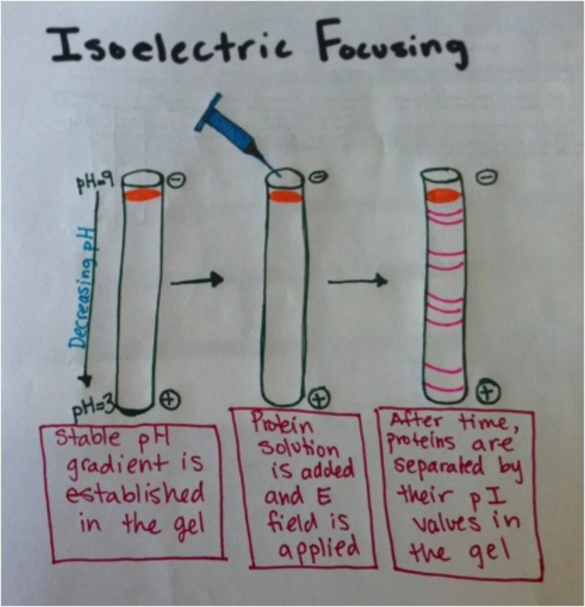
A gel is cast with a pH gradient. When an electric field is applied, a protein migrates toward the pole opposite its net charge. As it moves, it crosses pH zones that change its protonation state. When it reaches the pH that equals its isoelectric point (pI), the protein has net zero charge and stops moving. Each protein “focuses” at its own pI.
Isoelectric focusing. Each protein stops migrating when the local pH equals its pI. Sharp bands correspond to individual pI values. Credit: Wikimedia Commons, CC BY-SA
IEF is often used to detect small charge differences between protein variants - for example, distinguishing isoforms that differ by a single amino acid charge or by a post-translational modification.
2D Gel Electrophoresis - Charge Then Size
Combining IEF and SDS-PAGE gives 2D gel electrophoresis, the highest-resolution method for separating complex protein mixtures.
Run IEF in one dimension (horizontal), separating by pI.
Rotate the strip 90 degrees, apply SDS, and run SDS-PAGE in the perpendicular dimension (vertical), separating by size.
The result is a 2D “spot pattern” where each protein appears as a single dot at unique coordinates of (pI, molecular weight). Thousands of proteins can be resolved in one gel, and the pattern can be compared between healthy and diseased tissue to find differentially expressed proteins.
Detecting the Bands
After the gel is run, you need to visualize the proteins:
Coomassie blue or silver stain binds proteins nonspecifically and shows all bands.
Western blot: transfer gel contents to a membrane, then probe with antibodies to detect one specific protein. This is the standard technique for proving a specific protein is present.
What does SDS do to proteins before electrophoresis?
Click to reveal answer
SDS is a detergent that denatures proteins (unfolds them), coats them in negative charge proportional to length, and disrupts non-covalent interactions. The result is that every protein has a similar charge-to-mass ratio, so migration depends only on size. Without SDS, migration would depend on size, shape, and native charge all at once.
In isoelectric focusing, what determines where a protein stops moving?
Click to reveal answer
The protein stops migrating when the local pH of the gel equals the protein's isoelectric point (pI). At that pH, the protein has zero net charge, so the electric field no longer drives it forward. Different proteins stop at different pI values, producing sharp bands along the pH gradient.
Why would a researcher use a 2D gel instead of a standard SDS-PAGE?
Click to reveal answer
SDS-PAGE separates proteins by size only. If two proteins have very similar molecular weights but different pI values, they appear as a single band on SDS-PAGE. 2D gels add an IEF dimension, separating those same two proteins into distinct spots. This is how 2D gels resolve thousands of proteins in a complex lysate.
Once you have a pure protein, you can ask what amino acids are in it and in what order. Two main techniques, with mass spectrometry now dominating in the real world and Edman degradation classic enough to remain on the MCAT.
Edman Degradation - One Residue at a Time
Pehr Edman developed this method in 1950. A chemical reagent (phenyl isothiocyanate, PITC) reacts specifically with the N-terminal amino acid. A second step cleaves that amino acid off as a phenylthiohydantoin (PTH) derivative, which is then identified by HPLC (by comparing its retention time to known PTH standards). The remaining protein has a fresh N-terminus one residue shorter.
Repeat. Each cycle releases the next amino acid in the sequence. Cycle 1 gives residue 1, cycle 2 gives residue 2, and so on.
Limitations
Only works on the N-terminus. If the protein has a blocked N-terminus (common in eukaryotic proteins with N-terminal acetylation), Edman fails.
Becomes noisy beyond ~30-50 residues per run. To sequence a larger protein, you typically cleave it into fragments (with trypsin, CNBr, etc.), sequence each fragment, and then piece together the overlaps.
Each cycle takes ~30-60 minutes, so it is slow.
Edman degradation has largely been replaced by mass spectrometry in modern labs. It still appears on the MCAT because the principle - label-cleave-identify at the N-terminus - is an instructive chemistry example.
Mass Spectrometry
Mass spec measures mass-to-charge ratio (m/z). For proteins, the workflow is usually:
Digest the protein into peptides with a sequence-specific protease (often trypsin, which cuts after Arg or Lys).
Ionize the peptides and measure their m/z values.
Select one peptide at a time and fragment it inside the mass spec (tandem MS). The fragment pattern reveals the amino acid sequence.
A computer matches the observed masses to a database of all known protein sequences and identifies the protein.
MALDI-TOF and Electrospray
Two common ionization methods you should recognize:
MALDI (matrix-assisted laser desorption/ionization): peptides are embedded in a matrix that absorbs laser energy, releasing ions.
ESI (electrospray ionization): peptides are sprayed from a fine capillary under high voltage, generating droplets that evaporate to leave ions.
Both are then pushed through a mass analyzer. TOF (time-of-flight) analyzers separate ions by how long they take to cover a fixed distance - lighter ions move faster.
Why Mass Spec Wins
Mass spec can identify a protein from a tiny sample (femtomoles), can identify all proteins in a complex mixture (proteomics), and can detect post-translational modifications by the mass shift they add (phosphorylation = +80 Da, methylation = +14 Da, ubiquitination = +8564 Da). None of this is possible with Edman.
Total Amino Acid Composition
Sometimes you just want to know how many of each amino acid are in a protein, not the order. Acid hydrolysis (6 M HCl at 110°C for 24 hours) breaks every peptide bond. The resulting mixture of free amino acids can be quantified to give percent composition. Tryptophan is destroyed by acid hydrolysis and must be measured separately. This method gives composition but not sequence.
Quantifying Total Protein: The Bradford Assay
Before any sequencing or characterization experiment, you need to know how much protein you actually have. The Bradford assay is the MCAT-canonical method.
Alternative methods the MCAT occasionally mentions: BCA assay (cheaper in bulk, uses Cu+ reduction), Lowry assay (older, similar principle to BCA), and UV absorbance at 280 nm (which reads tryptophan and tyrosine absorbance, no dye needed).
Which end of a peptide does Edman degradation sequence, and what reagent is used?
Click to reveal answer
The N-terminus. Phenyl isothiocyanate (PITC, Edman's reagent) reacts with the N-terminal amino acid's free amino group. A subsequent cleavage releases that residue as a phenylthiohydantoin (PTH) derivative, which is identified by HPLC. The process exposes a new N-terminus for the next cycle.
Why is trypsin commonly used to digest proteins before mass spectrometry?
Click to reveal answer
Trypsin is a serine protease that cleaves peptide bonds C-terminal to lysine or arginine (unless the next residue is proline). This produces peptides of a convenient size (~8-20 residues) with predictable C-terminal residues, which simplifies mass spec analysis and improves identification from sequence databases.
How can mass spectrometry detect post-translational modifications like phosphorylation?
Click to reveal answer
Each modification adds a specific mass. Phosphorylation adds 80 Da (the mass of HPO3). If the observed peptide mass is 80 Da larger than the predicted mass for the unmodified sequence, the peptide is likely phosphorylated. Tandem MS can further localize which residue is phosphorylated by examining the fragment ions.
Knowing a protein’s amino acid sequence is a start, but function comes from 3D shape. Three techniques dominate structural biology. The MCAT focuses mostly on X-ray crystallography; NMR and cryo-EM often appear as passage context.
X-Ray Crystallography
The gold-standard method for atomic-resolution protein structures since the 1960s. The workflow:
Grow a crystal of the purified protein. This is often the hardest step - proteins do not always crystallize cleanly.
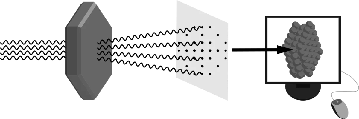
Diffract X-rays through the crystal. The regular lattice of protein molecules scatters X-rays into a characteristic spot pattern.
Compute an electron density map from the diffraction pattern (mathematically, an inverse Fourier transform).
Fit the amino acid sequence into the electron density to build the 3D model.
X-ray crystallography. A beam of X-rays diffracts off the regular lattice of protein molecules in a crystal, producing a pattern of spots whose intensities encode the 3D electron density of the protein. Credit: Wikimedia Commons, CC BY-SA
Strengths and Limits
Strength: atomic resolution (~1-2 Å) for well-ordered crystals. Shows every backbone and side-chain atom.
Limit: requires a crystal. Membrane proteins and flexible proteins often refuse to crystallize. The crystal also locks the protein in one conformation, so you may miss dynamic motion.
Nuclear Magnetic Resonance (NMR) Spectroscopy
NMR uses the magnetic properties of atomic nuclei (primarily 1H, 13C, 15N) in strong magnetic fields. The chemical environment of each nucleus shifts its resonance frequency slightly, and interactions between neighboring nuclei produce characteristic peaks. Computational methods convert NMR spectra into 3D structure constraints.
NMR works on proteins in solution, not crystals. This is a major advantage - you see the protein in a more natural environment and can also measure dynamics. The major limit is size: NMR becomes very difficult above ~30 kDa because peaks overlap and relaxation effects distort spectra.
Cryo-Electron Microscopy (Cryo-EM)
Cryo-EM captures thousands of pictures of individual protein molecules frozen in a thin layer of vitreous ice. Each picture is a different angle on the same molecule. Computer software aligns and averages the pictures to reconstruct a 3D density map, from which a model is built.
Cryo-EM does not need a crystal. It excels at large complexes (ribosomes, whole viruses, membrane proteins in their natural environment). Resolution has improved dramatically, and modern cryo-EM routinely achieves 2-3 Å, competitive with crystallography. The 2017 Nobel Prize in Chemistry recognized this “resolution revolution.”
Quick Comparison
Method
Sample state
Size range
Resolution
Sees dynamics?
X-ray crystallography
Crystalline solid
~5 kDa to megadalton complexes
~1-3 Å
No (static snapshot)
NMR
Solution
Usually < 30 kDa
~2-4 Å
Yes (time-resolved)
Cryo-EM
Vitreous ice (flash-frozen)
Large complexes (>150 kDa typical)
~2-4 Å modern
Partial (can capture different states)
What is the most difficult step in X-ray crystallography, and why?
Click to reveal answer
Growing a high-quality crystal of the protein. Many proteins - especially membrane proteins and flexible ones - do not form ordered crystals easily. Without a well-diffracting crystal, the technique cannot yield a structure. Entire subfields of structural biology focus just on crystallization strategies.
Why is NMR limited to relatively small proteins?
Click to reveal answer
As protein size grows, the number of NMR peaks increases and they overlap badly. Also, larger proteins tumble more slowly in solution, which broadens signals and reduces spectral quality. Above about 30 kDa, NMR becomes very difficult without specialized labeling schemes, although modern techniques have pushed the limit higher.
What major advantage does cryo-EM have over X-ray crystallography?
Click to reveal answer
Cryo-EM does not require a crystal. Proteins are imaged in a thin layer of vitreous ice, close to their native solution state. This makes it especially useful for large complexes, membrane proteins, and flexible systems that resist crystallization. Modern resolution rivals crystallography at 2-3 Å for well-behaved samples.