
Motor Proteins
Motor proteins convert chemical energy (ATP hydrolysis) into mechanical work. They literally walk along cellular tracks, carrying cargo or pulling on filaments to cause contraction. Three MCAT-relevant motor proteins, all walking different tracks.
| Motor | Walks on | Direction | Typical cargo/job |
|---|---|---|---|
| Myosin | Actin filaments | Plus end (most myosins) | Muscle contraction; vesicle transport |
| Kinesin | Microtubules | Plus end (toward periphery, anterograde) | Axonal transport outward from cell body |
| Dynein | Microtubules | Minus end (toward center, retrograde) | Axonal transport inward; cilia and flagella beating |
Myosin and Muscle Contraction
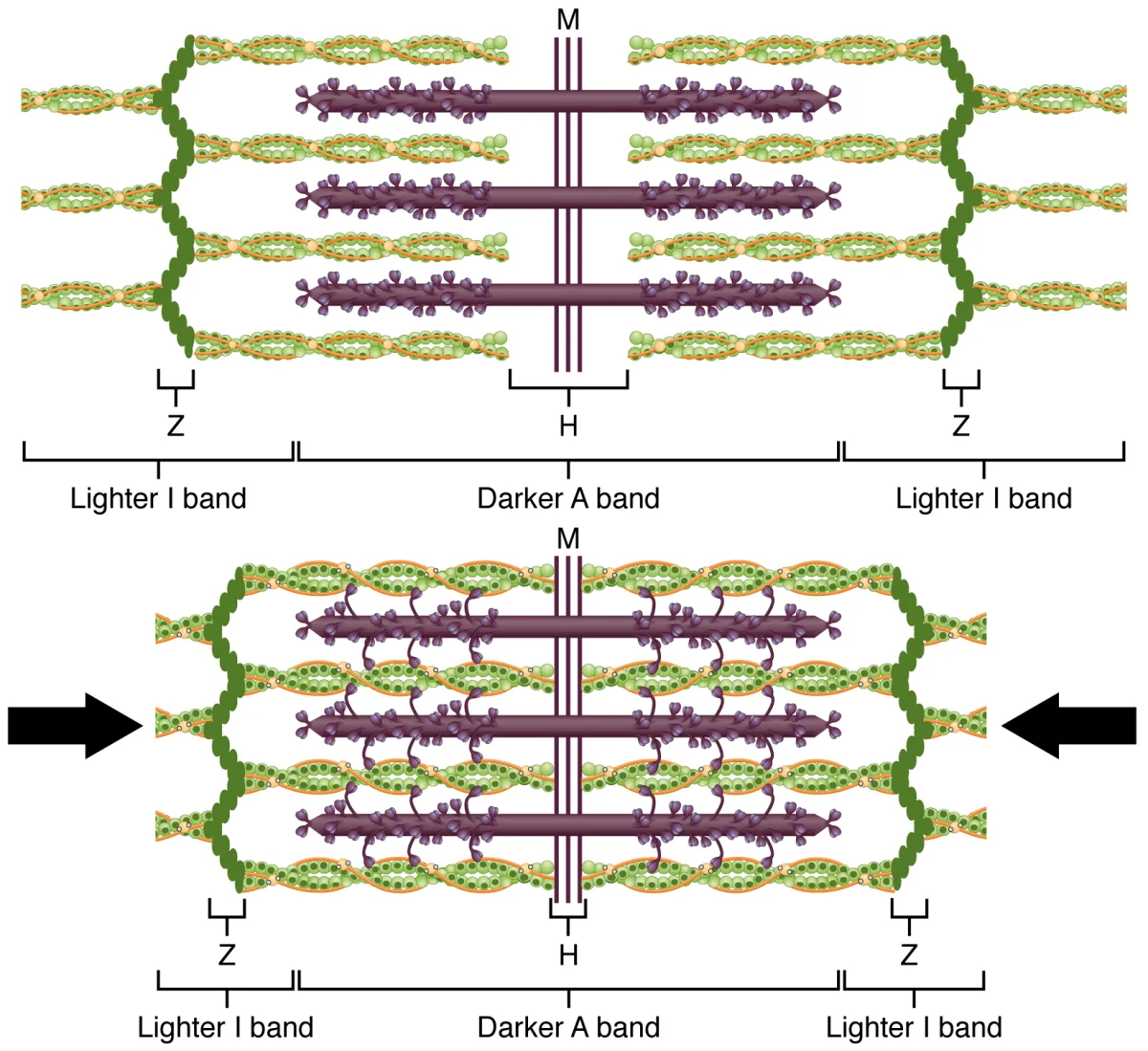
In skeletal muscle, myosin is organized into thick filaments. Actin forms thin filaments. Together they slide past one another to produce contraction. The unit of contraction is the sarcomere, bounded by two Z-discs.
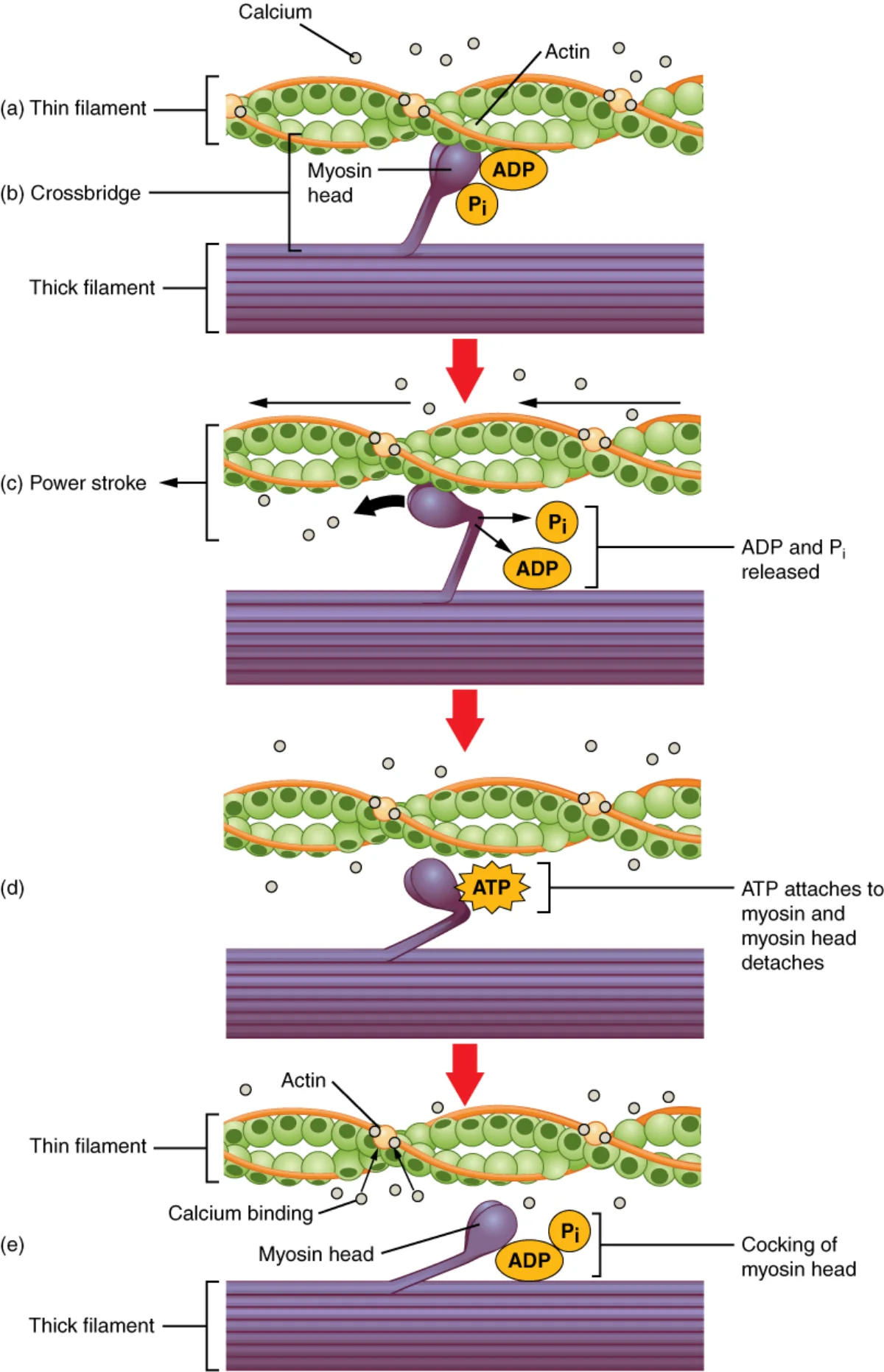
The cross-bridge cycle is the four-step ATP-driven process that makes one myosin head “walk” along actin:
- ATP binds the myosin head, releasing it from actin.
- ATP hydrolysis cocks the myosin head into a high-energy position (like a spring being loaded).
- Myosin binds actin and releases inorganic phosphate.
- Power stroke: myosin swings back to its original shape, pulling actin toward the center of the sarcomere. ADP leaves.
Rigor Mortis
After death, cells run out of ATP. Without ATP, myosin cannot release actin (step 1 of the cycle). Muscles seize up in a contracted state called rigor mortis. A few hours later, proteases begin degrading the myofibrils and the muscle relaxes again.
Kinesin
Kinesin is a two-headed motor that walks toward the plus end of microtubules. It has a heavy chain with motor domains, a stalk, and light chains that attach to cargo (vesicles, mitochondria, RNA granules). Each step hydrolyzes one ATP and covers about 8 nm.
The classic example is axonal transport: kinesin carries neurotransmitter vesicles and mitochondria down the axon, from the cell body to the synapse. Without kinesin, a long neuron could not supply its distant axon terminals with fresh proteins.
Dynein
Dynein is the opposite-direction partner. It walks toward the minus end of microtubules (back toward the cell body). It transports endocytosed material, recycles old organelles, and - in a completely different role - powers the beating of cilia and flagella.
In cilia and flagella, dynein molecules between adjacent microtubule pairs pull past one another, producing the wave-like bending that propels cells (sperm flagella, airway cilia). The axoneme structure - nine outer microtubule doublets plus two central singlets, “9+2” - is bound together by dynein arms and nexin links.