
Hemoglobin
Hemoglobin is the textbook example of a cooperative allosteric protein. It carries oxygen from the lungs to every tissue in your body. The MCAT tests hemoglobin in every exam cycle, so learn this section cold.
Structure
Hemoglobin is a tetramer of four subunits. Adult hemoglobin (HbA) has two alpha chains and two beta chains (α2β2). Each chain wraps around a heme group, and each heme holds one Fe2+ ion that binds one O2 molecule. So each hemoglobin tetramer carries four O2 molecules at full capacity.
The heme group is a planar porphyrin ring with an iron at its center. Only the Fe2+ (ferrous) form binds oxygen. Oxidation to Fe3+ (ferric) gives methemoglobin, which cannot carry O2.
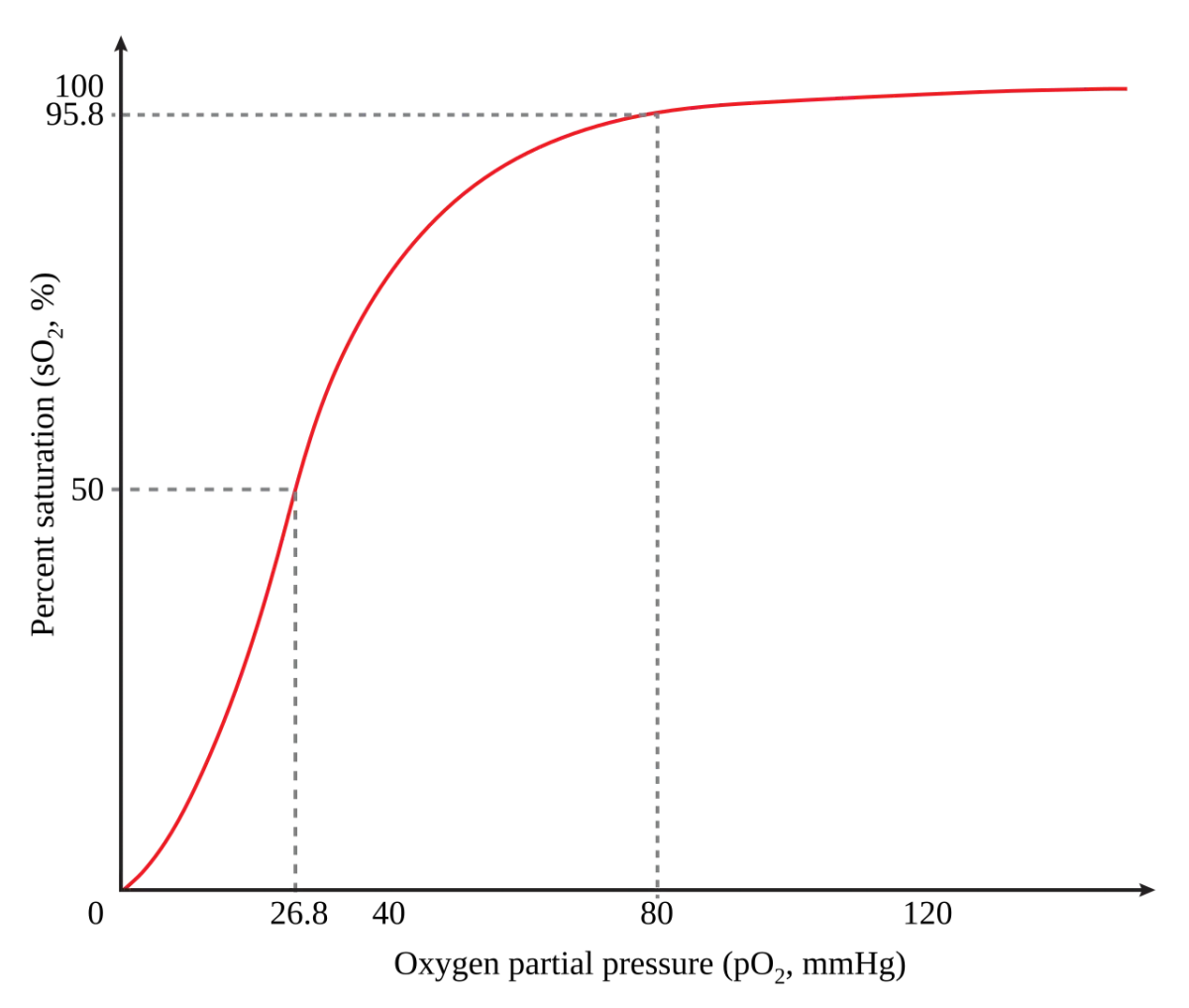
Cooperativity and the Sigmoidal Curve
When the first O2 binds a heme, it pulls its iron slightly into the plane of the porphyrin ring, which tugs on the surrounding protein and shifts the whole tetramer from the T (tense) state to the R (relaxed) state. In the R state, the other three hemes have much higher O2 affinity. Binding becomes progressively easier with each additional O2.
The result: hemoglobin releases most of its O2 over a narrow range of pO2, exactly where cells need it. At the high pO2 of the lungs (~100 mmHg), hemoglobin is almost fully saturated. At the low pO2 of working tissues (~20-40 mmHg), it rapidly dumps most of its O2.
T vs. R State
| Property | T (tense) state | R (relaxed) state |
|---|---|---|
| Oxygen affinity | Low | High |
| Stabilized by | Low pO2, high CO2, low pH, high 2,3-BPG | High pO2 |
| Physiologic location | Working tissues | Lungs |
Hemoglobin shuttles back and forth between T (unload) and R (load) many times per minute.
The Bohr Effect
Hemoglobin unloads more O2 where the tissue needs more - working muscles that are producing CO2 and lactic acid. That is the Bohr effect: increased CO2 and H+ (low pH) stabilize the T state, decreasing hemoglobin’s O2 affinity and favoring O2 release.
More CO2 means more H+ in the tissue. More H+ means hemoglobin releases O2 more readily. The exhausted muscle gets the oxygen it needs, exactly when it needs it. In the lungs, CO2 is exhaled, pH rises, and hemoglobin reloads with O2.
2,3-BPG
2,3-Bisphosphoglycerate (2,3-BPG) is a small molecule made from a glycolysis intermediate. It binds the central cavity of hemoglobin in the T state and stabilizes it, lowering O2 affinity. This is how red blood cells adjust hemoglobin’s affinity in response to chronic conditions.
- High altitude: low ambient O2 triggers increased 2,3-BPG. Right-shifts the curve so tissues can still extract O2 even when saturation is lower.
- Chronic lung disease: similar adaptation.
- Fetal hemoglobin (HbF) has two gamma chains instead of two beta chains. The gamma chains do not bind 2,3-BPG as tightly, so HbF has higher O2 affinity. The fetal curve is left-shifted relative to the adult curve, letting fetal hemoglobin steal O2 from maternal hemoglobin across the placenta.
Carbon Monoxide
CO binds hemoglobin ~250 times more tightly than O2 at the same heme iron, forming carboxyhemoglobin. Even low CO concentrations can saturate enough hemes to threaten life. Worse, when CO binds one heme, the remaining hemes shift to the R state and bind O2 too tightly - they refuse to release it at the tissues. Both loading impairment and unloading impairment occur simultaneously.
Treatment is 100 percent O2 (or hyperbaric O2), which mass-acts the CO out of the heme and restores normal binding.