Cells need to sense what is happening outside - hormone levels, neurotransmitters, tastes, smells, light. Receptors on the cell surface grab signaling molecules and transmit the signal inside. The MCAT focuses on two major receptor families (GPCRs and RTKs) and the second messengers they produce.
G-Protein Coupled Receptors (GPCRs)
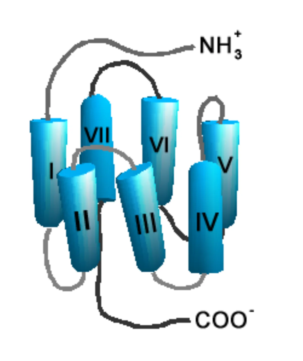
GPCRs are the largest family of cell-surface receptors. They have seven transmembrane alpha helices (so they are called 7TM receptors), an extracellular ligand-binding face, and an intracellular face that couples to a heterotrimeric G protein (alpha, beta, gamma subunits).
GPCR architecture. Seven helices thread through the membrane, creating a ligand-binding pocket on the outside and a G-protein coupling face on the inside. Credit: Wikimedia Commons, CC BY-SA
GPCR Activation Cycle
Ligand binds the extracellular face of the receptor.
The receptor changes shape, which activates the Gα subunit - it swaps GDP for GTP.
Gα separates from the βγ dimer and drifts along the inner membrane to a downstream enzyme (adenylyl cyclase, phospholipase C, etc.).
The downstream enzyme produces a second messenger (cAMP, IP3, DAG, Ca2+) that amplifies the signal inside the cell.
Gα hydrolyzes GTP to GDP (it has slow intrinsic GTPase activity), turns off, and reassociates with βγ.
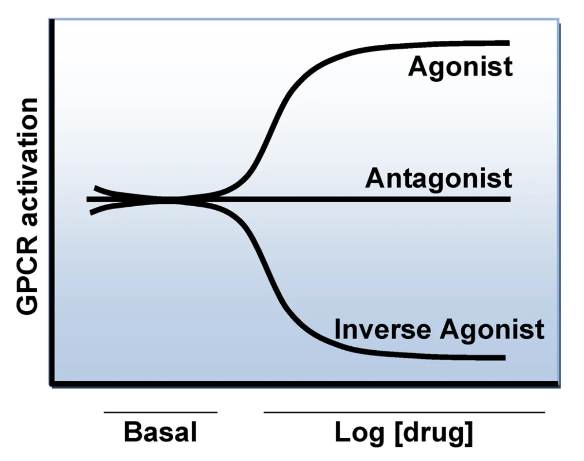
GPCR signaling cycle. Ligand binding triggers G-protein activation, which activates downstream enzymes until GTP is hydrolyzed back to GDP. Credit: Wikimedia Commons, CC BY-SA
The Main Second Messengers
Second messenger
Made by
Downstream effect
cAMP
Adenylyl cyclase (from ATP)
Activates protein kinase A (PKA)
cGMP
Guanylyl cyclase (from GTP)
Activates protein kinase G
IP3
Phospholipase C (from PIP2)
Opens Ca2+ channels on the ER
DAG
Phospholipase C (from PIP2)
Activates protein kinase C (PKC)
Ca2+
Released from ER by IP3
Binds calmodulin, activates many enzymes
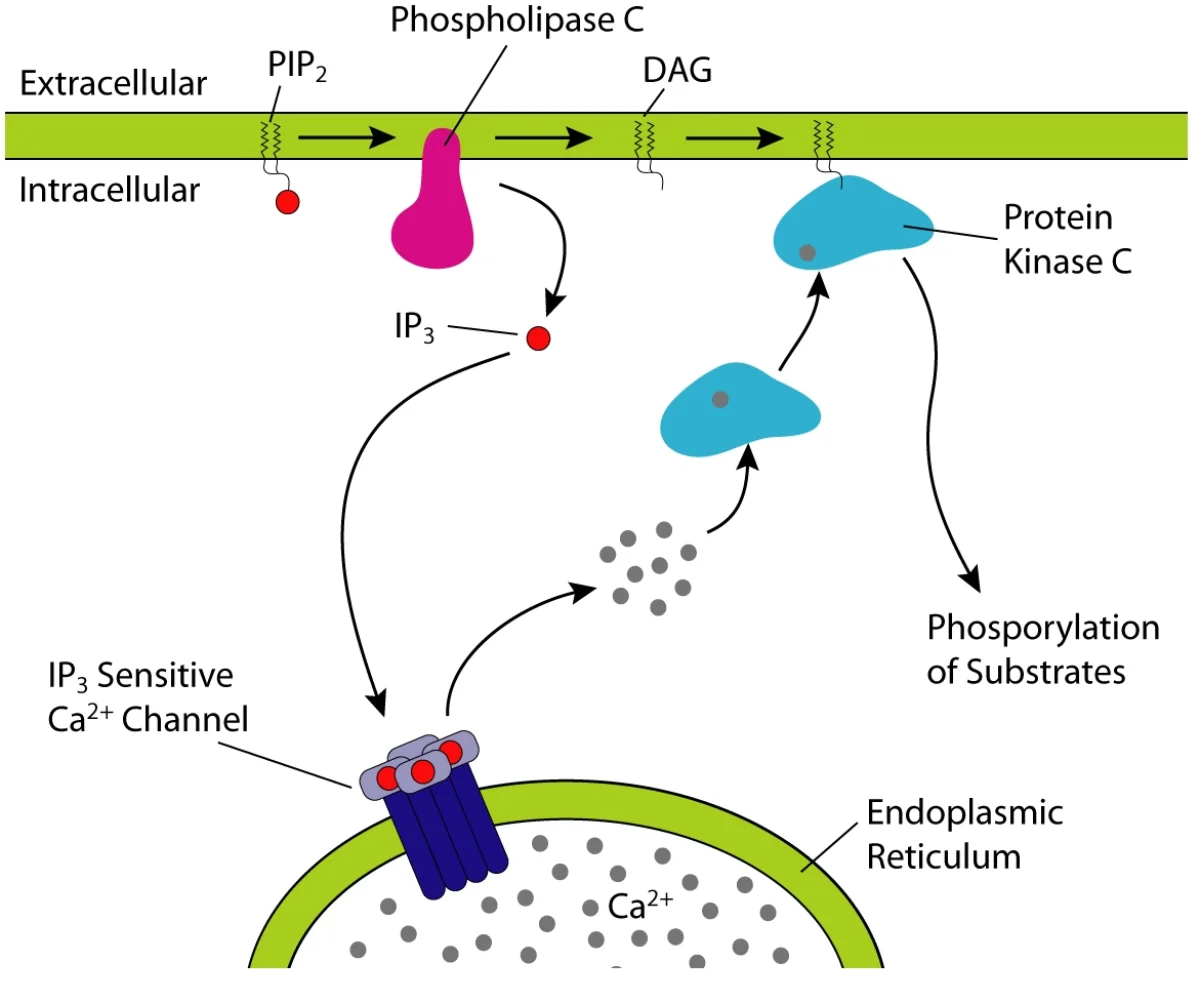
The IP3 / DAG signaling pathway. PLC cleaves membrane PIP2 into IP3 (water-soluble, triggers Ca2+ release from ER) and DAG (membrane-bound, activates PKC). This is the Gq-coupled receptor output. Credit: Wikimedia Commons, CC BY-SA
Different Gα Subtypes
The MCAT expects you to distinguish a few Gα flavors:
Gq: activates phospholipase C, generates IP3 and DAG (and raises cytoplasmic Ca2+). Example: alpha-1 adrenergic receptor.
Receptor Tyrosine Kinases (RTKs)
RTKs are single-pass transmembrane receptors whose intracellular domain is a kinase. When a ligand (usually a growth factor like insulin, EGF, or FGF) binds, two receptor molecules dimerize and cross-phosphorylate each other on tyrosine residues. The phosphorylated tyrosines recruit downstream signaling proteins, typically triggering the Ras/MAP kinase pathway.
RTKs control cell growth, differentiation, and survival. Many cancer-driving mutations affect RTKs or downstream components (EGFR mutations in lung cancer, HER2 amplification in breast cancer, BCR-ABL in chronic myeloid leukemia).
Receptor Ion Channels
Ligand-gated ion channels are receptors that double as ion channels. When neurotransmitter binds, the channel opens and ions flow. The nicotinic acetylcholine receptor (at the neuromuscular junction) and the GABA-A receptor (an inhibitory chloride channel in the brain) are classic examples.
How many transmembrane helices does a GPCR have, and what is the role of its G protein?
Click to reveal answer
Seven transmembrane alpha helices. The intracellular face couples to a heterotrimeric G protein (α, β, γ). When the receptor is activated, the Gα subunit exchanges GDP for GTP, dissociates from βγ, and activates a downstream effector enzyme (adenylyl cyclase, phospholipase C, etc.) that generates second messengers.
Which second messengers does a Gq-coupled receptor produce, and what do they do?
Click to reveal answer
Gq activates phospholipase C, which cleaves PIP2 into IP3 and DAG. IP3 binds receptors on the endoplasmic reticulum, releasing Ca2+ into the cytoplasm. DAG remains in the membrane and activates protein kinase C. The combined result: a calcium signal plus PKC phosphorylation of downstream targets.
How does a receptor tyrosine kinase activate itself when ligand binds?
Click to reveal answer
Ligand binding causes two receptor monomers to dimerize. The intracellular kinase domains then cross-phosphorylate each other on tyrosine residues. Those phosphotyrosines become docking sites for SH2-domain-containing signaling proteins, triggering downstream pathways like Ras/MAPK. Insulin, EGF, and FGF receptors all work this way.