Imagine a sports stadium with a single marble sitting on the 50-yard line. The field is otherwise empty. So are the stands, concourses, parking lot, surrounding blocks. That is the scale of an atom: almost entirely empty space, with virtually all the mass packed into an incomprehensibly tiny center.
The marble is the nucleus. The empty space around it is where the electrons live, smeared out in cloud-like orbitals. Even though the nucleus is ~100,000× smaller than the atom, it contains 99.97% of the atom’s mass. Get this scale right and the rest of atomic and nuclear physics is much easier to picture.
The Three Subatomic Particles
Every atom is built from just three particles:
Particle
Symbol
Charge
Mass (amu)
Location
Proton
p
+1
~1
Nucleus
Neutron
n
0
~1
Nucleus
Electron
e⁻
-1
~18361
Electron cloud
Protons and neutrons (collectively called nucleons) live in the nucleus. Electrons orbit far outside in the electron cloud. The electron’s mass is so small compared to the nucleons that it is essentially negligible for nuclear calculations.
Atomic Number and Mass Number
Two numbers define every nucleus:
Atomic number (Z) - the number of protons. This determines the element. Every carbon atom has Z = 6. Every oxygen atom has Z = 8. Change Z and you change the element entirely.
Mass number (A) - the total number of nucleons (protons + neutrons). A = Z + N, where N is the number of neutrons.
Nuclear Notation
The standard way to write a specific nucleus (nuclide) places the mass number as a superscript and the atomic number as a subscript, both to the left of the element symbol:
ZAX — for example, 612C is carbon-12 (6 protons, 6 neutrons) and 614C is carbon-14 (6 protons, 8 neutrons).
Since the element symbol already tells you Z (carbon is always 6), you will often see the shorthand carbon-12 or C-12. But on the MCAT, full nuclear notation appears frequently in decay equations, so practice reading it quickly.
Isotopes
Isotopes are atoms of the same element (same Z) with different numbers of neutrons (different N, therefore different A). Carbon-12 and carbon-14 are both carbon - they have identical chemical properties because they have the same number of electrons. But carbon-14 has two extra neutrons, making its nucleus unstable and radioactive.
Most elements exist as a mixture of stable isotopes in nature. The atomic mass listed on the periodic table (for example, 12.011 for carbon) is the weighted average of all naturally occurring isotopes based on their abundance.
Atomic Mass Units
The atomic mass unit (amu or u) is defined so that one carbon-12 atom has a mass of exactly 12 u. By this definition:
1 proton ~ 1.0073 u
1 neutron ~ 1.0087 u
1 electron ~ 0.00055 u
One amu equals 1.66 x 10⁻²⁷ kg. You will not need to convert between amu and kg often, but you should know the conversion exists - especially for mass defect and binding energy calculations later in this chapter.
Size Scales
Getting a feel for the relative sizes helps with intuition:
Atom diameter: ~10−10 m (1 angstrom)
Nucleus diameter: ~10−15 m (1 femtometer)
Ratio: the nucleus is about 100,000 times smaller than the atom itself
This means the atom is overwhelmingly empty space. If the nucleus were the size of a marble (1 cm), the electron cloud would extend roughly 100 meters in every direction - about the length of a football field.
An atom has a mass number of 37 and 17 protons. How many neutrons does it have? What element is it?
Click to reveal answer
20 neutrons. The element is chlorine (Cl). N = A - Z = 37 - 17 = 20 neutrons. Z = 17 identifies the element as chlorine. This isotope is chlorine-37.
Two atoms both have 8 protons. One has 8 neutrons and the other has 10. Are they the same element? What is the relationship between them?
Click to reveal answer
Yes, they are the same element - oxygen (Z = 8). They are isotopes of each other. Isotopes share the same atomic number (same element) but differ in neutron count. These are oxygen-16 and oxygen-18.
Picture a ladder. You can stand on the first rung, the second rung, or the third — but you can’t hover between rungs. Electrons in an atom work the same way: they can only exist at specific, fixed energy levels. They can jump between levels, but they can’t sit in between.
This is the core idea behind the Bohr model, and it explains one of the most surprising facts in chemistry: atoms emit and absorb light in specific colors (discrete spectral lines), not a continuous rainbow. The colors of fireworks, the orange of a sodium street lamp, the unique fingerprint of every element on a spectrometer — all come from this one quantization rule.
Quantized Energy Levels
In 1913, Niels Bohr proposed that electrons in hydrogen orbit the nucleus only at certain allowed radii, each corresponding to a specific energy. These orbits are labeled by the principal quantum number n, where n = 1, 2, 3, and so on.
Key values to know:
n
Energy (eV)
1
-13.6
2
-3.4
3
-1.51
4
-0.85
infinity
0 (free)
The ground state (n = 1) is the lowest energy level and the most stable. Any level above n = 1 is an excited state. The ionization energy of hydrogen - the energy needed to completely remove the electron from the ground state - is 13.6 eV.
Electron Transitions
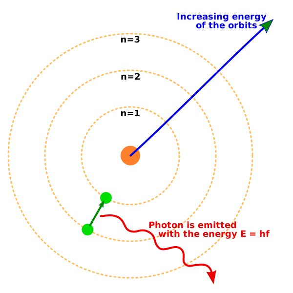
Electron transitions in the hydrogen atom. Drops to n = 1 produce ultraviolet photons (Lyman series), drops to n = 2 produce visible light (Balmer series), and drops to n = 3 produce infrared photons (Paschen series). Larger energy gaps correspond to shorter-wavelength, higher-energy photons. Credit: Wikimedia Commons, CC BY-SA 3.0
An electron can jump between energy levels by absorbing or emitting a photon whose energy exactly matches the difference between the two levels.
Absorption: an electron absorbs a photon and jumps to a higher level (nlow to nhigh).
Emission: an electron drops to a lower level and emits a photon (nhigh to nlow).
Example Calculation
What wavelength photon is emitted when a hydrogen electron drops from n = 3 to n = 2?
Energy difference: |E3 - E2| = |-1.51 - (-3.4)| = 1.89 eV
Convert to joules: 1.89 eV x 1.6 x 10−19 J/eV = 3.02 x 10⁻¹⁹ J
Wavelength: λ = hc / E = (6.63 x 10⁻³⁴)(3 x 10⁸) / (3.02 x 10⁻¹⁹) = 6.59 ×10⁻⁷ m = 659 nm
This is red light - part of the Balmer series (visible range), which we will explore in the next section.
The Heisenberg Uncertainty Principle
The Bohr model imagines electrons in neat circular orbits, but quantum mechanics tells us we cannot actually pin down an electron’s exact position and momentum at the same time.
This is not about imperfect instruments. It is a fundamental limit built into nature. An electron does not have a precise position and momentum simultaneously - it exists as a probability cloud around the nucleus.
For the MCAT, you need to understand the uncertainty principle conceptually. You will not be asked to perform calculations with it, but you should recognize that it explains why the Bohr model’s neat circular orbits are an oversimplification. Real electrons are better described by probability distributions (orbitals), which you studied in General Chemistry.
What is the energy of a hydrogen electron in the n = 3 state? How much energy is needed to ionize it from this state?
Click to reveal answer
E3 = -913.6 = -1.51 eV. Ionization requires 1.51 eV. Ionization means bringing the electron from its current level to E = 0 (free). Since it starts at -1.51 eV, you need to add 1.51 eV to reach zero.
An electron in hydrogen absorbs a photon and jumps from n = 1 to n = 4. What is the energy of the absorbed photon?
Click to reveal answer
E = |E4 - E1| = |-0.85 - (-13.6)| = 12.75 eV. The photon must carry exactly the energy difference between the two levels. This is in the ultraviolet range (high energy, short wavelength).
Watch a fireworks display: reds, greens, blues, and golds exploding across the sky. Each color comes from a different element — strontium for red, barium for green, copper for blue. The atoms get heated to extreme temperatures, their electrons jump to excited states, and as those electrons fall back down to lower levels, they release photons of very specific wavelengths.
Every element has its own unique fingerprint of colors — its emission spectrum. This is how astronomers identify what stars are made of without ever visiting them: they read the spectrum of light coming from the star and match the lines to known elements. The same principle drives flame tests in chemistry labs, neon signs, and spectroscopic medical diagnostics.
Three Types of Spectra
Continuous Spectrum
A hot, dense object (like a light bulb filament or the sun’s surface) emits light at all wavelengths, producing a smooth rainbow with no gaps. This is called a continuous spectrum or blackbody spectrum.
Emission Spectrum (Bright Lines)
A hot, low-density gas emits light at only specific wavelengths, producing bright colored lines on a dark background. Each line corresponds to a specific electron transition within the atom. Every element has a unique set of emission lines - an atomic fingerprint.
Absorption Spectrum (Dark Lines)
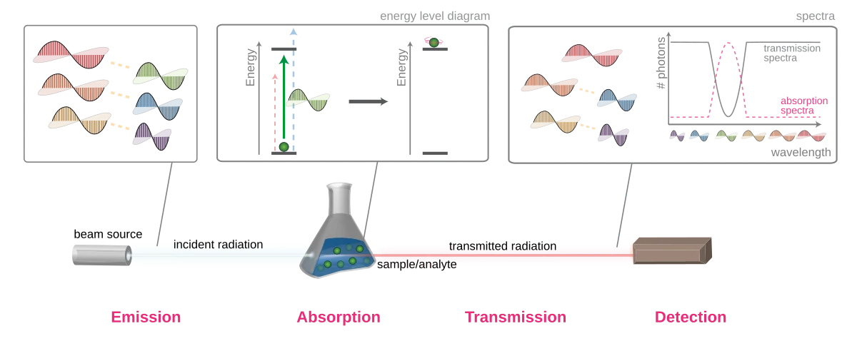
When continuous light passes through a cool gas, the gas absorbs photons at the exact wavelengths it would normally emit. The result is a continuous rainbow with dark lines at specific positions. The dark lines in an absorption spectrum appear at the same wavelengths as the bright lines in the emission spectrum of the same element.
The three types of spectra. A continuous spectrum contains all wavelengths. An emission spectrum shows bright lines at specific wavelengths where electrons drop to lower energy levels. An absorption spectrum shows dark lines at the same wavelengths where photons are absorbed by a cool gas. Credit: Wikimedia Commons, CC BY-SA 3.0
Hydrogen Spectral Series
Hydrogen’s emission lines are grouped into series based on the lower energy level the electron falls to:
Series
Final Level (n)
Region
Notes
Lyman
n = 1
Ultraviolet
Highest energy transitions
Balmer
n = 2
Visible
The one you can see with your eyes
Paschen
n = 3
Infrared
Lower energy transitions
Why Lyman Is UV and Balmer Is Visible
Transitions down to n = 1 involve the largest energy gaps (recall the energy levels crowd together at higher n). Large energy gaps mean high-frequency, short-wavelength photons - ultraviolet. Transitions to n = 2 involve moderate energy gaps, landing in the visible range. Transitions to n = 3 have the smallest gaps and produce low-energy infrared photons.
Within each series, the transition from the nearest level (n + 1 to n) produces the lowest-energy photon, and the transition from n = infinity to n produces the highest-energy photon (the series limit).
Example: Balmer Series
The Balmer series includes all transitions ending at n = 2:
n = 3 to n = 2: red light (656 nm)
n = 4 to n = 2: blue-green (486 nm)
n = 5 to n = 2: blue-violet (434 nm)
n = 6 to n = 2: violet (410 nm)
Notice the lines get closer together at shorter wavelengths, converging toward the series limit at 365 nm.
Connecting Spectra to Energy Levels
Every spectral line corresponds to one specific transition between two energy levels. If you know the energy levels of an atom, you can predict every possible spectral line. Conversely, measuring the spectral lines lets you work backward to determine the energy levels.
A hydrogen electron transitions from n = 4 to n = 1. Is a photon emitted or absorbed? Which series does this belong to? What region of the electromagnetic spectrum?
Click to reveal answer
A photon is emitted (electron drops to lower level). This is the Lyman series (ends at n = 1). The photon is in the ultraviolet region. Transitions to n = 1 always produce UV photons because the energy gap to the ground state is very large.
Why do different elements produce different colored flames in a flame test?
Click to reveal answer
Each element has a unique set of energy levels, so the electron transitions produce photons at different, characteristic wavelengths. Strontium's energy level spacing happens to produce red photons, copper produces blue-green, and so on. The emission spectrum is an atomic fingerprint.
Imagine trying to knock a coconut out of a tree. Throw a hundred ping-pong balls at it: nothing happens. Each one is too light to dislodge the coconut, no matter how many you throw. Now throw a single baseball hard enough — it knocks the coconut right down.
The photoelectric effect works the same way. Shining dim ultraviolet light on a metal can eject electrons. But flooding the metal with bright red light does nothing — because each red photon carries too little energy, and you cannot combine photon energies. This single result was so weird that it didn’t fit classical wave theory of light, and Einstein’s 1905 explanation (using photons) won him the Nobel Prize. The photoelectric effect is a centerpiece of how the MCAT tests the particle nature of light.
🎯 Predict First
Red light shines on a metal and ejects no electrons. You make the red light ten times brighter. What happens?
Test your prediction with the simulation below. Start below the threshold and crank the intensity: more photons arrive, but nothing comes off the plate. Then slide the frequency past the threshold and watch electrons fly off, getting faster as the light gets bluer while intensity only changes how many there are.
When light shines on a metal surface, electrons can be ejected. These ejected electrons are called photoelectrons. Classical wave theory predicted that any frequency of light should work, as long as the intensity is high enough. But experiments showed something completely different:
Below a certain threshold frequency (f0), no electrons are ejected regardless of intensity.
Above f0, electrons are ejected immediately - even at extremely low intensity.
Increasing the intensity of light above f0 increases the number of ejected electrons, but not their speed.
Increasing the frequency above f0 increases the maximum kinetic energy of the ejected electrons.
These results made no sense under the wave model of light. Einstein explained them in 1905 by proposing that light comes in discrete packets - photons - each carrying energy E = hf.
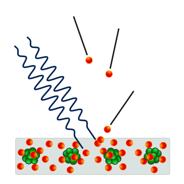
The photoelectric effect. A photon with sufficient energy (above the threshold frequency) strikes a metal surface and ejects an electron. The ejected electron’s maximum kinetic energy equals the photon energy minus the work function: KE = hf - φ. Credit: Wikimedia Commons, CC BY-SA 3.0
Einstein’s Photoelectric Equation
The work function (φ) is a property of the metal - it is the minimum energy an electron needs to escape the surface. Different metals have different work functions. For example, cesium has a low work function (~2.1 eV) while platinum has a high one (~5.6 eV).
Threshold Frequency
The threshold frequency is the minimum frequency that can eject electrons. At this frequency, the photon carries just enough energy to overcome the work function, with nothing left over for kinetic energy:
Intensity vs. Frequency
This distinction trips up many students, so here it is clearly:
| What you change | What happens |
|----------------|--------------|
| Increase frequency (above f0) | Each photon carries more energy, so ejected electrons are faster (higher KE_max) |
| Increase intensity (above f0) | More photons hit the surface per second, so more electrons are ejected (higher current), but each electron has the same KE_max |
| Increase intensity (below f0) | Nothing happens - more photons, but each one still lacks sufficient energy |
Graphical Representation
A graph of KE_max vs. frequency is a straight line:
Slope = h (Planck’s constant) - the same for all metals
x-intercept = f0 (threshold frequency) - different for each metal
y-intercept = -φ (negative work function)
For frequencies below f0, the graph sits at KE_max = 0 (no electrons ejected). Different metals produce parallel lines shifted left or right depending on their work function.
A metal has a work function of 4.0 eV. Light with photon energy 6.0 eV strikes the surface. What is the maximum kinetic energy of the ejected electrons?
Click to reveal answer
KE_max = hf - φ = 6.0 - 4.0 = 2.0 eV. The photon’s energy minus the work function gives the leftover kinetic energy. If the photon energy were less than 4.0 eV, no electrons would be ejected at all.
You double the intensity of light hitting a metal surface (frequency is above threshold). What happens to the number of ejected electrons and their maximum kinetic energy?
Click to reveal answer
The number of ejected electrons doubles (twice as many photons), but the maximum kinetic energy stays the same. Each photon still carries the same energy (hf), so each electron gets the same KE_max = hf - φ. Intensity affects quantity of photons, not energy per photon.
You already know that light behaves as both a wave (interference, diffraction) and a particle (photoelectric effect, photons). In 1924, Louis de Broglie proposed a radical inverse: if light can act like a particle, then particles should act like waves.
He was right. Electrons, protons, even baseballs have a wavelength. The catch: for anything bigger than a subatomic particle, that wavelength is so absurdly small that wave behavior is completely undetectable. A baseball’s de Broglie wavelength is about 10−19 times smaller than an atomic nucleus — there’s no experiment that could ever measure it. But for electrons, the wavelengths are atomic-scale, big enough to produce diffraction patterns. That’s the whole basis of electron microscopy.
De Broglie Wavelength
Every moving particle has an associated wavelength:
Putting Numbers to It
An electron moving at 106 m/s has a de Broglie wavelength of about 7 x 10⁻¹⁰ m - comparable to the spacing between atoms in a crystal. This means electrons can diffract off crystal lattices, just like X-rays. This was confirmed experimentally by Davisson and Germer in 1927.
A 0.15 kg baseball moving at 40 m/s has a de Broglie wavelength of about 10−34 m. That is roughly 1019 times smaller than the nucleus of an atom. Wave effects at this scale are utterly unmeasurable. This is why baseballs follow Newton’s laws, not quantum mechanics.
Electron Diffraction
Because electrons have wavelengths on the order of atomic spacings, they can diffract when passing through narrow slits or crystal lattices. This creates interference patterns - bright and dark bands - just like light diffracting through a double slit. Electron diffraction is direct proof that matter has wave properties.
Electron microscopes exploit this. By using electrons instead of visible light, electron microscopes achieve much higher resolution because electron wavelengths can be made much shorter than the wavelength of visible light (400-700 nm).
The Pauli Exclusion Principle
As you learned in General Chemistry, electrons in atoms are described by four quantum numbers: n (principal), l (angular momentum), ml (magnetic), and ms (spin). The Pauli exclusion principle places a strict limit on how electrons fill these states:
Without the Pauli exclusion principle, every electron would drop to the n = 1 ground state, all atoms would be roughly the same size, and chemistry as we know it would not exist. The periodic table, chemical bonding, and the diversity of molecular structures all depend on this one rule.
Connecting the Concepts
Wave-particle duality, the Heisenberg uncertainty principle (Section 9.2), and the Pauli exclusion principle together form the foundation of quantum mechanics. For the MCAT, the key takeaways are:
All matter has wave properties, but they are only significant for very small particles.
You cannot simultaneously know a particle’s exact position and momentum.
No two electrons in an atom can share the same quantum state.
These principles explain atomic structure, spectral lines, and why the periodic table looks the way it does.
An electron and a proton are both moving at the same speed. Which has the longer de Broglie wavelength, and by approximately how much?
Click to reveal answer
The electron has the longer wavelength, by a factor of about 1836. Since λ = h/(mv) and the proton is ~1836 times heavier than the electron, at the same speed the proton's wavelength is 1836 times shorter. Lighter particles have longer wavelengths.
Why can an orbital hold a maximum of two electrons?
Click to reveal answer
Because of the Pauli exclusion principle. An orbital is defined by three quantum numbers (n, l, ml). The only remaining quantum number is spin (ms), which has only two possible values: +21 and -21. So at most two electrons can share the same orbital, and they must have opposite spins.
Some nuclei are unstable. They have too many protons, too many neutrons, or simply too much energy, and they spontaneously transform into more stable configurations by ejecting particles or energy. This is radioactive decay — and it’s happening right now in the ground beneath your feet, in the bananas in your kitchen, and in trace amounts of carbon-14 inside your own body.
The process is entirely random for any individual nucleus — you cannot predict when one specific atom will decay. But for large collections of atoms, the statistics are remarkably precise (next section: half-life). The MCAT focuses on three main types of decay (alpha, beta, gamma) and the conservation rules that govern them.
Why Do Nuclei Decay?
The nucleus holds protons tightly packed together, and protons repel each other via the electromagnetic force. The strong nuclear force overcomes this repulsion and holds the nucleus together, but it only works at extremely short range (about 10−15 m). When the balance between these forces tips unfavorably - too many protons for the neutrons to stabilize, or too many neutrons for the size of the nucleus - the nucleus becomes unstable and decays.
Alpha Decay
In α decay, the nucleus ejects an α particle - a helium-4 nucleus containing 2 protons and 2 neutrons.
Result: Z decreases by 2, A decreases by 4. The parent element transforms into an element two positions to the left on the periodic table.
Example: 92238U→90234Th+24He
Alpha particles are heavy and doubly charged (+2). They are the least penetrating form of radiation - a sheet of paper or the dead outer layer of skin stops them. However, if an α emitter is inhaled or ingested, it becomes extremely dangerous because all that energy is deposited in a tiny area of living tissue.
Beta-Minus Decay
In β-minus decay, a neutron inside the nucleus converts into a proton, emitting an electron (β particle) and an antineutrino.
n→p+e−+νˉe
Result: Z increases by 1 (one more proton), A stays the same (total nucleons unchanged). The element shifts one position to the right on the periodic table.
Example: 614C→714N+e−+νˉe
Beta-minus decay occurs in neutron-rich nuclei. The nucleus has too many neutrons relative to protons, so it converts one to restore balance.
Beta-Plus Decay (Positron Emission)
In β-plus decay, a proton converts into a neutron, emitting a positron (the antimatter counterpart of an electron) and a neutrino.
p→n+e++νe
Result: Z decreases by 1 (one fewer proton), A stays the same. The element shifts one position to the left on the periodic table.
Example: 611C→511B+e++νe
Beta-plus decay occurs in proton-rich nuclei. The positron, once emitted, quickly encounters an electron and both annihilate, producing two γ rays. This annihilation is the basis of PET (positron emission tomography) scanning.
Gamma Decay
Gamma decay is the emission of a high-energy photon (γ ray) from an excited nucleus. It often follows α or β decay, when the daughter nucleus is left in an excited state.
Result: Z stays the same, A stays the same. Only energy is released - no particles, no change in identity. The nucleus simply drops to a lower energy state.
Gamma rays are the most penetrating form of radiation. They require thick lead or concrete to stop.
Summary Table
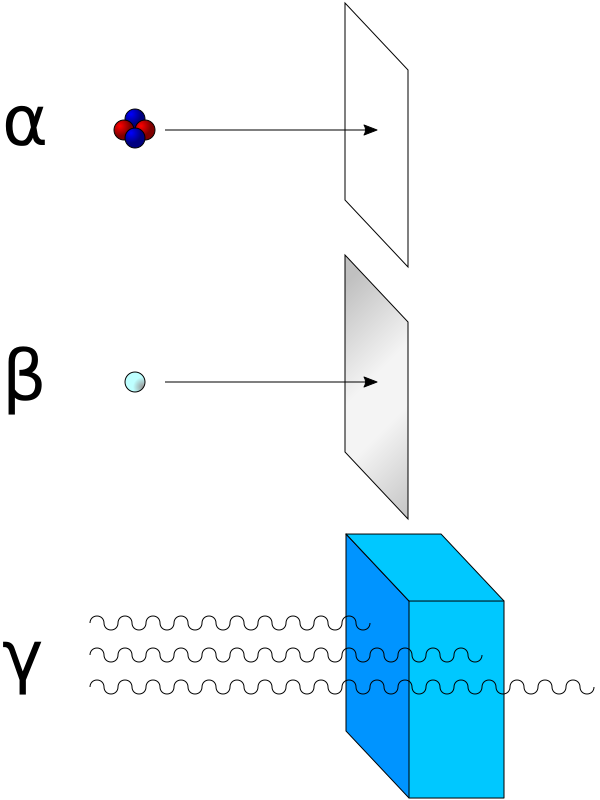
The three main types of radioactive decay and their penetrating power. Alpha particles (helium nuclei) are stopped by paper. Beta particles (electrons or positrons) penetrate paper but are stopped by aluminum. Gamma rays (high-energy photons) require thick lead or concrete to stop. Credit: Wikimedia Commons, CC BY-SA 3.0
Decay Type
Particle Emitted
Change in Z
Change in A
Penetration
Alpha
24He
-2
-4
Lowest (paper stops it)
Beta-minus
e−+νˉe
+1
0
Medium (aluminum stops it)
Beta-plus
e++νe
-1
0
Medium (then annihilation)
Gamma
photon (γ ray)
0
0
Highest (lead/concrete)
A nucleus undergoes β-minus decay. How do the atomic number (Z) and mass number (A) change?
Click to reveal answer
Z increases by 1, A stays the same. A neutron converts to a proton (increasing Z by 1) while emitting an electron and an antineutrino. The total number of nucleons (A) is unchanged because one neutron simply became one proton.
Fluorine-18 (Z = 9) undergoes positron emission. What is the daughter nucleus?
Click to reveal answer
Oxygen-18 (Z = 8, A = 18). Positron emission (β-plus decay) decreases Z by 1 (9 to 8 = oxygen) while A remains 18. A proton converted to a neutron, plus a positron and a neutrino were emitted.
Balancing nuclear equations is like balancing a checkbook — what goes in must equal what comes out. In chemical equations, you balance atoms. In nuclear equations, you balance two quantities: mass number (A) and charge (Z).
If you can add and subtract single-digit numbers, you can solve every nuclear equation the MCAT throws at you. The trick is just being systematic about which numbers go where.
Conservation Laws
Two quantities are always conserved in nuclear reactions:
Mass number (A) - the total number of nucleons (protons + neutrons) on the left side equals the total on the right side.
Atomic number (Z) - the total charge (proton count) on the left side equals the total on the right side.
Common Particles in Nuclear Equations
Before we practice, here are the particles you will encounter, with their A and Z values:
Particle
Symbol
A
Z
Proton
11p
1
1
Neutron
01n
1
0
Electron (β-minus)
−10e
0
-1
Positron (β-plus)
+10e
0
+1
Alpha particle
24He
4
2
Gamma photon
00γ
0
0
Practice: Identifying Unknown Products
Example 1: Alpha Decay of Radium-226
88226Ra→?+24He
Balance A: 226 = A + 4, so A = 222
Balance Z: 88 = Z + 2, so Z = 86
Z = 86 is radon (Rn). Answer: 86222Rn
Example 2: Beta-Minus Decay of Iodine-131
53131I→?+−10e+νˉe
Balance A: 131 = A + 0, so A = 131
Balance Z: 53 = Z + (-1), so Z = 54
Z = 54 is xenon (Xe). Answer: 54131Xe
Example 3: Identify the Missing Particle
84210Po→82206Pb+?
Balance A: 210 = 206 + A, so A = 4
Balance Z: 84 = 82 + Z, so Z = 2
A = 4, Z = 2 is an α particle (24He).
Decay Chains
Unstable nuclei rarely reach stability in a single step. A parent nucleus decays to a daughter, which may also be unstable and decay further, forming a decay chain. The chain continues until a stable nucleus is produced.
The most famous example is the uranium-238 decay chain, which involves 14 steps (8 α decays and 6 β decays) before reaching stable lead-206. You do not need to memorize the entire chain, but you should be able to work through any individual step.
Tracking Multiple Decays
If a nucleus undergoes two α decays and one β-minus decay:
Total change in A: 2 x (-4) = -8
Total change in Z: 2 x (-2) + 1 x (+1) = -3
So if you start with 92238U and apply two alphas and one β-minus, you end at A = 230, Z = 89 (actinium-230).
Electron Capture
One additional process worth knowing: in electron capture, the nucleus absorbs an inner-shell electron, converting a proton to a neutron. The effect on Z and A is identical to β-plus decay (Z decreases by 1, A unchanged), but no positron is emitted. Instead, the atom emits X-rays as outer electrons fill the vacancy left by the captured electron.
Thorium-232 (Z = 90) undergoes α decay. What are the mass number and atomic number of the daughter nucleus?
Click to reveal answer
A = 228, Z = 88 (radium-228). Alpha decay reduces A by 4 (232 - 4 = 228) and Z by 2 (90 - 2 = 88). The daughter is radium.
A nucleus undergoes three α decays and two β-minus decays. What is the total change in A and Z?
Click to reveal answer
A decreases by 12. Z decreases by 4. Three α decays: A changes by 3 x (-4) = -12, Z changes by 3 x (-2) = -6. Two β-minus decays: A changes by 0, Z changes by 2 x (+1) = +2. Net: A = -12, Z = -6 + 2 = -4.
Imagine 1000 popcorn kernels in a hot pan. After one minute, roughly half have popped — about 500 unpopped left. Another minute, half of the remaining pop — now about 250. After another minute, ~125. Each “round” cuts the remaining count in half.
Radioactive decay works exactly the same way. After each half-life, exactly half the remaining unstable nuclei have decayed. The MCAT loves this topic because the math is dead simple (just halving), and the underlying physics ties to carbon-14 dating, drug half-lives in biology, medical imaging tracers, and any first-order kinetic process you learned about in chemistry.
🎯 Predict First
A radioactive sample has a half-life of 3 seconds. After 6 seconds (two half-lives), how much of the sample remains?
The simulation below lets you watch this process in real time. Each circle represents a single radioactive atom with an independent, random chance of decaying each moment - yet despite the randomness, the overall curve follows a smooth, predictable exponential. Hit “Start Decay” and watch the law of large numbers in action.
The half-life (t1/2) is the time it takes for half of a radioactive sample to decay. After n half-lives, the fraction of the original sample remaining is:
The Half-Life Table
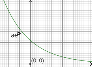
Radioactive decay follows an exponential curve. After each half-life, exactly half of the remaining radioactive atoms have decayed. The curve never reaches zero but approaches it asymptotically. Credit: Wikimedia Commons, CC BY-SA 3.0
This table is worth memorizing - it covers nearly every MCAT half-life question:
Divide the total time by the half-life: n = t / t1/2
Step 2: Apply the Fraction
Multiply the initial amount by (21)^n.
Example
A sample contains 800 mg of iodine-131 (t1/2 = 8 days). How much remains after 24 days?
n = 24 / 8 = 3 half-lives
Amount remaining = 800 x (1/2)3 = 800 x 81 = 100 mg
First-Order Kinetics
Radioactive decay is a first-order process, which means the rate of decay is proportional to the amount of radioactive material present. The more atoms you have, the more decays per second - but the fraction that decays per unit time stays constant.
This is the same first-order kinetics you learned in chemistry. The decay constant (λ) relates to the half-life:
Semi-Log Plots
On a normal (linear) graph, radioactive decay produces a curved exponential line that swoops downward. But if you plot the natural log of the amount remaining (ln N) versus time, you get a straight line with slope = -λ.
Activity
The activity of a radioactive sample is the number of decays per second, measured in becquerels (Bq) or curies (Ci). Activity follows the same half-life pattern as the number of atoms:
Activity = λ x N
Since N decreases by half each half-life, so does the activity. A freshly prepared sample is most active, and the activity drops exponentially over time.
A radioactive isotope has a half-life of 6 hours. You start with 240 mg. How much remains after 18 hours?
Click to reveal answer
30 mg. n = 618 = 3 half-lives. Amount remaining = 240 x (1/2)3 = 240 x 81 = 30 mg.
A sample has decayed to 161 of its original amount. How many half-lives have passed?
Click to reveal answer
4 half-lives. (21)⁴ = 161. Each half-life cuts the remaining amount in half: 1 to 21 to 41 to 81 to 161.
Here’s a strange fact: weigh two protons and two neutrons separately. Now weigh the helium-4 nucleus they form. The nucleus weighs less than the sum of its parts.
Where did the missing mass go? It was converted into the energy that glues the nucleus together. This “missing mass” is called the mass defect, and the energy it represents — via Einstein’s E=mc2 — is the nuclear binding energy.
This single phenomenon is the reason nuclear reactions release millions of times more energy than chemical reactions, and it’s why atomic bombs and nuclear reactors exist.
Mass Defect
The mass defect (Δm) is the difference between the mass of the individual nucleons and the actual mass of the assembled nucleus:
E = mc2
Einstein’s famous equation connects the mass defect to binding energy:
A useful conversion: 1 amu of mass corresponds to 931.5 MeV of energy. This allows you to skip the c2 calculation entirely - just multiply the mass defect in amu by 931.5 to get the binding energy in MeV.
Example
Helium-4 has a mass defect of about 0.0304 amu. Its binding energy is:
E = 0.0304 x 931.5 MeV/amu = 28.3 MeV
This means you would need to supply 28.3 MeV to completely disassemble a helium-4 nucleus into two free protons and two free neutrons.
Binding Energy per Nucleon
The total binding energy tells you how much energy holds the entire nucleus together, but to compare the stability of different nuclei, we need the binding energy per nucleon (BE/A). This is simply the total binding energy divided by the number of nucleons.
The Binding Energy per Nucleon Curve
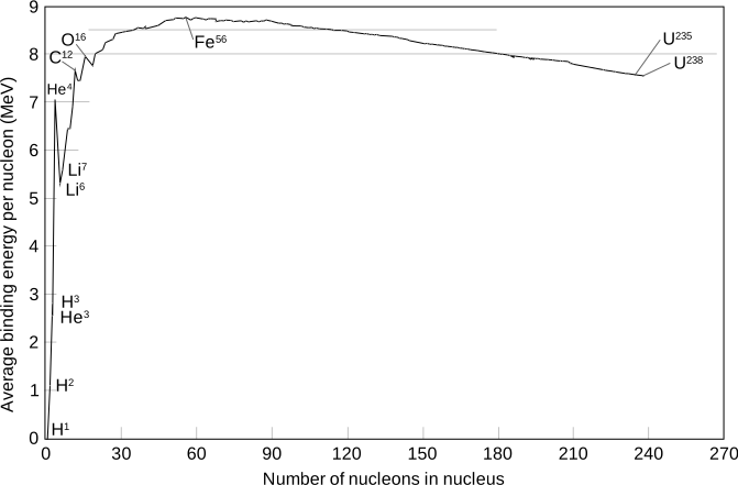
The binding energy per nucleon curve. Iron-56 sits at the peak (~8.8 MeV/nucleon), making it the most stable nucleus. Fusion of light nuclei (left of iron) and fission of heavy nuclei (right of iron) both release energy by moving toward the iron peak. Credit: Wikimedia Commons, CC BY-SA 3.0
If you plot binding energy per nucleon (y-axis) against mass number A (x-axis), you get a curve that rises steeply for light nuclei, peaks at iron-56, and then gradually decreases for heavy nuclei.
This curve is the single most important diagram in nuclear physics for the MCAT. It explains everything:
Light nuclei (left of iron): have relatively low BE/A. Combining them (fusion) moves up the curve toward iron, releasing energy.
Heavy nuclei (right of iron): also have lower BE/A than iron. Splitting them (fission) moves toward iron from the other direction, also releasing energy.
Iron-56 sits at the peak: it is the most tightly bound nucleus. You cannot extract energy by either fusing or splitting iron.
Why Nuclear Reactions Release So Much Energy
Chemical reactions involve rearranging electrons and breaking/forming chemical bonds, with energies on the order of a few eV per reaction. Nuclear reactions involve rearranging nucleons and tapping into the mass defect, with energies on the order of MeV per reaction - roughly a million times more energy per event.
This enormous energy difference is why a small amount of nuclear fuel can power a city, while burning the same mass of coal would barely heat a building.
A nucleus has a mass defect of 0.5 amu. What is its binding energy in MeV?
Click to reveal answer
About 465.8 MeV. Binding energy = Δm x 931.5 MeV/amu = 0.5 x 931.5 = 465.75 MeV. This is the energy you would need to supply to completely disassemble the nucleus into individual protons and neutrons.
Why does fusing two light nuclei release energy, while fusing two iron nuclei does not?
Click to reveal answer
Light nuclei have low binding energy per nucleon. Fusing them produces a heavier nucleus with higher BE/A, and the difference is released as energy. Iron-56 already sits at the peak of the binding energy curve. Fusing two iron nuclei would produce a heavier nucleus with lower BE/A - this would require energy input rather than release it.
In the last section you learned that both fission and fusion release energy — because both move nuclei toward the iron-56 peak on the binding energy curve. But the two processes couldn’t be more different in practice. Fission splits heavy nuclei apart and powers nuclear reactors and atomic bombs. Fusion fuses light nuclei together and powers every star in the universe — including the sun, right now, turning hydrogen into helium.
Here’s how each one actually works, where it occurs, and why one is hard to do on Earth while the other happens naturally in billions of stars.
Nuclear Fission
Fission is the splitting of a heavy nucleus into two or more lighter nuclei, typically accompanied by the release of neutrons and a large amount of energy.
How Fission Works
A slow (thermal) neutron is absorbed by a heavy nucleus (commonly uranium-235 or plutonium-239).
The nucleus becomes excited and unstable.
The nucleus splits into two medium-sized daughter nuclei, typically releasing 2-3 additional neutrons.
The mass of the products is slightly less than the mass of the reactants - the missing mass appears as kinetic energy (about 200 MeV per fission event).
Chain Reactions
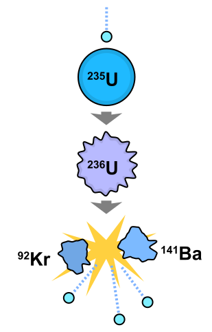
A nuclear fission chain reaction. A neutron strikes a uranium-235 nucleus, causing it to split into daughter nuclei and release additional neutrons. Each released neutron can trigger further fission events, creating a self-sustaining chain reaction. Credit: Wikimedia Commons, CC BY-SA 3.0
The 2-3 neutrons released by each fission event can strike other uranium nuclei and trigger further fissions. If enough fissile material is present, this creates a self-sustaining chain reaction.
Subcritical: on average, fewer than one neutron per fission causes another fission. The reaction dies out.
Critical: exactly one neutron per fission causes another fission. The reaction is self-sustaining at a constant rate. This is how nuclear power plants operate.
Supercritical: more than one neutron per fission causes further fissions. The reaction grows exponentially. This is how nuclear weapons work.
Control Rods
In a nuclear reactor, control rods (made of materials that absorb neutrons, such as boron or cadmium) are inserted between fuel rods to regulate the chain reaction. Pushing the rods in absorbs more neutrons, slowing the reaction. Pulling them out allows more neutrons to cause fissions, speeding it up. At steady operation, the reactor is kept exactly critical.
Nuclear Fusion
Fusion is the combining of two light nuclei into a heavier nucleus, releasing energy. This is the process that powers every star in the universe.
The Sun’s Fusion Cycle
The dominant process in the sun is the proton-proton chain, which effectively fuses four hydrogen nuclei (protons) into one helium-4 nucleus:
411H→24He+2e++2νe+energy
The mass of four protons is greater than the mass of one helium-4 nucleus. The difference (~0.029 amu per event, or about 26.7 MeV) is released as kinetic energy and radiation.
Why Fusion Is Hard on Earth
Fusion requires temperatures of millions of degrees to overcome Coulomb repulsion between positively charged nuclei. At these temperatures, matter exists as plasma. Containing this plasma is the central engineering challenge - no solid container can withstand those temperatures. Current approaches use magnetic confinement (tokamaks) or inertial confinement (lasers).
Comparing Fission and Fusion
Feature
Fission
Fusion
Process
Heavy nucleus splits
Light nuclei combine
Fuel
Uranium-235, Plutonium-239
Hydrogen isotopes (deuterium, tritium)
Direction on BE curve
Heavy nuclei move left toward iron
Light nuclei move right toward iron
Energy per event
~200 MeV
~26.7 MeV (per 4H to He)
Energy per kilogram of fuel
High
Even higher
Conditions
Neutron bombardment
Extreme temperature (~107 K)
Waste
Radioactive fission products
Helium (non-radioactive)
Why does fusion require extremely high temperatures, while fission can be triggered by slow neutrons?
Click to reveal answer
Fusion requires overcoming the electrostatic (Coulomb) repulsion between two positively charged nuclei. Extreme temperatures give the nuclei enough kinetic energy to get close enough for the strong force to take over. Fission uses neutrons, which carry no charge and therefore face no Coulomb barrier - they can approach and enter a nucleus even at low speeds.
In a nuclear reactor, what is the role of control rods, and what happens if they are fully removed?
Click to reveal answer
Control rods absorb neutrons to regulate the chain reaction rate. Inserting them further slows the reaction; pulling them out allows more neutrons to cause fissions. If fully removed, the reactor would become supercritical - the reaction rate would increase exponentially, potentially leading to a meltdown.
Imagine you’re sorting coins by weight, but you can’t touch them. Instead, you roll them down a ramp (giving them all the same kinetic energy), then blow a fan across their path as they fly out. The heavier coins resist the wind and fly farther before curving; the lighter coins curve sharply. By measuring where each coin lands, you know its mass.
A mass spectrometer does essentially the same thing — except the “coins” are ions, the “ramp” is an electric field, and the “fan” is a magnetic field. The device is one of the most important tools in chemistry, biochemistry, forensics, and pharmaceutical research. Watson and Crick used mass spectrometry data (among other things) when figuring out DNA’s structure; modern proteomics and drug-testing labs depend on it every day.
How a Mass Spectrometer Works
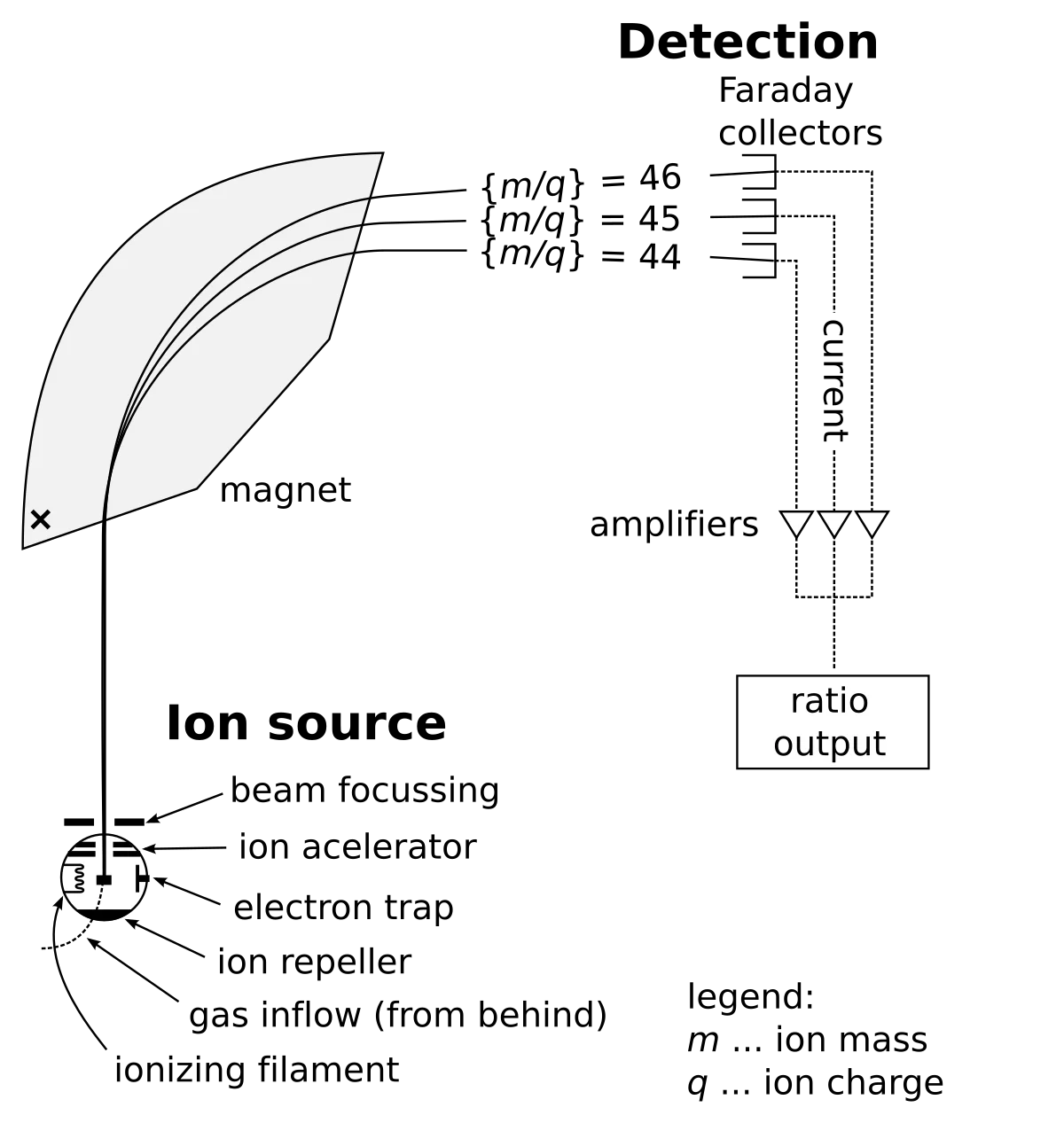
Schematic of a mass spectrometer. Ions are produced in the ionization source, accelerated through a voltage, deflected by a magnetic field into circular paths (heavier ions curve less), and detected at different positions based on their mass-to-charge ratio. Credit: Wikimedia Commons, CC BY-SA 3.0
A mass spectrometer separates ions by their mass-to-charge ratio (m/q). The process has four stages:
1. Ionization
The sample is ionized - atoms or molecules are stripped of one or more electrons (or, less commonly, gain electrons) to become charged. Common methods include electron bombardment and electrospray ionization. The result is a beam of ions with charge q.
2. Acceleration
The ions pass through a potential difference (voltage V), which accelerates them. The kinetic energy gained equals the work done by the electric field:
qV=21mv2
All ions with the same charge gain the same kinetic energy, but lighter ions end up moving faster than heavier ions.
3. Deflection
The ions enter a uniform magnetic field (B) directed perpendicular to their velocity. The magnetic force provides centripetal acceleration, bending the ions into a circular path:
qvB=rmv2
Solving for the radius:
4. Detection
A detector (photographic plate or electronic detector) records where each ion strikes. Ions with different m/q values land at different positions, separated by their radius of curvature.
Velocity Selector (Optional Stage)
Some mass spectrometers include a velocity selector before the magnetic deflection stage. A velocity selector uses crossed electric and magnetic fields. Only ions with a specific velocity pass through undeflected:
v=BE
where E is the electric field strength and B is the magnetic field strength. All other ions are deflected into the walls. This ensures that all ions entering the magnetic deflection region have the same speed, simplifying the analysis.
Deriving m/q
Combining the acceleration and deflection equations (for a mass spectrometer with a velocity selector where all ions have speed v=E/B1):
From deflection: r=mv/(qB2)
Rearranging:
qm=vrB2=ErB1B2
Without a velocity selector, combining qV=21mv2 with r=mv/(qB):
qm=2Vr2B2
You do not need to memorize these combined equations, but you should be able to derive them by combining the two base formulas.
Applications
Isotope identification: separating isotopes of the same element (same Z, different A) based on mass differences.
Molecular weight determination: identifying unknown compounds by their molecular mass.
Forensic and environmental analysis: detecting trace amounts of specific substances.
Pharmaceutical development: confirming drug purity and structure.
Two singly-charged ions enter a mass spectrometer with the same velocity. Ion A has twice the mass of ion B. How do their radii of deflection compare?
Click to reveal answer
Ion A has twice the radius of ion B. Since r = mv/(qB), and both ions have the same v, q, and B, the radius is directly proportional to mass. Doubling the mass doubles the radius. Ion A curves more gently and lands farther from the entrance point.
In a mass spectrometer, if you increase the magnetic field strength while keeping everything else the same, what happens to the radius of each ion's path?
Click to reveal answer
The radius decreases. Since r = mv/(qB), increasing B (in the denominator) decreases r. A stronger magnetic field bends the ions more tightly, and all ions land closer to the entrance.
Nuclear and atomic physics aren’t just abstract theory — they’re the foundation of the most important tools in modern medicine. Every time a doctor orders an X-ray, CT scan, PET scan, or MRI, they’re applying the principles from this chapter: photon energy, electron transitions, radioactive decay, electromagnetic induction.
The MCAT loves these technologies as passage topics because they neatly tie the physics of the last 11 sections to real clinical practice. This section walks through each modality (X-ray, CT, PET, MRI), explains the physics behind radiation shielding and the inverse square law, and covers basic dosimetry.
X-Rays
X-rays are high-energy photons with wavelengths between about 0.01 and 10 nm, placing them between ultraviolet light and γ rays on the electromagnetic spectrum. They penetrate soft tissue but are absorbed by dense materials like bone and metal, which is why X-ray images show bones as bright white against darker soft tissue.
PET Scans
Positron Emission Tomography (PET) directly uses the nuclear physics you learned in Section 9.6. A patient is injected with a tracer molecule labeled with a positron-emitting isotope, most commonly fluorine-18 attached to a glucose analog (FDG).
Here is the physics chain:
Fluorine-18 undergoes β-plus decay, emitting a positron.
The positron travels a very short distance before encountering an electron.
The positron and electron annihilate, converting their combined mass into two γ rays that fly off in exactly opposite directions (180 degrees apart).
A ring of detectors around the patient records these coincident γ rays. By tracing the lines between pairs of detectors, the computer reconstructs where each annihilation occurred.
MRI
Magnetic Resonance Imaging uses no ionizing radiation at all. Instead, it exploits the magnetic properties of hydrogen nuclei (protons), which are abundant in water and fat throughout the body.
Radiation Shielding
Different types of radiation require different shielding because of their vastly different penetrating abilities:
Radiation
Stopped by
Why
Alpha (helium nuclei)
Sheet of paper, skin
Large, heavy, doubly-charged - interacts strongly with matter, loses energy quickly
Beta (electrons/positrons)
Thin aluminum sheet (~mm)
Lighter and singly-charged - penetrates farther than α but still interacts frequently
Gamma (photons)
Thick lead or concrete
No charge, no mass - interacts weakly with matter, can travel through many centimeters of material
Inverse Square Law for Radiation
The intensity of radiation from a point source decreases with the square of the distance:
This has big practical implications: simply stepping back from a radioactive source sharply reduces your exposure. Moving from 1 meter to 3 meters away reduces intensity by a factor of 9.
Dosimetry Concepts
In a PET scan, fluorine-18 emits a positron. What happens to the positron, and what is detected by the scanner?
Click to reveal answer
The positron annihilates with a nearby electron, producing two 511 keV γ rays that travel in opposite directions. The PET scanner's ring of detectors identifies coincident γ ray pairs and uses their trajectories to reconstruct where in the body the annihilation occurred.
A technician stands 2 meters from a γ source and receives a dose rate of 100 mSv/hr. What is the dose rate at 6 meters?
Click to reveal answer
About 11.1 mSv/hr. By the inverse square law: I2=I1(r1/r2)2=100×(2/6)2=100×(1/3)2=100/9=11.1 mSv/hr. Tripling the distance reduced the intensity by a factor of 9.