
Special Senses
The special senses - vision, hearing, equilibrium, taste, and smell - are served by complex receptor organs concentrated in the head. Unlike general senses (touch, temperature, pain) that use relatively simple receptors distributed throughout the body, the special senses rely on highly specialized structures that convert very specific forms of energy into neural signals. The MCAT tests the anatomy of these organs, the mechanism of signal transduction in each, and the neural pathways that carry sensory information to the brain.
Vision: Eye Anatomy
The eye is a fluid-filled sphere designed to focus light onto the retina, a thin layer of neural tissue at the back of the eye that contains photoreceptors.
Outer layer (fibrous tunic):
- Sclera: the white, protective outer coat of the eye. It maintains eye shape and provides attachment points for extraocular muscles.
- Cornea: the transparent anterior portion of the outer layer. It is the primary refractive structure, bending incoming light rays to begin the focusing process.
Middle layer (vascular tunic/uvea):
- Choroid: a pigmented, highly vascular layer that nourishes the retina and absorbs stray light.
- Ciliary body: contains the ciliary muscle, which controls the shape of the lens for focusing (accommodation). It also produces aqueous humor.
- Iris: the colored muscular ring that controls pupil diameter, regulating the amount of light entering the eye.
Inner layer (neural tunic):
- Retina: the innermost layer containing the photoreceptors (rods and cones), bipolar cells, and ganglion cells. It is where light is converted into neural signals.
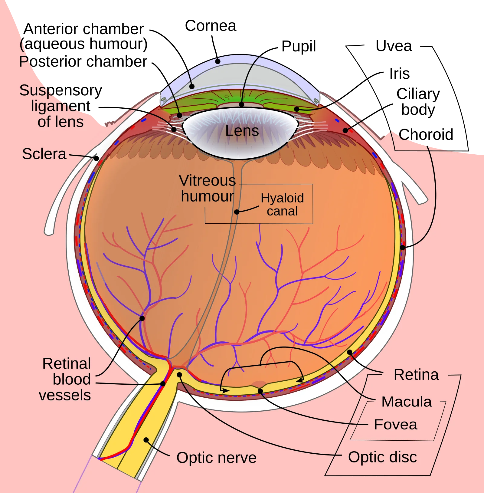
The lens sits behind the iris and fine-tunes the focusing of light onto the retina. It is held in place by suspensory ligaments (zonular fibers) attached to the ciliary body. When the ciliary muscle contracts, the suspensory ligaments loosen, and the elastic lens rounds up to focus on near objects. When the ciliary muscle relaxes, the ligaments pull taut and flatten the lens for distance vision. This process is called accommodation.
Aqueous humor fills the anterior and posterior chambers (in front of the lens) and is continuously produced and drained. If drainage is blocked, intraocular pressure rises, which can damage the optic nerve. Vitreous humor is the gel-like substance filling the large posterior cavity behind the lens.
Vision: Rods, Cones, and Phototransduction
The retina contains two types of photoreceptors, each optimized for different conditions.
Rods (~120 million per eye):
- Extremely sensitive to light - responsible for scotopic (dim-light) vision
- Produce images in shades of grey only (no color discrimination)
- Concentrated in the peripheral retina
- Contain the photopigment rhodopsin (retinal + opsin)
- Many rods converge onto a single ganglion cell, increasing sensitivity but decreasing acuity
Cones (~6 million per eye):
- Require brighter light - responsible for photopic (daylight) vision and color vision
- Three subtypes based on their peak sensitivity: red (long wavelength), green (medium wavelength), and blue (short wavelength)
- Concentrated in the fovea centralis, a small pit in the center of the retina that produces the sharpest image
- Fewer cones converge per ganglion cell, giving high acuity but lower sensitivity
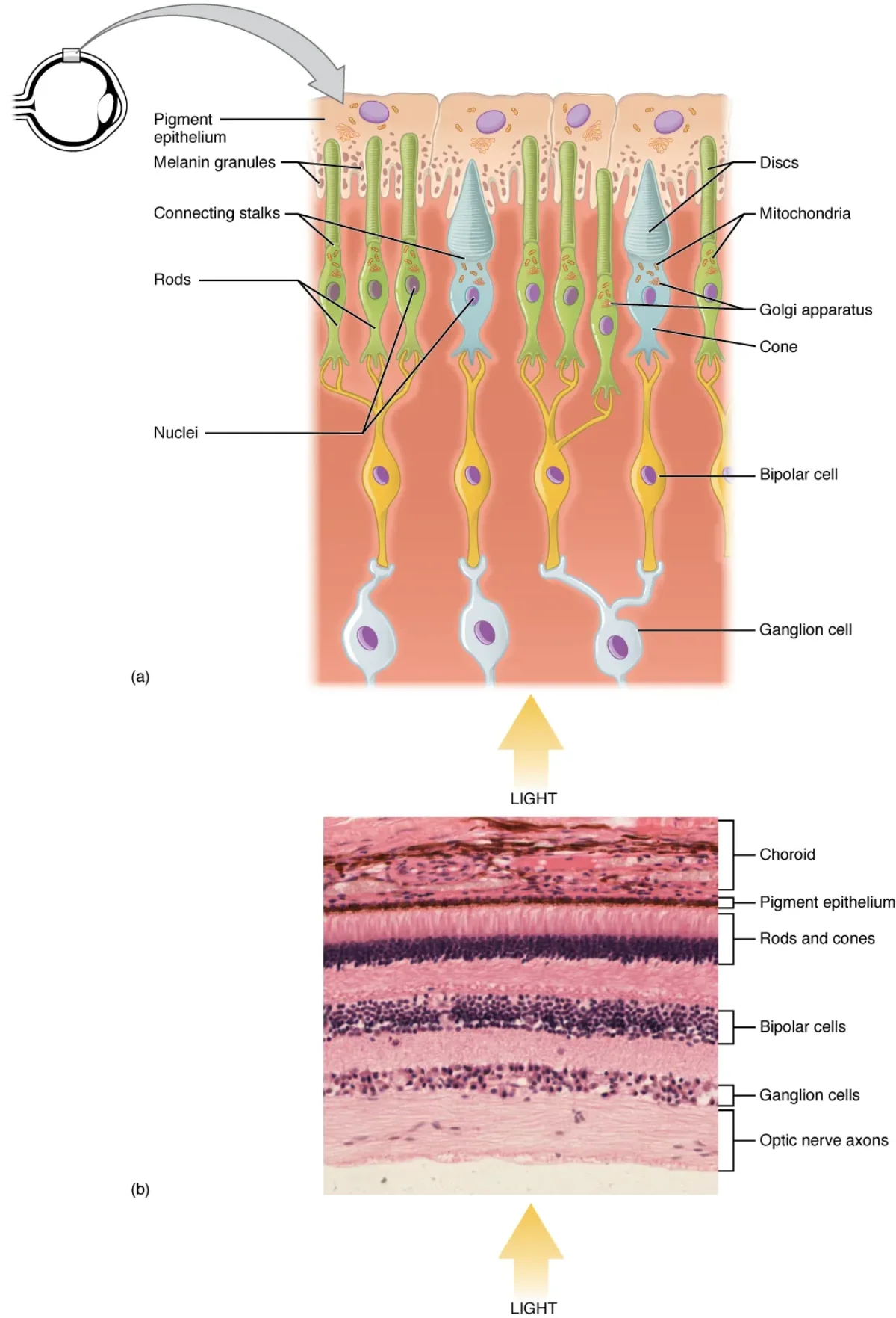
Phototransduction is the process by which light energy is converted to a neural signal. In the dark, photoreceptors are partially depolarized and continuously release glutamate. When light strikes rhodopsin (in rods) or photopsin (in cones), retinal changes from the cis to trans configuration, activating a G-protein signaling cascade that closes cGMP-gated sodium channels, causing the photoreceptor to hyperpolarize and reduce glutamate release. This change in neurotransmitter release is detected by bipolar cells and ultimately transmitted to ganglion cells.
Vision: The Visual Pathway
The visual pathway from retina to cortex involves several processing steps:
- Photoreceptors (rods/cones) synapse on bipolar cells
- Bipolar cells synapse on ganglion cells (whose axons form the optic nerve)
- The optic nerve (CN II) exits the eye at the optic disc (the blind spot - no photoreceptors here)
- At the optic chiasm, fibers from the nasal (medial) half of each retina cross to the opposite side, while fibers from the temporal (lateral) half remain ipsilateral
- The optic tracts carry the reorganized fibers to the lateral geniculate nucleus (LGN) of the thalamus
- From the LGN, fibers project to the primary visual cortex in the occipital lobe
Because of the partial crossing at the optic chiasm, each hemisphere receives visual information from the contralateral visual field. The left visual cortex processes the right visual field, and vice versa.
Refractive errors:
- Myopia (nearsightedness): the eyeball is too long or the lens is too curved, causing the focal point to fall in front of the retina. Corrected with concave (diverging) lenses.
- Hyperopia (farsightedness): the eyeball is too short or the lens is too flat, causing the focal point to fall behind the retina. Corrected with convex (converging) lenses.
Hearing: Ear Anatomy
The ear is divided into three anatomical regions, each playing a distinct role in converting sound waves into neural signals.
Outer ear: The pinna (auricle) funnels sound waves into the external auditory canal, which directs them toward the tympanic membrane.
Middle ear: The tympanic membrane (eardrum) vibrates in response to sound waves. These vibrations are transmitted through three tiny bones called ossicles - the malleus (hammer), incus (anvil), and stapes (stirrup) - which amplify the sound energy by approximately 20-fold. The stapes footplate pushes against the oval window, transferring vibrations into the fluid-filled inner ear.
The middle ear also connects to the pharynx via the Eustachian (auditory) tube, which equalizes air pressure on both sides of the tympanic membrane.
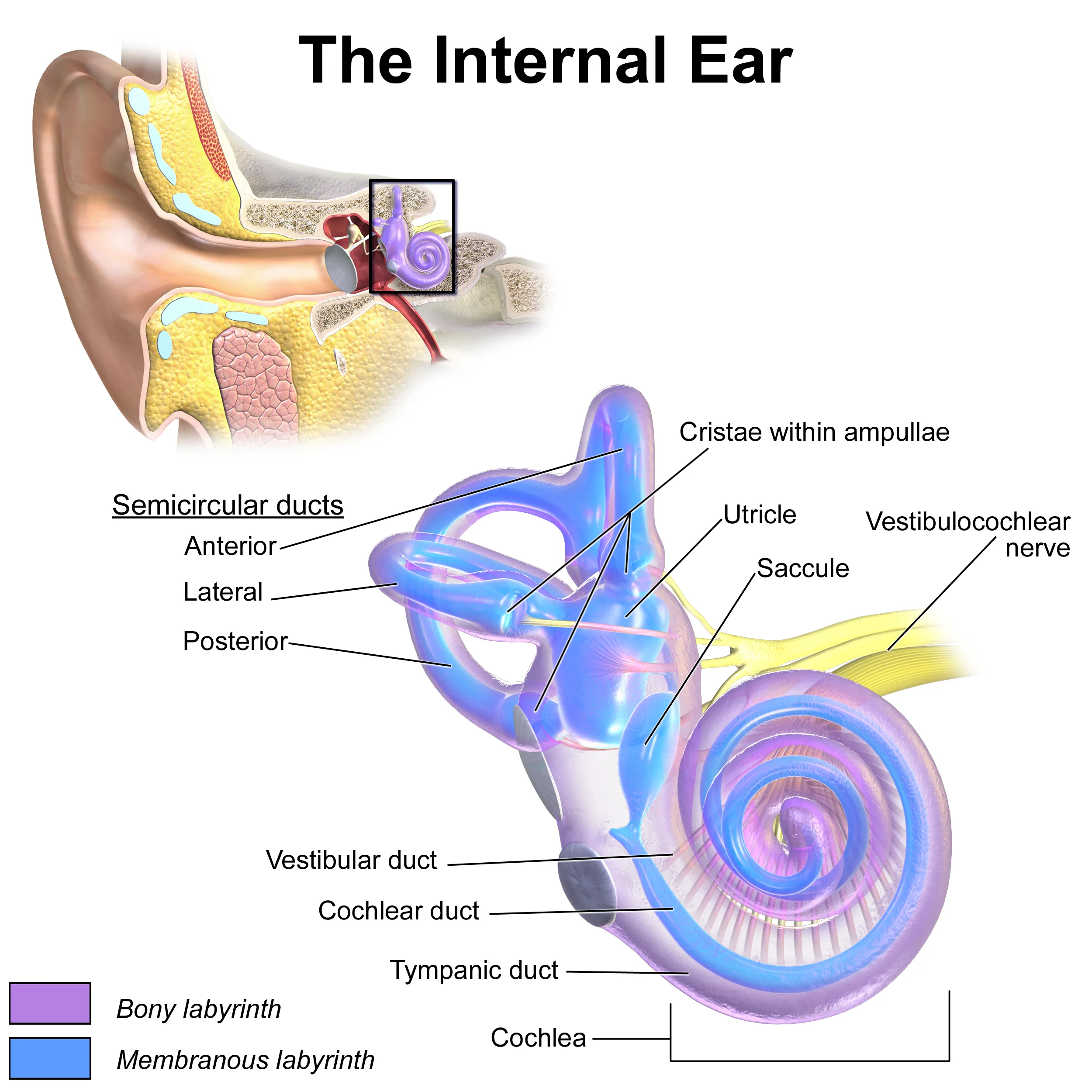
Inner ear: The inner ear contains both the hearing organ (cochlea) and the balance organs (semicircular canals, utricle, and saccule). It is filled with fluid and embedded within the temporal bone.
Hearing: Sound Transduction in the Cochlea
The cochlea is a snail-shaped, fluid-filled structure divided into three chambers: the scala vestibuli, scala media (cochlear duct), and scala tympani. The organ of Corti sits on the basilar membrane within the scala media and contains the hair cells - the mechanoreceptor cells responsible for hearing.
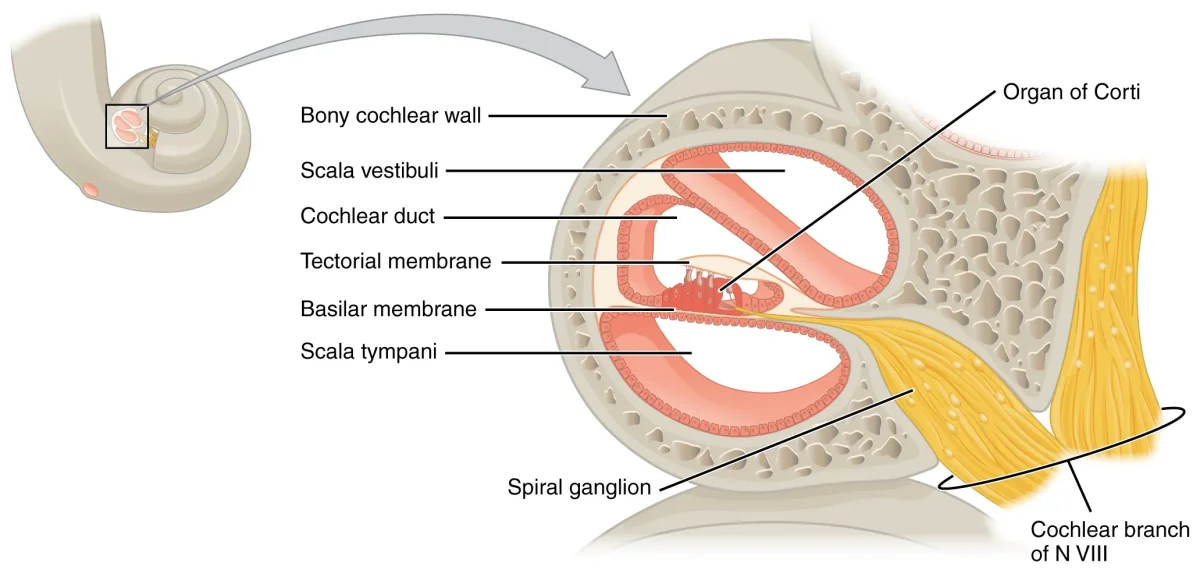
The transduction process follows a precise sequence:
- Sound waves vibrate the tympanic membrane
- Ossicles amplify the vibration and transmit it to the oval window
- Pressure waves travel through the cochlear fluid (perilymph)
- The basilar membrane vibrates in response
- Hair cells on the basilar membrane are displaced, and their stereocilia bend against the tectorial membrane
- Bending of stereocilia opens mechanically gated ion channels, depolarizing the hair cell
- The hair cell releases neurotransmitter onto the cochlear branch of CN VIII (vestibulocochlear nerve)
- The signal travels to the auditory cortex in the temporal lobe
The Vestibular System: Balance and Equilibrium
The vestibular apparatus is located in the inner ear, adjacent to the cochlea. It detects head position and movement, allowing you to maintain balance and coordinate eye movements.
Semicircular canals (three per ear): These three fluid-filled rings are oriented in three perpendicular planes (anterior, posterior, and lateral). They detect rotational (angular) acceleration. When the head rotates, the fluid (endolymph) lags behind due to inertia. This deflects a gelatinous structure called the cupula within the ampulla at the base of each canal, bending the hair cells embedded within it.
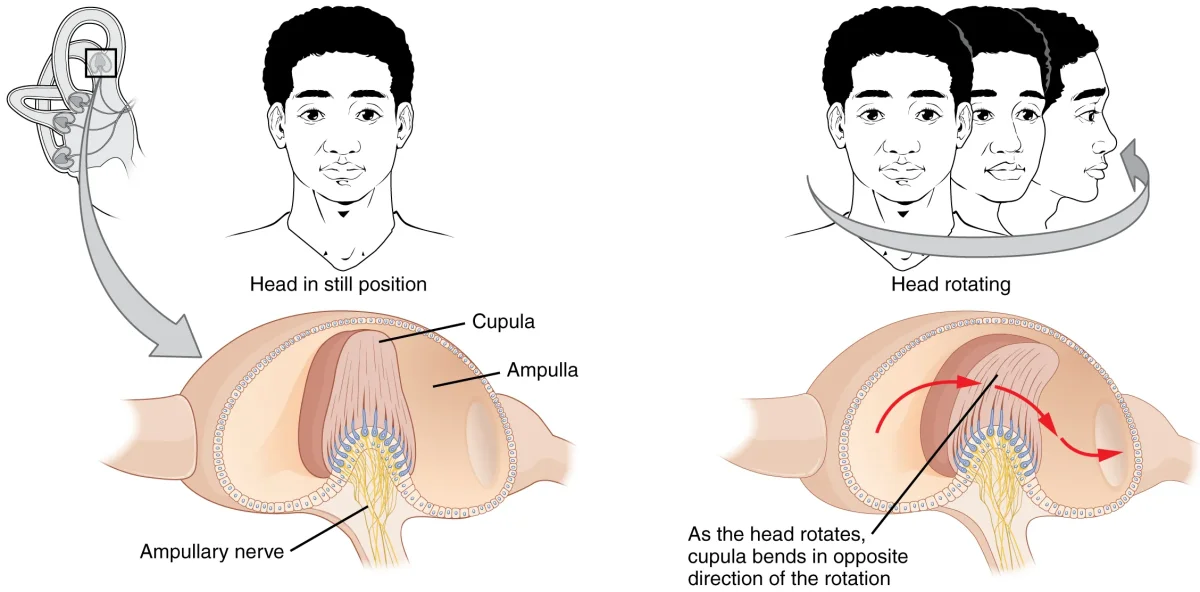
Utricle and saccule: These two chambers detect linear acceleration and the static position of the head relative to gravity. They contain a gelatinous layer topped with calcium carbonate crystals called otoliths (literally “ear stones”). When the head tilts or undergoes linear acceleration, gravity or inertial force shifts the otoliths, bending the underlying hair cells.
All vestibular signals travel via the vestibular branch of CN VIII to the brainstem and cerebellum.
Taste (Gustation)
Taste receptors are located within taste buds, which are found on papillae (raised bumps) on the tongue surface, as well as on the soft palate, epiglottis, and pharynx. Each taste bud contains 50-100 taste receptor cells.
There are five basic taste modalities:
| Taste | Stimulus | Biological Significance |
|---|---|---|
| Sweet | Sugars, some amino acids | Energy-rich food |
| Salty | Na+ and other ions | Electrolyte balance |
| Sour | H+ (acids) | Potentially spoiled food |
| Bitter | Alkaloids, toxins | Potentially poisonous substances |
| Umami | Glutamate, amino acids | Protein-rich food |
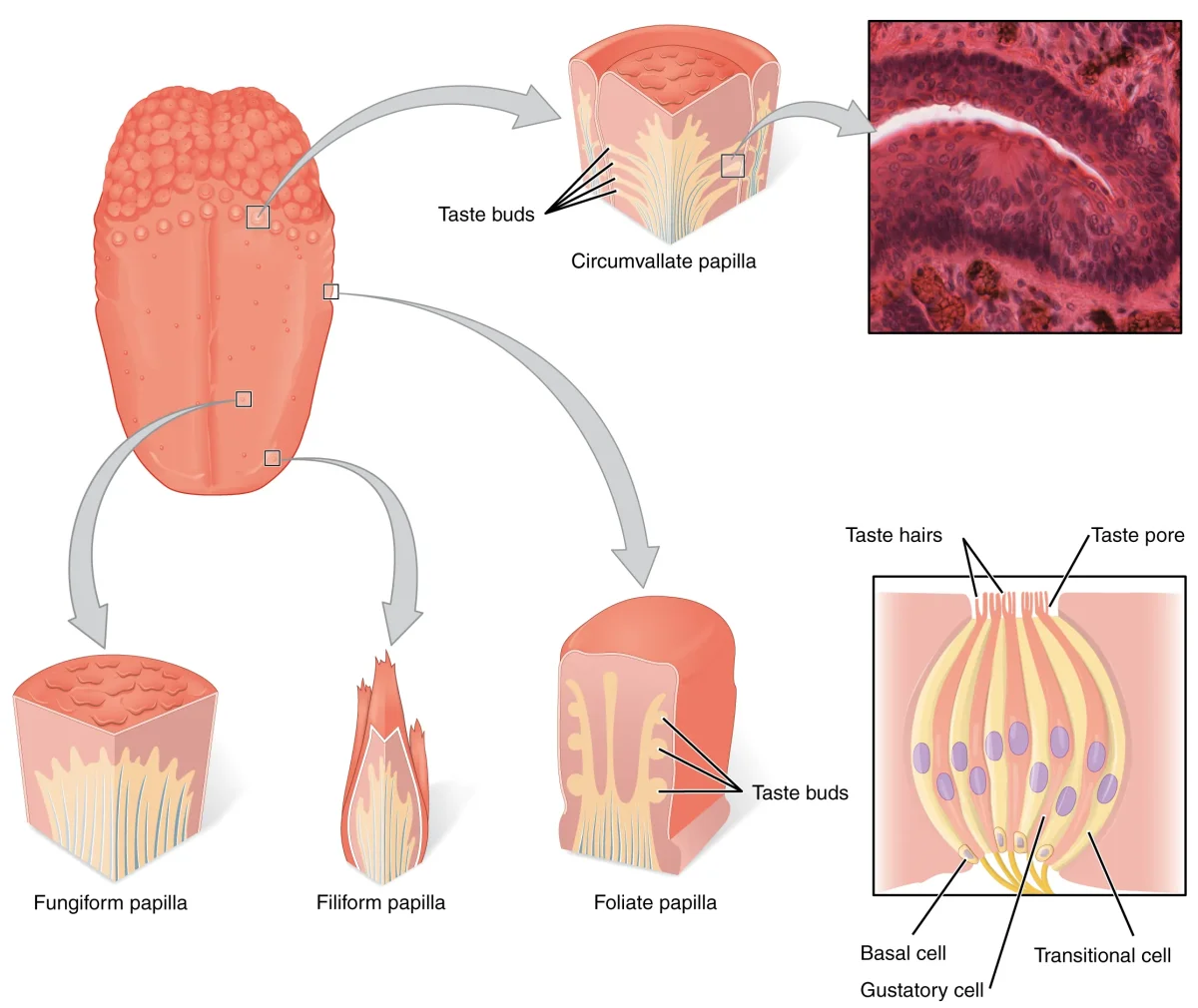
Taste signals are carried by three cranial nerves: CN VII (facial) for the anterior two-thirds of the tongue, CN IX (glossopharyngeal) for the posterior one-third, and CN X (vagus) for the epiglottis region. All taste information synapses in the solitary nucleus of the medulla, passes through the thalamus, and reaches the gustatory cortex in the insula.
Smell (Olfaction)
Olfactory receptor neurons are located in the olfactory epithelium high in the nasal cavity. Each receptor neuron expresses a single type of odorant receptor protein on its cilia. Humans have approximately 400 different odorant receptor types, and each odorant activates a specific combination of receptors, allowing us to distinguish thousands of different smells.
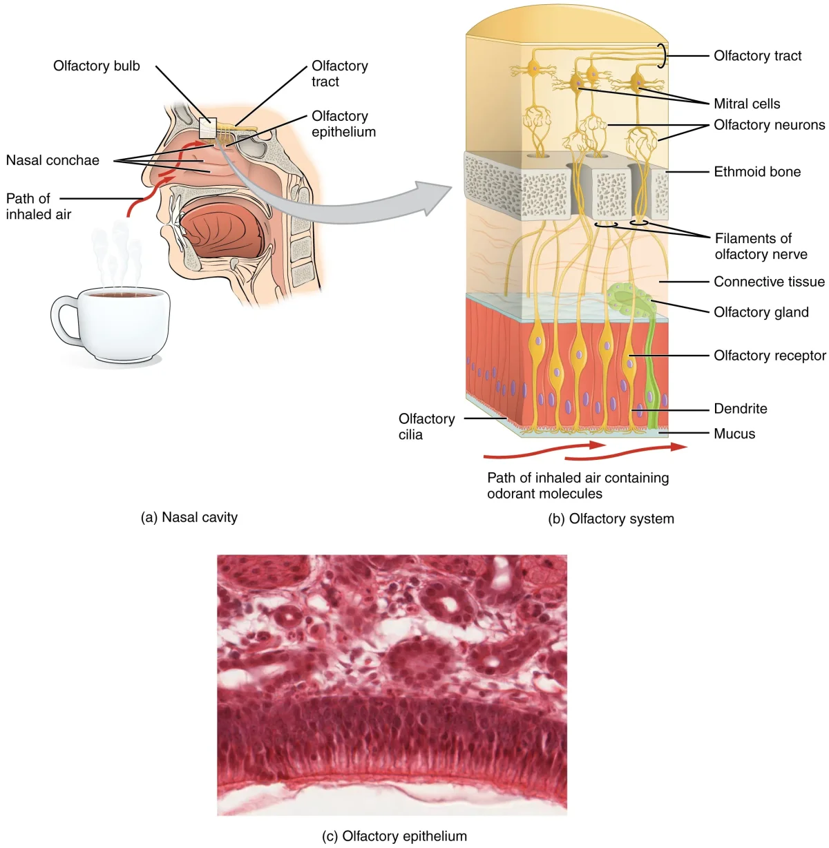
The axons of olfactory receptor neurons pass through tiny holes in the cribriform plate of the ethmoid bone and synapse in the olfactory bulb. From the olfactory bulb, the signal travels via the olfactory tract (CN I) to the olfactory cortex (piriform cortex) and the limbic system (amygdala, hippocampus).
Summary of Special Sense Pathways
| Sense | Receptor | Cranial Nerve | Thalamic Relay? | Cortical Destination |
|---|---|---|---|---|
| Vision | Rods, cones | CN II (Optic) | Yes (LGN) | Occipital lobe |
| Hearing | Hair cells (cochlea) | CN VIII (Vestibulocochlear) | Yes (MGB) | Temporal lobe |
| Balance | Hair cells (vestibular) | CN VIII (Vestibulocochlear) | No (to cerebellum/brainstem) | - |
| Taste | Taste receptor cells | CN VII, IX, X | Yes (VPM) | Insula (gustatory cortex) |
| Smell | Olfactory receptor neurons | CN I (Olfactory) | No | Piriform cortex, limbic system |