A fatty acid is a long hydrocarbon chain with a carboxylic acid group at one end. Most natural fatty acids have an even number of carbons (typically 14-24) because they are built two carbons at a time during fatty acid synthesis.
Saturated vs. Unsaturated
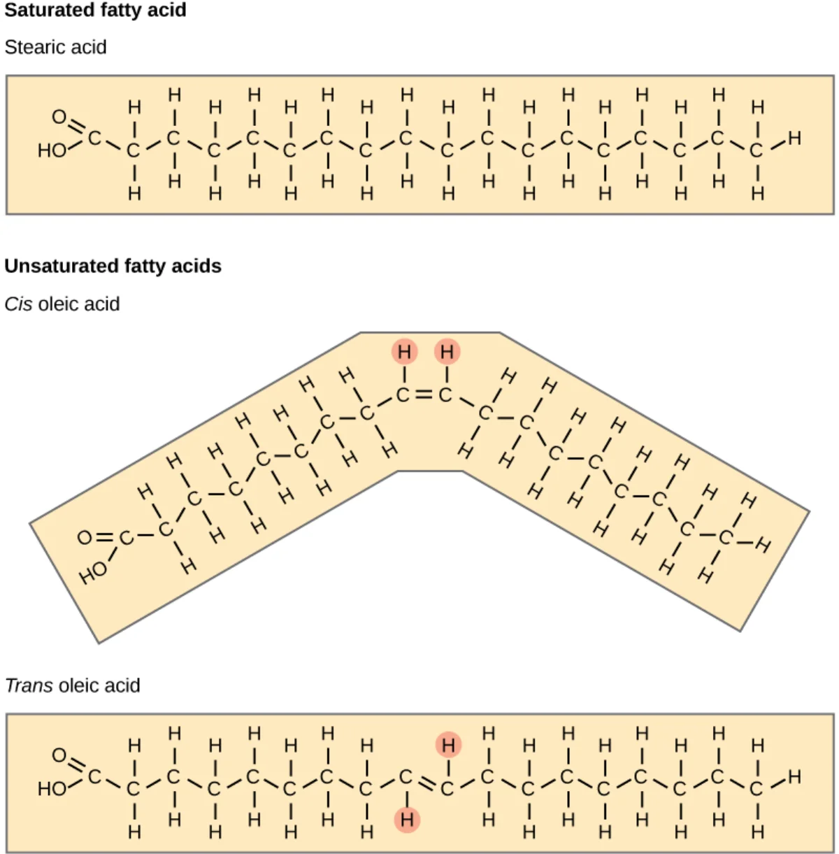
Saturated fatty acid: no carbon-carbon double bonds. The chain is a straight rod. Packs tightly → solid at room temperature (butter, lard, tropical oils).
Unsaturated fatty acid: one or more C=C double bonds. Each cis double bond puts a kink in the chain. Kinked chains cannot pack tightly → liquid at room temperature (olive oil, canola oil, fish oil).
Saturated fatty acids (top) pack tightly into solids. Unsaturated fatty acids (bottom) have kinks from cis double bonds and remain liquid. Credit: OpenStax Biology 2e, CC BY 4.0
Cis vs. Trans
Naturally occurring unsaturated fatty acids are almost always cis, which puts a sharp bend in the chain.
Trans fats are mostly industrial - produced by partial hydrogenation of vegetable oils. The trans double bond does NOT kink the chain, so trans fats behave more like saturated fats (solid, pack tightly). This is why trans fats raise LDL and lower HDL, contributing to cardiovascular disease. Most developed countries now restrict artificial trans fats.
Omega Numbering and Essential Fatty Acids
Fatty acids can be numbered two ways:
Delta numbering starts from the carboxyl (C1). A delta-9 double bond is between C9 and C10.
Omega numbering starts from the methyl end. Omega-3 means the first double bond is 3 carbons from the omega (methyl) end.
Alpha-linolenic acid (ALA), an 18-carbon omega-3 fatty acid. Blue numbers count carbons from the carboxyl end (delta numbering); red numbers count from the methyl (omega) end. The first double bond sits three carbons from the omega end - that is what "omega-3" means. An omega-6 fatty acid (like linoleic acid) would instead have its first double bond six carbons from the omega end. Credit: Wikimedia Commons, CC BY-SA
Humans cannot introduce double bonds past C9 from the carboxyl end, so we cannot synthesize omega-3 or omega-6 fatty acids. These are essential fatty acids and must come from the diet. Omega-3s (like alpha-linolenic acid, EPA, DHA) are found in fish and flaxseed. Omega-6s (like linoleic acid) are in most vegetable oils.
Why is butter solid at room temperature but olive oil liquid?
Click to reveal answer
Butter is rich in saturated fatty acids with straight, rod-like chains that pack tightly into a solid. Olive oil is rich in cis-unsaturated fatty acids (mainly oleic acid) whose cis double bond introduces a kink. Kinked chains cannot pack tightly, so the lipid is liquid at room temperature.
What does "omega-3 fatty acid" mean?
Click to reveal answer
The first double bond is 3 carbons from the methyl (omega) end of the fatty acid chain. Omega-3s include alpha-linolenic acid, EPA, and DHA. They are essential because humans cannot introduce double bonds past C9 from the carboxyl end and thus cannot synthesize them.
Why are trans fats considered unhealthy compared to cis unsaturated fats?
Click to reveal answer
A trans double bond keeps the chain straight, so trans unsaturated fatty acids pack tightly and behave like saturated fats. Dietarily, they raise LDL cholesterol and lower HDL, worsening cardiovascular risk. Cis double bonds kink the chain and prevent tight packing, preserving fluidity and the healthier metabolic profile of unsaturated fats.
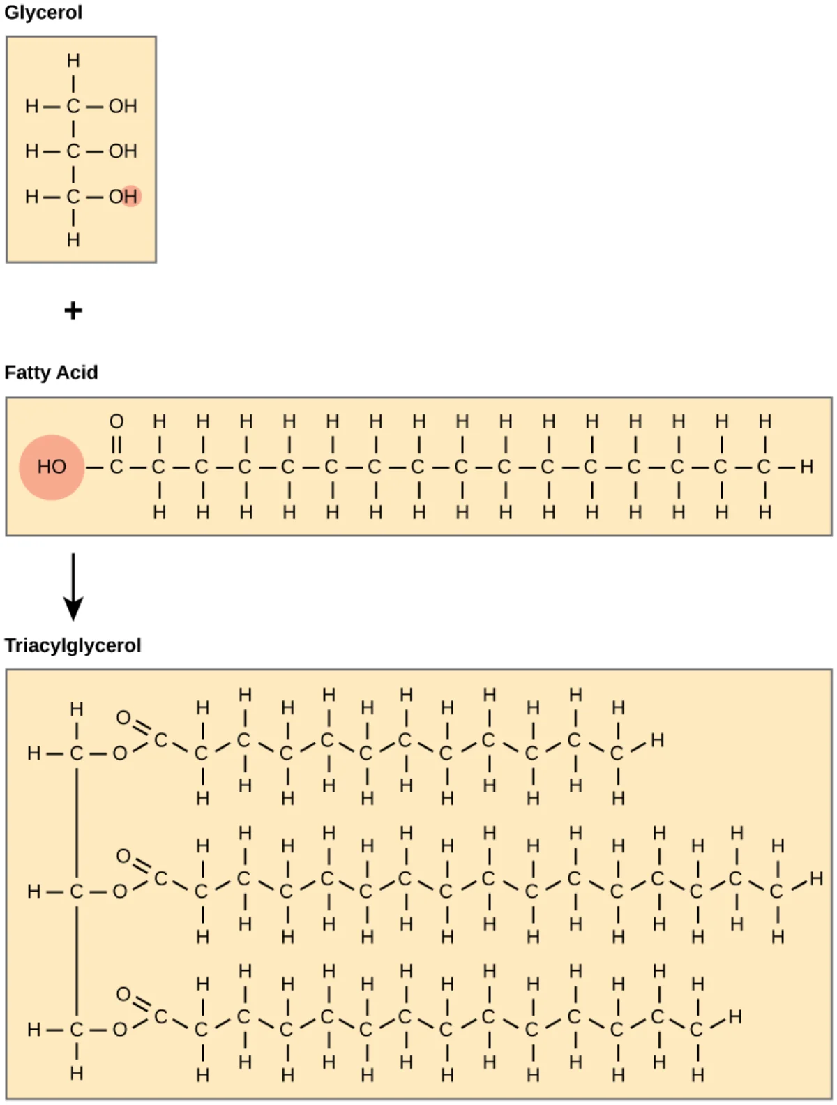
A triacylglycerol (triglyceride, TAG) is glycerol with three fatty acids attached by ester bonds. This is the body’s main form of stored energy.
Triacylglycerol. Glycerol (left) plus three fatty acids linked by ester bonds. The three fatty acids can be the same (simple TAG) or different (mixed TAG). Credit: OpenStax Biology 2e, CC BY 4.0
Why Fat is Energy-Dense
Fat yields about 9 kcal per gram.
Carbohydrate and protein yield about 4 kcal per gram.
Fat stores more than twice the energy per gram because its carbons are more reduced. Fatty acid chains are long stretches of C-H bonds with few oxygens. Each C-H bond is a high-energy electron waiting to enter the electron transport chain. Carbohydrates are already partially oxidized (lots of C-O bonds), so they have fewer electrons per gram to donate.
Fat is also stored without water. Glycogen is stored bound to water (about 2 g water per gram glycogen). A gram of fat stores pure energy with no extra water weight.
Storage and Release
Triglycerides are stored in adipocytes (fat cells) as a single large droplet. Mobilization happens in response to low-energy signals:
Glucagon and epinephrine activate hormone-sensitive lipase (HSL) in adipose via cAMP and PKA. HSL hydrolyzes triglycerides into glycerol and free fatty acids, which are released into blood.
Insulin opposes this. In the fed state, insulin inhibits HSL and activates lipoprotein lipase at capillaries, promoting fat storage.
The liberated fatty acids travel bound to albumin to tissues that need fuel. Glycerol goes to the liver, where it enters glycolysis as dihydroxyacetone phosphate (via glycerol kinase).
Why does fat yield about 9 kcal/g while carbohydrate and protein yield only about 4 kcal/g?
Click to reveal answer
Fatty acid chains are highly reduced - they are long stretches of C-H bonds with few oxygens. More C-H bonds mean more electrons to feed the electron transport chain, producing more NADH/FADH2 and therefore more ATP per gram. Carbohydrates are already partially oxidized (C-O bonds), so they release less energy per gram.
Which hormone activates hormone-sensitive lipase to mobilize fat from adipose tissue?
Click to reveal answer
Glucagon and epinephrine both activate HSL via the cAMP/PKA pathway in adipocytes. Insulin opposes this action, dephosphorylating (inactivating) HSL in the fed state. The net result is that fasting and stress mobilize stored fat, while the fed state stores it.
How does glycerol released from triglyceride breakdown enter energy metabolism?
Click to reveal answer
Glycerol travels to the liver, is phosphorylated by glycerol kinase to glycerol-3-phosphate, and then oxidized to dihydroxyacetone phosphate (DHAP). DHAP is an intermediate in glycolysis, so it can enter glycolysis or gluconeogenesis. Only about 5% of the energy in a triglyceride molecule comes from the glycerol backbone - most comes from the three fatty acid tails via beta-oxidation.
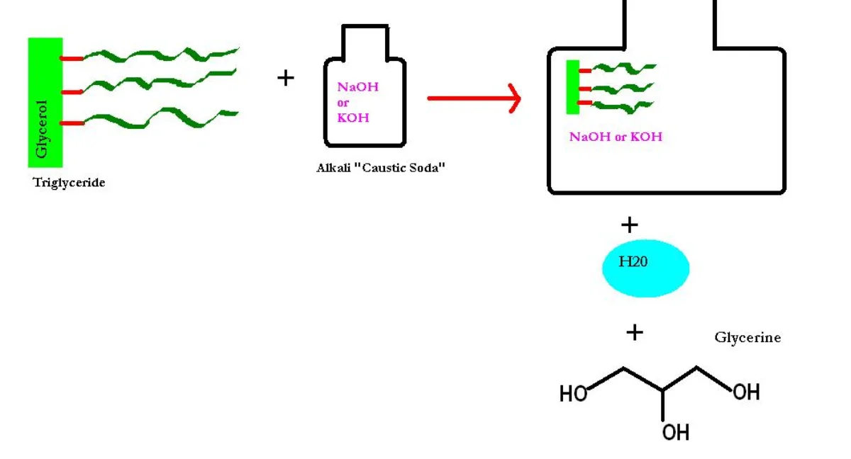
Saponification is the base-catalyzed hydrolysis of a triglyceride (or any ester) into its alcohol and the salt of its carboxylic acid. For a triacylglycerol, the products are glycerol and three fatty acid salts. The fatty acid salts are soap.
The lipid family tree
Lipid structure
1
Scroll sideways to see the whole map.
Saponifiable: contains an ester Non-saponifiable: no ester to cut The property they all share Amphipathic, so it builds membranes
What saponification actually isBase hydrolysis of an ester. Add NaOH to a triacylglycerol and you get glycerol plus three fatty acid salts, which are soap. Only lipids with an ester bond can do this, which is exactly why the tree splits there.
Why fat stores more energyFatty acids are almost entirely reduced carbon and hydrogen, so there is a great deal of oxidation left to do. They are also stored anhydrous, while glycogen drags about twice its own weight in water. Roughly 9 kcal per gram against 4.
The one definition that covers all of themA lipid is not defined by a structure, it is defined by a property: insoluble in water, soluble in organic solvent. That is why this family contains molecules as different as a soap, a hormone, and a vitamin.
Every lipid is here because of what it will not dissolve in, not because of what it is made of. The first split, saponifiable or not, is the only structural question worth asking, and it predicts whether a molecule stores energy or carries a message.Saponification. Triglyceride + NaOH (or KOH) → glycerol + 3 fatty acid salts. The salts are soap: amphipathic molecules with hydrophobic tails and ionic hydrophilic heads. Credit: Wikimedia Commons, CC BY-SA
Strong base (usually NaOH for hard soaps, KOH for soft soaps) attacks each ester carbonyl, cleaving the ester and leaving the carboxylate anion. Sodium (or potassium) counterions form the salt.
Why Soap Cleans
A soap molecule is amphipathic. One end is ionic (the carboxylate COO-), which loves water. The other end is a long hydrocarbon tail, which loves grease. When soap is added to water containing oil, the molecules arrange into micelles - little spheres with hydrophobic tails pointing inward (trapping oil) and hydrophilic heads facing out (in water). The oil is now suspended in water and can be rinsed away.
Saponification vs. Hydrolysis
Ester hydrolysis can happen in acidic or basic conditions. In the body, lipases (hydrolases) break ester bonds without base - they use water and acid/base catalysis within the active site. The pure-chemistry saponification reaction uses excess strong base. Both give the same products.
What products form when a triglyceride is saponified?
Click to reveal answer
Glycerol and three fatty acid salts (soap). The base (usually NaOH or KOH) hydrolyzes each ester bond on the glycerol backbone, producing glycerol and three free fatty acid anions that pair with the sodium or potassium counterion.
Why does soap remove oily dirt from a surface?
Click to reveal answer
Soap molecules are amphipathic: the carboxylate head is hydrophilic and the long hydrocarbon tail is hydrophobic. In water, soap molecules surround oil droplets with their hydrophobic tails pointing into the oil and their hydrophilic heads facing outward. The resulting micelle is water-soluble and can be rinsed away with the oil trapped inside.
Why does saponification go essentially to completion under strong base?
Click to reveal answer
The carboxylic acid product is deprotonated to the carboxylate anion under strong base. Carboxylate is stabilized (delocalized negative charge over both oxygens) and very unlikely to revert to the starting ester. The irreversible deprotonation pulls the equilibrium all the way toward products, making the reaction effectively one-way.
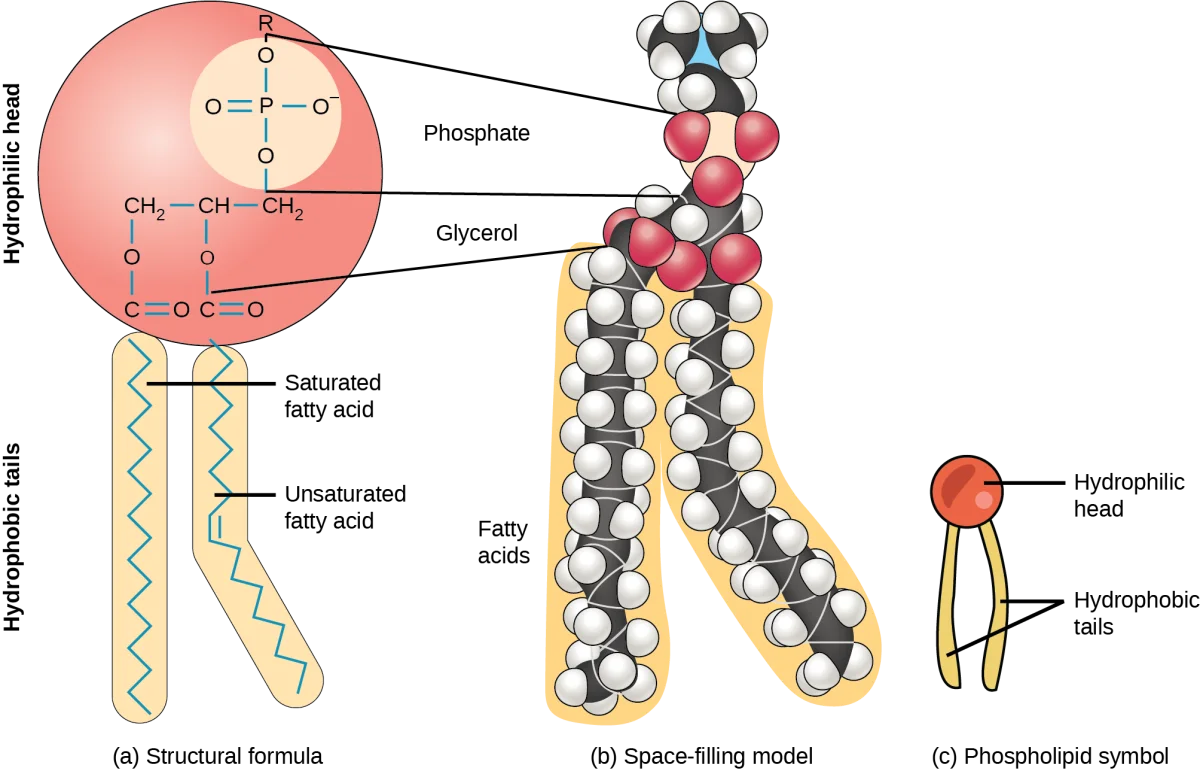
Phospholipids are the main structural lipids of cell membranes. A phospholipid looks like a triglyceride with one modification: instead of a third fatty acid, it has a phosphate group with a polar head group.
Structure
A phospholipid has:
A glycerol backbone.
Two fatty acid tails on C1 and C2 (one saturated, one unsaturated is common).
A phosphate on C3, linked to a polar head group (choline, ethanolamine, serine, inositol, or glycerol).
Phosphatidylcholine, the most abundant phospholipid. Glycerol + 2 fatty acids + phosphate + choline head group. Credit: OpenStax Biology 2e, CC BY 4.0
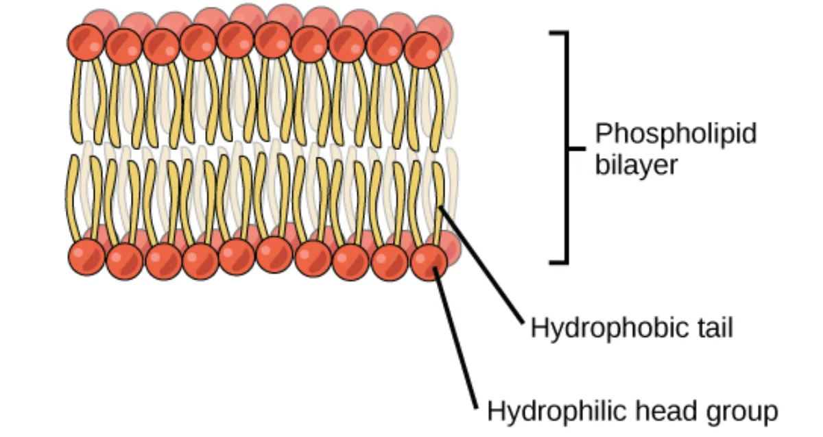
The phosphate head is hydrophilic (charged). The two fatty acid tails are hydrophobic. That makes phospholipids amphipathic - one end loves water, the other hates it.
The Bilayer
In water, amphipathic molecules assemble to hide their hydrophobic parts from water. For phospholipids, the lowest-energy arrangement is a bilayer: two layers with tails facing inward (touching each other, away from water) and heads facing outward (in contact with water on both sides).
The phospholipid bilayer. Hydrophilic heads face water on both sides; hydrophobic tails meet in the middle. This is the architecture of every cell membrane. Credit: OpenStax Biology 2e, CC BY 4.0
Common Head Groups
Different head groups give phospholipids slightly different properties and locations:
Phospholipid
Head group
Notes
Phosphatidylcholine (lecithin)
Choline
Most abundant; outer leaflet
Phosphatidylethanolamine
Ethanolamine
Abundant; inner leaflet
Phosphatidylserine
Serine
Inner leaflet normally; flipped to outer leaflet in apoptosis as an “eat me” signal
Phosphatidylinositol
Inositol
Inner leaflet; PIP2 is a signaling precursor cleaved to IP3 and DAG
Bilayer Fluidity
Membrane fluidity depends on:
Fatty acid saturation: more unsaturation (kinks) → more fluid. Saturated tails pack tightly → less fluid.
Chain length: longer tails interact more → less fluid.
Cholesterol: at high temperature it reduces fluidity (holds things in place). At low temperature it prevents the bilayer from becoming too rigid (keeps things moving). It is a bidirectional stabilizer.
Temperature: higher temperature → more kinetic motion → more fluid.
Why do phospholipids spontaneously form a bilayer in water?
Click to reveal answer
Phospholipids are amphipathic - they have a hydrophilic phosphate head and two hydrophobic fatty acid tails. To minimize the free energy of the system, they arrange so their tails are hidden from water (facing each other) and their heads are exposed to water (facing both aqueous compartments). The bilayer arrangement satisfies this condition with minimal energy cost.
What signal does phosphatidylserine give when it appears on the outer leaflet of a cell membrane?
Click to reveal answer
"Eat me." Phosphatidylserine is normally confined to the inner leaflet. During apoptosis, flippases and scramblases move PS to the outer leaflet. Macrophages recognize external PS as a signal to phagocytose the dying cell, so the cell is cleared without triggering an inflammatory response.
How does the degree of fatty acid saturation affect membrane fluidity?
Click to reveal answer
More unsaturated fatty acids mean more cis double bonds, which put kinks in the tails. Kinked tails cannot pack tightly, so the membrane is more fluid at a given temperature. Saturated tails pack tightly and produce a more rigid, gel-like membrane. Organisms living in cold environments often incorporate more unsaturated fatty acids into their membranes to stay fluid.
Sphingolipids are the second major class of membrane lipids (after glycerophospholipids). They are built on a sphingosine backbone rather than glycerol. They are especially abundant in nervous tissue - myelin is up to 30 percent sphingolipid.
The Building Blocks
Sphingosine: an 18-carbon amino alcohol with a trans double bond. Not a glycerol.
Ceramide: sphingosine plus a fatty acid attached via an amide bond to the C2 amine. Ceramide is the parent compound for all sphingolipids.
Sphingomyelin: ceramide + phosphocholine head group. A phospholipid of the sphingosine family.
Cerebrosides: ceramide + one sugar (glucose or galactose).
Sphingomyelin: sphingosine + fatty acid (amide-linked) + phosphocholine head. It is the main sphingolipid of the myelin sheath. Credit: Wikimedia Commons, CC BY-SA
Sphingolipids in Myelin and Signaling
Myelin wraps axons to speed electrical conduction. It is built of concentrated lipid layers with a high sphingolipid content, including sphingomyelin and glycosphingolipids. Damage to myelin (demyelination) slows nerve conduction - the clinical hallmark of multiple sclerosis.
Sphingolipids are also bioactive signaling molecules. Ceramide, sphingosine, and sphingosine-1-phosphate (S1P) regulate apoptosis, cell proliferation, and immune cell trafficking. S1P is the target of an approved multiple sclerosis drug class (S1P receptor modulators).
Lysosomal Storage Diseases
Sphingolipids are degraded in lysosomes by a chain of enzymes. A defect in any single enzyme causes the specific sphingolipid upstream to accumulate and damage the cell. These sphingolipidoses are classic genetic lysosomal storage diseases.
Disease
Enzyme deficient
Accumulated lipid
Notes
Tay-Sachs
Hexosaminidase A
GM2 ganglioside
Cherry-red macula, neurodegeneration in infancy
Gaucher
Glucocerebrosidase
Glucocerebroside
Most common; can be mild or severe
Niemann-Pick
Sphingomyelinase
Sphingomyelin
Severe neurological form in infants
Krabbe
Galactocerebrosidase
Galactocerebroside
Demyelinating
Fabry
Alpha-galactosidase A
Ceramide trihexoside
X-linked; angiokeratomas, kidney disease
What is the backbone of a sphingolipid, and how does it differ from a glycerophospholipid?
Click to reveal answer
Sphingolipids are built on sphingosine, an 18-carbon amino alcohol with a trans double bond. Glycerophospholipids are built on glycerol. Sphingosine has only one free hydroxyl available for a head group (plus its own built-in hydrocarbon tail), so sphingolipids pack slightly differently than glycerophospholipids but serve similar membrane roles.
Why are sphingolipids especially abundant in myelin?
Click to reveal answer
Myelin is an insulating wrap around axons, made of concentrated lipid bilayers with very few embedded proteins. Sphingolipids, particularly sphingomyelin and glycosphingolipids, stack into this dense electrical insulator. Demyelinating diseases like multiple sclerosis disrupt this sphingolipid-rich architecture.
What is the unifying mechanism behind sphingolipidoses like Tay-Sachs, Gaucher, and Niemann-Pick?
Click to reveal answer
Each is caused by a defect in a single lysosomal hydrolase in the sphingolipid degradation pathway. The specific sphingolipid upstream of the broken step accumulates inside lysosomes, eventually damaging the cell. The specific accumulated lipid and clinical picture depend on which enzyme is missing. Tay-Sachs: hexosaminidase A → GM2 ganglioside. Gaucher: glucocerebrosidase → glucocerebroside. Niemann-Pick: sphingomyelinase → sphingomyelin.
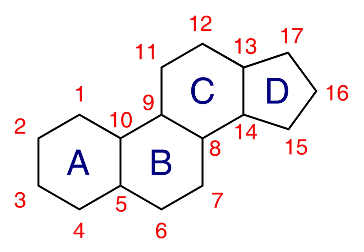
Cholesterol is the starting material for every steroid hormone, bile acids, and vitamin D. It is also a major component of cell membranes. All steroids share the same four-ring nucleus.
The Four-Ring Skeleton
The steroid nucleus: three six-membered rings (A, B, C) fused to one five-membered ring (D). Carbons are numbered 1-17. Every steroid in your body is built on this scaffold. Credit: Wikimedia Commons, CC BY-SA
Three six-membered rings (A, B, C).
One five-membered ring (D).
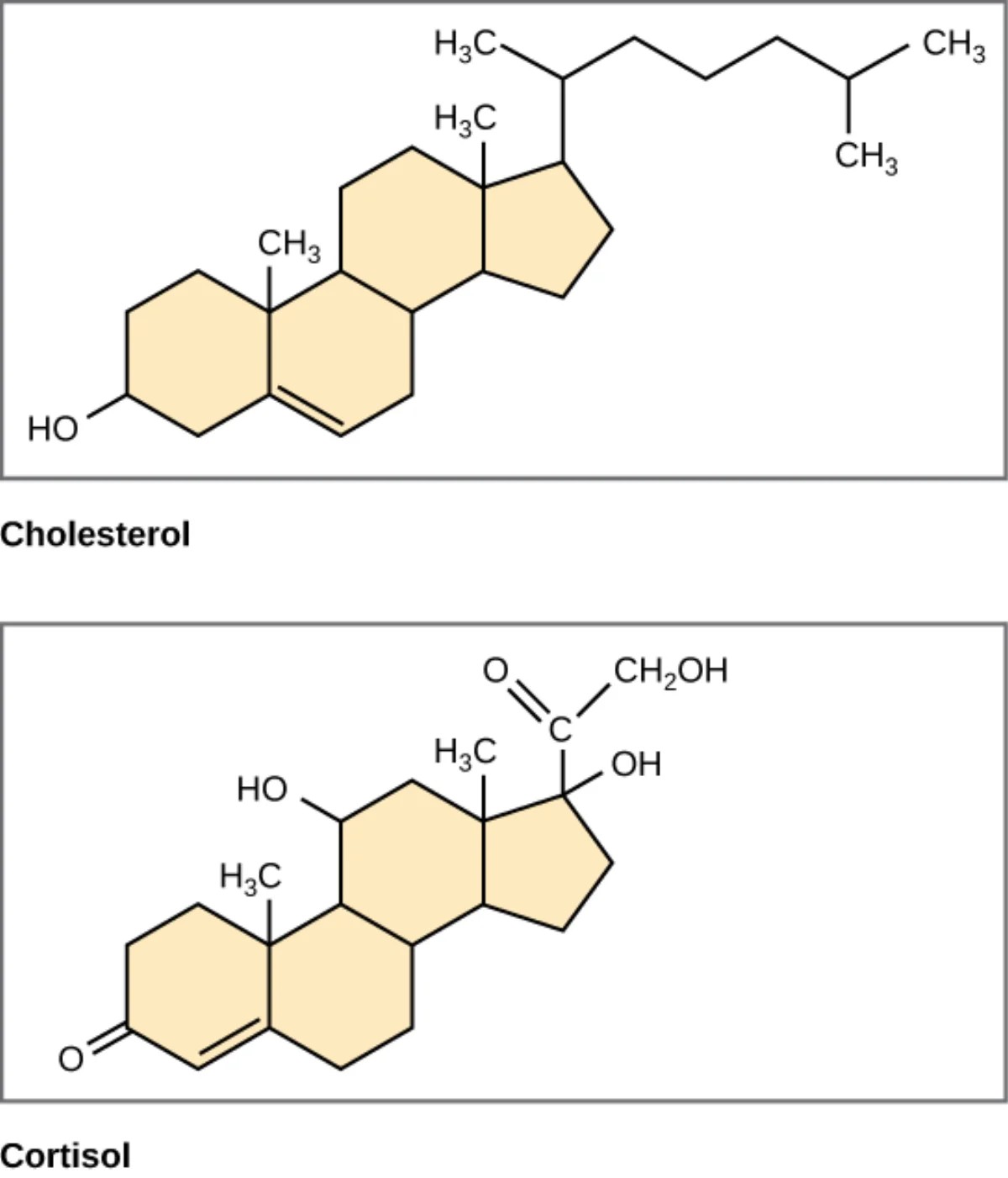
Various modifications (side chains, double bonds, -OH groups) on this core give each specific steroid.
Cholesterol
Cholesterol: steroid nucleus + hydroxyl at C3 + double bond between C5 and C6 + branched side chain at C17. Credit: Wikimedia Commons, CC BY-SA
Cholesterol inserts into the phospholipid bilayer with its -OH near the phosphate heads and its ring system parallel to the fatty acid tails. It acts as a bidirectional stabilizer of membrane fluidity:
At high temperature, cholesterol reduces fluidity by restricting motion of nearby fatty acid tails.
At low temperature, cholesterol prevents the bilayer from becoming too rigid by disrupting tight packing.
Most cells can make cholesterol themselves; the liver makes most of the body’s supply. HMG-CoA reductase is the rate-limiting enzyme of cholesterol synthesis. Statins inhibit HMG-CoA reductase.
The Major Steroid Hormones
All steroid hormones are synthesized from cholesterol in the adrenal cortex, gonads, or placenta. The five MCAT-relevant classes:
Cholesterol (left) and cortisol (right) share the same four-ring steroid scaffold. Every steroid hormone is a modification of cholesterol. Credit: OpenStax Biology 2e, CC BY 4.0
Cholesterol is oxidized in the liver to bile acids (cholic acid, chenodeoxycholic acid). Bile acids are amphipathic - they emulsify dietary fats in the small intestine, increasing the surface area accessible to pancreatic lipase. Without bile acids, fat absorption is poor (which is why patients with bile duct obstruction develop fatty stools, steatorrhea).
Vitamin D
Vitamin D is a “steroid hormone” technically, despite being called a vitamin. UV light on skin converts 7-dehydrocholesterol to cholecalciferol (vitamin D3). Two hydroxylations (one in liver, one in kidney) make active 1,25-dihydroxyvitamin D, which regulates calcium absorption and bone mineralization.
What four-ring structure is shared by all steroids?
Click to reveal answer
Three six-membered rings fused to one five-membered ring (labeled A, B, C, D). This steroid nucleus is the backbone of cholesterol, cortisol, testosterone, estradiol, aldosterone, bile acids, and vitamin D. Every steroid differs only in which functional groups decorate this core.
How does cholesterol affect membrane fluidity at high vs. low temperature?
Click to reveal answer
Cholesterol is a bidirectional stabilizer. At high temperature, it reduces fluidity by restricting fatty acid tail motion. At low temperature, it prevents the membrane from packing into a rigid gel by disrupting tight tail-tail interactions. Either way, cholesterol keeps membrane fluidity closer to a middle range.
How do steroid hormones produce their cellular effects, and how is this different from peptide hormones?
Click to reveal answer
Steroid hormones are lipid-soluble, so they diffuse across the plasma membrane freely. They bind intracellular (often nuclear) receptors that directly regulate gene transcription. Effects are slow (hours to days) but long-lasting. Peptide hormones bind surface receptors and trigger rapid second-messenger cascades - fast, short-lived effects.
Terpenes are a huge family of natural products built from repeating 5-carbon units called isoprene (2-methyl-1,3-butadiene). The number of isoprene units classifies the terpene.
The Isoprene Rule
Every terpene is assembled from isoprene units, head-to-tail. The number of units gives the class name:
Class
Isoprene units
Carbons
Example
Monoterpene
2
10
Menthol, limonene (citrus)
Sesquiterpene
3
15
Farnesol
Diterpene
4
20
Retinol (vitamin A), phytol
Triterpene
6
30
Squalene (cholesterol precursor)
Tetraterpene
8
40
Beta-carotene
Polyterpene
Many
Large
Rubber
Terpene vs. Terpenoid
Terpene: pure hydrocarbon made of isoprene units.
Terpenoid: a terpene with added oxygens or rearrangements (which is most of them in real biology).
These terms are often used interchangeably, but technically “terpenoid” is broader.
MCAT-Relevant Examples
Squalene (C30) is the direct precursor of cholesterol. Cyclization of squalene builds the four-ring steroid skeleton.
Beta-carotene (C40) is a tetraterpene found in carrots, spinach, and other orange/green vegetables. It is cleaved into two molecules of vitamin A (retinol, C20) in your intestines.
Vitamin A (retinol) (C20) is a diterpene alcohol. Its aldehyde form (retinal) is the visual pigment in rhodopsin; light-induced cis-to-trans isomerization of retinal is the first step of vision.
Coenzyme Q (ubiquinone) has a long polyprenyl side chain (isoprenoid), giving it lipid solubility to shuttle electrons through the inner mitochondrial membrane.
What is the structural building block of a terpene, and how many carbons does it have?
Click to reveal answer
Isoprene (2-methyl-1,3-butadiene), a 5-carbon unit. Terpenes are built by joining isoprene units head-to-tail. The number of isoprenes determines the class: monoterpene (2 isoprenes = 10 carbons), sesquiterpene (3 = 15), diterpene (4 = 20), triterpene (6 = 30, e.g., squalene), and so on.
What is the difference between a terpene and a terpenoid?
Click to reveal answer
Terpenes are pure hydrocarbons built from isoprene units. Terpenoids (sometimes called isoprenoids) are terpene derivatives with added oxygens, functional groups, or rearranged skeletons. Most biologically important "terpenes" you encounter are technically terpenoids. The terms are often used interchangeably.
Which terpene is the direct precursor to cholesterol?
Click to reveal answer
Squalene - a 30-carbon triterpene. Cyclization of squalene builds the four-ring steroid nucleus, from which cholesterol and all other steroids are derived. Mevalonate, upstream of squalene, is the product of HMG-CoA reductase - the enzyme blocked by statins.
Waxes are simple esters: one long-chain fatty acid + one long-chain alcohol. Both partners are long hydrocarbon chains, so waxes are hydrophobic, inert, and stable.
The fatty acid is usually 14-36 carbons. The alcohol is usually 16-36 carbons. Two very long tails = solid at room temperature and extremely water-repellent.
Examples
Beeswax (triacontanyl palmitate and related esters) - comb of bees.
Lanolin - on sheep wool, used in skin creams.
Carnauba wax - from palm leaves, used to polish cars and surfboards.
Plant cuticle - a waxy layer on leaves and fruit that prevents water loss.
Ear wax (cerumen) - a mixture of wax esters, sebum, and dead cells that traps debris in the ear canal.
Function
Waxes are waterproofing and protective. The hydrocarbon chains form a tight, hydrophobic coating that water cannot penetrate.
What chemical groups make up a wax?
Click to reveal answer
A wax is a single ester between a long-chain fatty acid and a long-chain alcohol. Both chains are long hydrocarbon tails (typically 14-36 carbons each), making waxes extremely hydrophobic and water-repellent.
Why does a leaf have a waxy cuticle?
Click to reveal answer
The cuticle wax forms a hydrophobic coating on the leaf surface that prevents uncontrolled water loss. Without the waxy layer, the leaf would dehydrate quickly. Many fruits (apples, grapes) have similar cuticular waxes that can often be seen as a whitish bloom on the surface.
Fats are hydrophobic; blood is water. Shipping fat through blood requires a carrier particle: a lipoprotein. Each lipoprotein is a tiny ball with a hydrophobic core of triglycerides and cholesteryl esters, surrounded by a shell of phospholipids, free cholesterol, and proteins called apolipoproteins.
The five main classes differ by density, size, and cargo.
Lipoprotein structure: hydrophobic lipid core inside a phospholipid/protein shell. Apolipoproteins (e.g., apoA, apoB, apoE) identify the particle to receptors in tissues. Credit: Wikimedia Commons, CC BY-SA
The Five Classes
Class
Made by
Main cargo
Density
Role
Chylomicron
Intestine
Dietary triglycerides
Lowest (biggest)
Transports dietary fat from gut to tissues
VLDL
Liver
Endogenous triglycerides
Low
Liver → tissues (fat delivery)
IDL
Plasma (VLDL → IDL)
Intermediate
Intermediate
Short-lived; becomes LDL
LDL
Plasma (VLDL → IDL → LDL)
Cholesterol
Higher
Delivers cholesterol to tissues
HDL
Liver and intestine
Cholesterol
Highest (smallest)
Brings cholesterol BACK to liver (reverse transport)
What Each Class Does
Chylomicrons
Made in the intestine from dietary fat. They carry triglycerides through lymph and blood to tissues. Lipoprotein lipase on capillary endothelium cleaves triglycerides in chylomicrons, releasing free fatty acids to adipose and muscle. The remaining “chylomicron remnant” is cleared by the liver.
VLDL
Made in the liver from endogenous triglycerides (assembled from fatty acids and carbohydrates). VLDL carries fat to tissues similarly to chylomicrons, via lipoprotein lipase. As triglycerides are removed, VLDL shrinks and gets denser, becoming IDL, then LDL.
LDL
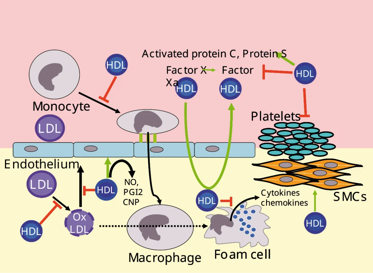
LDL is the cholesterol-delivery particle. Cells take up LDL via the LDL receptor (a classic example of receptor-mediated endocytosis). Dysfunctional LDL receptors (as in familial hypercholesterolemia) cause high blood LDL and early cardiovascular disease. Oxidized LDL is a major player in atherosclerotic plaque formation - it is taken up by macrophages that become foam cells in the artery wall.
HDL
HDL scavenges cholesterol from peripheral tissues (including atherosclerotic plaques) and returns it to the liver for disposal or reuse - reverse cholesterol transport. HDL is “good” because the more HDL, the more cholesterol can be cleared from tissues.
Apolipoproteins
The proteins on the surface identify the particle to receptors. A few you should recognize:
apoB-48: on chylomicrons. Made by intestine.
apoB-100: on VLDL/IDL/LDL. Made by liver. Binds the LDL receptor.
apoA-I: on HDL. Activates LCAT, the enzyme that esterifies free cholesterol for transport.
apoC-II: activates lipoprotein lipase (on chylomicrons and VLDL).
apoE: mediates clearance of chylomicron remnants and IDL by the liver.
Why is HDL called "good cholesterol" and LDL called "bad cholesterol"?
Click to reveal answer
LDL delivers cholesterol to peripheral tissues. High LDL means more cholesterol deposited in artery walls, leading to atherosclerosis. HDL performs reverse cholesterol transport: it scavenges cholesterol from tissues (including plaques) and returns it to the liver for clearance. High HDL is associated with a lower risk of heart disease.
Which lipoprotein transports dietary triglycerides from the intestine, and which transports endogenous triglycerides from the liver?
Click to reveal answer
Chylomicrons transport dietary triglycerides from the intestine. VLDL transports endogenous triglycerides (made in the liver from fatty acids and sugars). Both use lipoprotein lipase on capillary endothelium to release fatty acids to adipose and muscle tissue.
Why are lipoproteins necessary for fat transport in blood?
Click to reveal answer
Fats are hydrophobic and cannot dissolve in aqueous blood. Lipoproteins are amphipathic carriers - a hydrophobic lipid core surrounded by a shell of phospholipids, cholesterol, and apolipoproteins. The shell faces water, the core carries the fat, and apolipoproteins signal which tissue should receive the cargo.
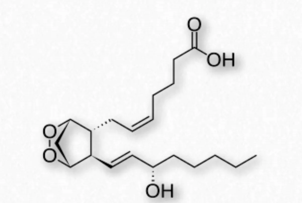
Eicosanoids are lipid signaling molecules made from arachidonic acid, a 20-carbon omega-6 polyunsaturated fatty acid. They do not travel far - they act locally on nearby cells and are rapidly degraded. Three main classes: prostaglandins, thromboxanes, and leukotrienes.
Prostaglandin H2, the precursor to all series-2 prostaglandins and thromboxanes. It contains the characteristic five-membered ring derived from arachidonic acid cyclization. Credit: Wikimedia Commons, CC BY-SA
The Arachidonic Acid Branch Point
Phospholipase A2 cleaves arachidonic acid from membrane phospholipids in response to cell damage or hormonal stimulation. Arachidonic acid then goes down one of two pathways:
COX pathway (cyclooxygenase): produces prostaglandins and thromboxanes.
Inhibits platelet aggregation, vasodilation (made by endothelium)
Thromboxane A2 (TXA2)
Promotes platelet aggregation, vasoconstriction (made by platelets)
Leukotriene B4
Neutrophil chemotaxis
Leukotriene C4, D4, E4
Bronchoconstriction, increased vascular permeability (role in asthma)
Prostacyclin and thromboxane are opposites - this balance determines whether a blood vessel clots or stays open.
Why NSAIDs Matter
NSAIDs (nonsteroidal anti-inflammatory drugs) block COX. No COX activity means no prostaglandin/thromboxane production, so:
Less PGE2 → less pain, less fever, less inflammation (therapeutic).
Less TXA2 → less platelet aggregation (why low-dose aspirin is used to prevent heart attack and stroke).
Less gastric PGE2 → less mucus protecting the stomach → NSAIDs can cause ulcers.
Examples: ibuprofen, naproxen, aspirin. Aspirin irreversibly acetylates COX; most others are reversible inhibitors.
Corticosteroids (like prednisone) inhibit phospholipase A2 indirectly (via lipocortin/annexin-1), so they block BOTH the COX and LOX pathways. That is why corticosteroids have broader anti-inflammatory effects than NSAIDs, but also more side effects.
What fatty acid is the precursor to most eicosanoids, and from where is it released?
Click to reveal answer
Arachidonic acid, a 20-carbon omega-6 polyunsaturated fatty acid. It is released from membrane phospholipids by phospholipase A2. Two enzymes branching from arachidonic acid - cyclooxygenase (COX) and lipoxygenase (LOX) - produce prostaglandins/thromboxanes and leukotrienes respectively.
How do NSAIDs reduce pain, fever, and inflammation?
Click to reveal answer
NSAIDs inhibit cyclooxygenase (COX), blocking the production of prostaglandins and thromboxanes. Less PGE2 means less activation of pain receptors and the hypothalamic fever response. Less TXA2 means less platelet activation. Aspirin specifically acetylates COX irreversibly; most other NSAIDs are reversible inhibitors.
Why do corticosteroids have broader anti-inflammatory effects than NSAIDs?
Click to reveal answer
NSAIDs inhibit only COX, so they reduce prostaglandins and thromboxanes while leaving leukotrienes unaffected. Corticosteroids indirectly inhibit phospholipase A2 (the enzyme that releases arachidonic acid in the first place), which shuts down BOTH COX and LOX pathways. That makes corticosteroids more comprehensive but also more prone to systemic side effects.
Four vitamins are fat-soluble: A, D, E, K. They are stored in body fat and the liver. Because they are not excreted readily, excess intake can be toxic (unlike water-soluble vitamins, where excess is urinated out). Absorption requires bile acids, so fat malabsorption (e.g., from pancreatic or biliary disease) can lead to deficiency.
Vitamin A (Retinol)
Role: vision (retinal is the chromophore in rhodopsin), growth, immune function, epithelial maintenance.
Deficiency: night blindness (the first sign), xerophthalmia (dry, damaged cornea), impaired immunity.
Sources: liver, eggs, dairy, carotenoids from orange/green vegetables (beta-carotene is cleaved into retinol).
Vitamin D (Cholecalciferol)
Role: calcium and phosphate homeostasis, bone mineralization. Technically a steroid hormone.
Deficiency: rickets in children (bowing of growing bones), osteomalacia in adults.
Sources: sunlight converts 7-dehydrocholesterol to cholecalciferol in skin; also in fatty fish, egg yolks, fortified milk.
Biochemically, vitamin D is activated by two hydroxylations: first in the liver (25-hydroxylation) and then in the kidney (1-alpha-hydroxylation) to produce the active 1,25-dihydroxyvitamin D. This active form binds nuclear receptors and upregulates genes for calcium absorption in the gut.
Vitamin E (Tocopherol)
Role: membrane antioxidant. Quenches lipid peroxyl radicals in membranes, protecting unsaturated fatty acids from oxidative damage.
Deficiency: hemolytic anemia (red cell membranes oxidize and lyse), neuromuscular symptoms (rare in developed countries).
Sources: vegetable oils, nuts, seeds.
Vitamin K (Phylloquinone)
Role: cofactor for gamma-carboxylation of clotting factors II, VII, IX, X, and proteins C and S. Without vitamin K, these factors cannot bind calcium and clot formation fails.
Deficiency: bleeding, bruising. Newborns are routinely given vitamin K at birth because they have limited stores and no gut bacteria yet.
Sources: leafy green vegetables, made by gut flora.
Warfarin, an anticoagulant, blocks vitamin K recycling (vitamin K epoxide reductase), leading to non-functional clotting factors and reduced coagulation.
Which four vitamins are fat-soluble, and why is it important that they are?
Click to reveal answer
Vitamins A, D, E, and K. Because they are fat-soluble, they require bile acids for intestinal absorption and can be stored in body fat and the liver. Excess can be toxic (unlike water-soluble vitamins, which are excreted in urine). Fat malabsorption (from pancreatic disease, biliary obstruction, etc.) causes deficiency of all four as a group.
What is the biochemical role of vitamin K, and how does warfarin interfere with it?
Click to reveal answer
Vitamin K is a cofactor for gamma-carboxylation of glutamate residues in clotting factors II, VII, IX, X (and proteins C and S). The gamma-carboxylation is required for the factors to bind calcium and participate in clotting. Warfarin blocks the recycling of vitamin K (inhibits vitamin K epoxide reductase), depleting the active form and producing non-functional clotting factors - the basis for warfarin's anticoagulant effect.
A patient with long-standing cystic fibrosis develops night blindness, bowing of the legs, and easy bruising. What is the unifying cause?
Click to reveal answer
Fat-soluble vitamin deficiency due to fat malabsorption. CF causes pancreatic insufficiency - not enough lipase and other digestive enzymes reach the gut. Dietary fat cannot be absorbed, so the fat-soluble vitamins go with it. Night blindness (vitamin A), bowing legs/osteomalacia (vitamin D), and easy bruising (vitamin K) are all classic. Vitamin E deficiency may also contribute.