
Transport & Amplification
We have covered what hormones do and where they come from. Now we need to address a question students often overlook: how do hormones actually get from their gland of origin to their target cell? And once they arrive, how does a single hormone molecule produce a massive cellular response?
The answers - carrier protein transport and signal amplification cascades - explain some of the most elegant and heavily tested physiology on the MCAT.
Hormone Transport in the Blood
How a hormone travels in the blood depends entirely on its solubility:
Water-soluble hormones (peptide hormones and catecholamines) dissolve freely in blood plasma. They do not need carrier proteins. They travel quickly, are rapidly degraded by plasma enzymes, and have short half-lives (minutes). This is why their effects are fast but brief.
Lipid-soluble hormones (steroid hormones and thyroid hormones) cannot dissolve in the aqueous blood. They must bind to carrier proteins for transport:
- Albumin - a general-purpose carrier that binds many hormones loosely and nonspecifically
- Specific binding globulins - highly specific carriers:
- Cortisol-binding globulin (CBG/transcortin) for cortisol
- Sex hormone-binding globulin (SHBG) for testosterone and estrogen
- Thyroxine-binding globulin (TBG) for T3 and T4
Free vs. Bound Hormone: A Critical Distinction
Only free (unbound) hormone is biologically active. The hormone bound to a carrier protein is essentially in storage - it cannot bind to receptors or enter target cells. The carrier protein serves as a reservoir and a protective shell that prevents the hormone from being degraded or excreted.
This creates an equilibrium:
Bound hormone (inactive reservoir) ⇌ Free hormone (active)
At any moment, only a small percentage of total hormone is free - typically 1-10%. But as free hormone is used up or degraded, more bound hormone dissociates from carriers to replenish the free pool. This buffer system smooths out fluctuations and extends the effective half-life of lipid-soluble hormones.
Signal Amplification: From One Molecule to Thousands
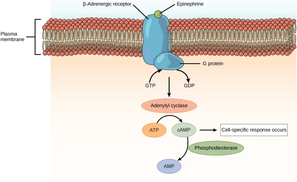
One of the most remarkable features of peptide hormone signaling is the amplification cascade. A single hormone molecule binding to a single receptor can ultimately produce millions of product molecules inside the cell. Here is how:
Step 1: Hormone binds receptor - one hormone molecule activates one receptor
Step 2: Receptor activates G-protein - one activated receptor can activate ~100 G-proteins (the receptor acts catalytically, activating one G-protein after another before the hormone dissociates)
Step 3: G-protein activates adenylyl cyclase - each G-protein activates one adenylyl cyclase enzyme
Step 4: Adenylyl cyclase produces cAMP - each adenylyl cyclase produces ~100 cAMP molecules per second
Step 5: cAMP activates Protein Kinase A (PKA) - each cAMP activates PKA
Step 6: PKA phosphorylates target proteins - each PKA molecule can phosphorylate many substrate proteins
At each step, one molecule activates many downstream molecules. The amplification is multiplicative:
1 hormone - 1 receptor - ~100 G-proteins - ~100 adenylyl cyclases - ~10,000 cAMP molecules - ~10,000 PKA activations - ~100,000+ phosphorylated proteins
This is why epinephrine works at picomolar concentrations ( M) - you need vanishingly small amounts of hormone because each molecule generates a massive intracellular response.
Other Second Messenger Systems
The cAMP pathway is the most common, but other second messenger systems exist:
IP3/DAG pathway - Some GPCRs activate phospholipase C (PLC), which cleaves PIP2 into IP3 and DAG. IP3 triggers Ca2+ release from the ER. DAG activates protein kinase C (PKC). This pathway is used by some hormones like GnRH and oxytocin.
Receptor tyrosine kinase (RTK) pathway - Used by insulin, growth factors, and IGF-1. The receptor itself has kinase activity - hormone binding causes the receptor to autophosphorylate, creating docking sites for intracellular signaling proteins (like IRS for insulin). This activates the Ras-MAPK pathway (cell growth) and the PI3K-Akt pathway (metabolic effects like GLUT4 translocation).
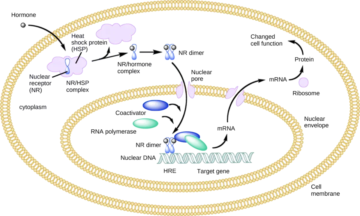
Intracellular receptor pathway - Steroid hormones and thyroid hormones use this pathway. No second messengers needed - the hormone directly binds a nuclear receptor that acts as a transcription factor. The “amplification” here comes from gene transcription: one activated gene can produce hundreds of mRNA copies, each translated into hundreds of protein copies.
Hormone Half-Life and Clinical Implications
A hormone’s half-life determines how quickly its effects can change:
| Hormone Type | Typical Half-Life | Why |
|---|---|---|
| Catecholamines | 1-2 minutes | Free in plasma, rapidly degraded by COMT and MAO |
| Peptide hormones | 4-40 minutes | Free in plasma, degraded by plasma proteases |
| Thyroid hormones | 1-7 days | Bound to TBG, protected from degradation |
| Steroid hormones | 60-90 minutes (cortisol) | Bound to carriers, but still cleared by liver |
Master Hormone Table
| Gland | Hormone | Type | Target | Key Actions |
|---|---|---|---|---|
| Anterior pituitary | GH | Peptide | Liver, bone | Growth via IGF-1, raises blood glucose |
| Anterior pituitary | TSH | Peptide | Thyroid | Stimulates T3/T4 production |
| Anterior pituitary | ACTH | Peptide | Adrenal cortex | Stimulates cortisol production |
| Anterior pituitary | FSH | Peptide | Gonads | Follicle growth / spermatogenesis |
| Anterior pituitary | LH | Peptide | Gonads | Ovulation, testosterone production |
| Anterior pituitary | Prolactin | Peptide | Mammary glands | Milk production |
| Posterior pituitary | ADH | Peptide | Kidneys | Water reabsorption |
| Posterior pituitary | Oxytocin | Peptide | Uterus, breast | Contractions, milk letdown |
| Thyroid | T3/T4 | Amino acid-derived | Most cells | Increase metabolic rate |
| Thyroid C cells | Calcitonin | Peptide | Bone, kidney | Lowers blood calcium |
| Parathyroid | PTH | Peptide | Bone, kidney, gut | Raises blood calcium |
| Adrenal cortex | Aldosterone | Steroid | Kidney | Na+ retention, K+ excretion |
| Adrenal cortex | Cortisol | Steroid | Most cells | Raises glucose, anti-inflammatory |
| Adrenal cortex | DHEA | Steroid | Peripheral tissues | Sex hormone precursor |
| Adrenal medulla | Epinephrine | Amino acid-derived | Heart, lungs, liver | Fight-or-flight response |
| Pancreas (beta) | Insulin | Peptide | Liver, muscle, fat | Lowers blood glucose |
| Pancreas (alpha) | Glucagon | Peptide | Liver | Raises blood glucose |
| Testes | Testosterone | Steroid | Many tissues | Male development, anabolic |
| Ovaries | Estrogen | Steroid | Many tissues | Female development, endometrial growth |
| Ovaries | Progesterone | Steroid | Uterus | Maintains endometrium, pregnancy |
| Pineal | Melatonin | Amino acid-derived | Brain | Circadian rhythm, sleep |
| Kidney | EPO | Peptide | Bone marrow | Red blood cell production |
| Kidney | Renin | Enzyme | Angiotensinogen | Initiates RAAS cascade |
| Heart | ANP | Peptide | Kidney, vessels | Lowers blood pressure, Na+ excretion |