Imagine you are cutting a pizza into smaller and smaller slices, but the pizza never gets bigger. That is cleavage. The fertilized egg divides rapidly - 1 cell becomes 2, then 4, then 8, then 16 - but the total mass stays the same. Each new cell is just a smaller piece of the original pie.
This section covers everything from the moment sperm meets egg to the moment the embryo burrows into the uterine wall and starts building the structures it needs to survive. Every step here is high-yield for the MCAT.
Fertilization
Fertilization typically occurs in the ampulla of the fallopian tube - the widest section, closest to the ovary. Hundreds of millions of sperm begin the journey, but only a few hundred reach the egg.
To reach the egg’s membrane, a sperm must penetrate two barriers:
Corona radiata - a layer of granulosa (follicle) cells surrounding the egg, providing nutrients and protection
Zona pellucida - a thick glycoprotein shell encasing the egg itself
The sperm breaks through using the acrosome reaction. The acrosomal apparatus - a specialized, membrane-bound vesicle at the tip of the sperm head - contains hydrolytic enzymes (including hyaluronidase and acrosin). When the sperm contacts the zona pellucida, the acrosomal membrane fuses with the sperm’s plasma membrane, releasing these enzymes. They digest the glycoproteins of the zona pellucida, clearing a path to the egg’s plasma membrane.
Once one sperm fuses with the egg membrane, two things happen immediately to prevent polyspermy (more than one sperm entering the egg). Polyspermy would create a triploid (or higher) embryo that cannot develop normally.
Fast block to polyspermy: The egg’s membrane rapidly depolarizes (shifts from a negative to a positive resting potential), electrically repelling other sperm within seconds. This is a transient change - it buys time for the permanent block.
Slow block to polyspermy (cortical reaction): A wave of calcium ions (Ca2+) is released from the endoplasmic reticulum inside the egg. This calcium signal triggers cortical granules - vesicles just below the egg’s membrane - to fuse with the plasma membrane and release their contents into the space between the membrane and the zona pellucida. These enzymes harden the zona pellucida into the fertilization membrane, permanently blocking additional sperm.
The sperm and egg nuclei (pronuclei) then merge, forming the zygote - the first cell of the new organism, containing the full diploid (2n = 46) chromosome count.
Twins: Dizygotic and Monozygotic
Twinning is a natural experiment in embryonic development - and a concept the MCAT uses to test your understanding of early cleavage and implantation.
Dizygotic (fraternal) twins form when two separate eggs are fertilized by two separate sperm during the same cycle. Each twin has its own placenta, its own chorion, and its own amnion. Genetically, they are as similar as any two siblings (sharing roughly 50% of their DNA). They are always dichorionic-diamniotic (di/di).
Monozygotic (identical) twins form when a single fertilized egg splits into two embryos. They are genetically identical (or near-identical). The timing of the split determines the membrane arrangement - and this is what the MCAT really cares about:
Timing of Split
Membrane Configuration
What Happens
Days 1-3 (before morula)
Dichorionic-diamniotic (di/di)
Split before trophoblast differentiates; each twin gets its own chorion and amnion
Days 4-8 (after trophoblast forms)
Monochorionic-diamniotic (mono/di)
Twins share one chorion (and placenta) but have separate amnions
Days 8-12 (after amnion forms)
Monochorionic-monoamniotic (mono/mono)
Twins share both chorion and amnion; higher risk
After day 12
Conjoined twins
Incomplete separation; twins are physically connected
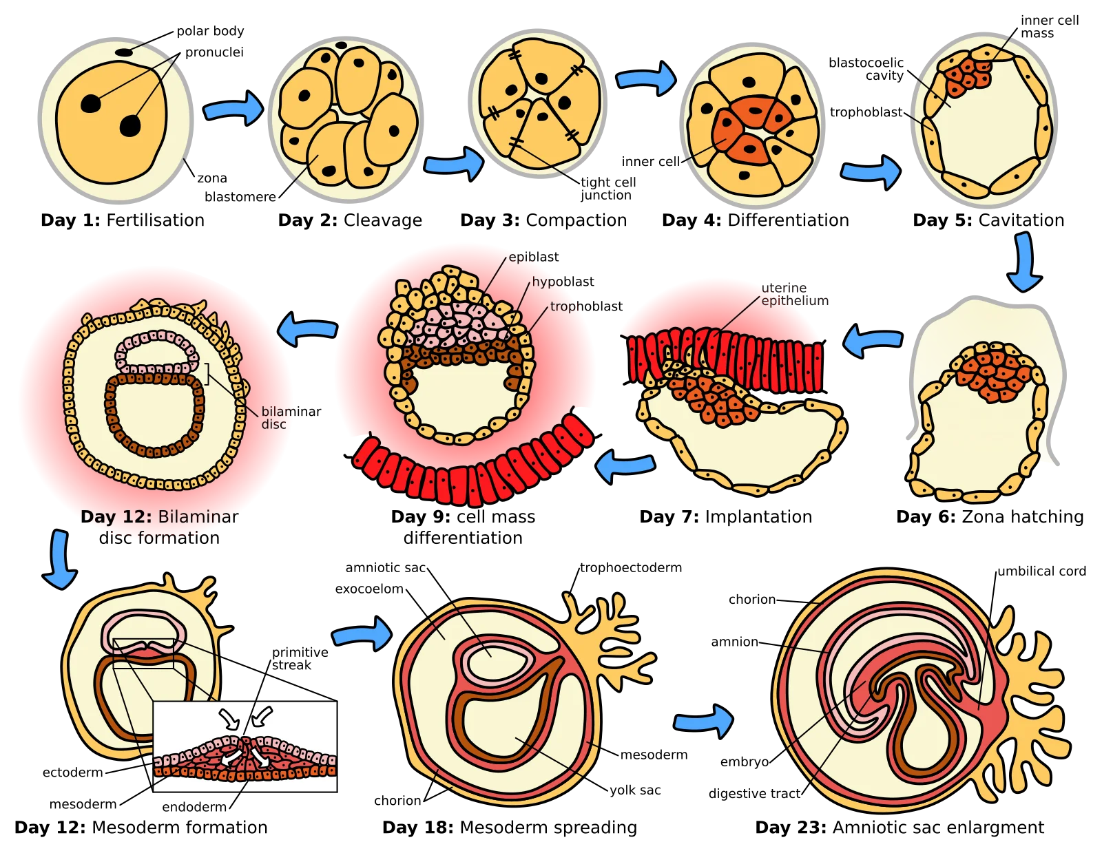
Early embryonic development: fertilization → cleavage → morula → blastocyst → implantation. Focus on: how cell number increases but total size stays the same during cleavage, and the inner cell mass vs. trophoblast distinction at the blastocyst stage. Credit: Wikimedia Commons, CC BY-SA 3.0
Cleavage: Dividing Without Growing
After fertilization, the zygote begins cleavage - a series of rapid mitotic divisions as it travels down the fallopian tube toward the uterus. This journey takes about 5-6 days.
The key feature of cleavage is that cells divide but the embryo does not grow. Each division produces smaller and smaller cells called blastomeres. Two ratios change during cleavage, and both are high-yield:
Increased nuclear-to-cytoplasmic (N:C) ratio: The zygote starts as one giant cell with a relatively small nucleus. As cleavage produces smaller cells, each cell’s nucleus takes up a larger proportion of the total cell volume. This gives the nucleus better control over cellular activity.
Increased surface area-to-volume ratio: Smaller cells have more membrane surface relative to their volume. This allows more efficient exchange of gases, nutrients, and waste products - essential for cells that lack a blood supply.
There are two types of cleavage that the MCAT distinguishes:
Type
Cell Fate
Key Feature
Example
Indeterminate
Each blastomere can become a complete organism
Explains identical twins
Humans, vertebrates
Determinate
Each blastomere is committed to a specific fate early
Cells cannot develop independently
Most invertebrates
Identical twins form when blastomeres from indeterminate cleavage separate early - each cell retains the ability to build an entire person. In organisms with determinate cleavage, losing a single blastomere means the embryo is missing an essential part.
The Morula
At around the 16-cell stage (about 3 days after fertilization), the embryo is a solid ball of cells called the morula (Latin for “little mulberry” - it looks like a mulberry fruit under the microscope).
The morula is still traveling down the fallopian tube at this point. No internal cavity has formed yet - it is a solid cluster of blastomeres compacted tightly together. The process of compaction flattens outer cells, which begin forming tight junctions with one another. This creates a distinction between outer cells and inner cells for the first time.
The Blastocyst (Blastula)
As the morula continues dividing, fluid-filled spaces appear between cells and merge to form a central cavity called the blastocoel. The morula has now become a blastula.
In mammals, we call this stage the blastocyst. It has two distinct regions:
Trophoblast: The outer ring of cells that will form the placenta and other extraembryonic structures. These cells will never become part of the baby.
Inner cell mass (ICM): A clump of cells on one side of the blastocoel that will become the embryo itself. These are the cells that contain the potential to form every tissue in the body.
Implantation
Around day 6-7 after fertilization, the blastocyst reaches the uterus and implants into the endometrium (uterine lining). The endometrium must be in the secretory phase (prepared by progesterone from the corpus luteum) to be receptive to implantation. Timing matters - if the blastocyst arrives too early or too late, implantation may fail.
During implantation:
The trophoblast cells invade the endometrial lining, burrowing into the tissue
The trophoblast differentiates into two layers: the inner cytotrophoblast and the outer syncytiotrophoblast (a multinucleated layer that directly contacts maternal blood)
The trophoblast forms the chorion, which develops finger-like projections called chorionic villi that anchor the embryo and vastly increase the surface area for nutrient and gas exchange with maternal blood
The placenta forms from the chorion (fetal tissue) combined with the decidua basalis (maternal uterine tissue)
Once implantation occurs, the trophoblast begins secreting human chorionic gonadotropin (hCG), which maintains the corpus luteum and its production of progesterone. This is the hormone detected by pregnancy tests.
The Umbilical Cord
The umbilical cord connects the developing fetus to the placenta and contains:
Two umbilical arteries - carry deoxygenated blood and waste FROM the fetus TO the placenta
One umbilical vein - carries oxygenated blood and nutrients FROM the placenta TO the fetus
This is the opposite of what you would normally expect: arteries typically carry oxygenated blood. But remember, arteries are defined by direction (away from the heart), not oxygen content. The fetal heart pumps blood to the placenta via the arteries, and the placenta returns oxygenated blood via the vein.
Extraembryonic Membranes
Four membranes develop to support the embryo. These are not part of the embryo’s body but are essential for its survival:
Membrane
Function
Chorion
Outermost membrane; forms the fetal part of the placenta; enables gas and nutrient exchange with maternal blood
Amnion
Creates the amniotic sac; filled with amniotic fluid that cushions the fetus, maintains temperature, and allows movement
Yolk sac
Produces the earliest blood cells and blood vessels; transfers nutrients to the embryo before the placenta is functional
Allantois
Involved in early waste removal; its blood vessels become the umbilical arteries and vein; contributes to bladder development
Summary: The Timeline
Time After Fertilization
Stage
Key Event
0 hours
Zygote
Sperm and egg nuclei fuse; diploid cell forms
Day 1-3
Cleavage
Rapid mitotic divisions; blastomere number increases
Day 3
Morula
Solid ball of ~16 cells; compaction begins
Day 4-5
Blastocyst
Blastocoel forms; trophoblast and ICM differentiate
Day 6-7
Implantation
Blastocyst burrows into endometrium
Day 7+
Placentation
Chorion and chorionic villi develop; placenta forms
A researcher separates a single blastomere from a 4-cell human embryo. Can this cell develop into a complete organism? Why or why not?
Click to reveal answer
Yes. Humans undergo indeterminate cleavage, meaning each blastomere retains the potential to develop into a complete organism during early division stages. This is why identical twins (and triplets) can form from separated blastomeres.
What are the fast and slow blocks to polyspermy, and how do they differ?
Click to reveal answer
Fast block: Membrane depolarization (electrical, occurs within seconds, transient). Slow block: Cortical reaction - calcium release triggers cortical granules to harden the zona pellucida into the fertilization membrane (chemical, takes about a minute, permanent). Both prevent more than one sperm from entering the egg.
What is the difference between the trophoblast and the inner cell mass?
Click to reveal answer
The trophoblast is the outer layer of the blastocyst that forms the placenta and other extraembryonic structures. The inner cell mass (ICM) is the cluster of cells inside the blastocyst that develops into the embryo itself. The trophoblast feeds and supports; the ICM builds the baby.
Monozygotic twins that split on day 5 will have what membrane configuration? What about a split on day 10?
Click to reveal answer
Day 5 split: Monochorionic-diamniotic (mono/di) - they share a chorion and placenta but have separate amnions (split occurred after trophoblast formed but before amnion formed). Day 10 split: Monochorionic-monoamniotic (mono/mono) - they share both chorion and amnion (split occurred after both membranes formed).
How many arteries and veins are in the umbilical cord, and what does each carry?
Click to reveal answer
Two umbilical arteries carry deoxygenated blood and waste away from the fetus to the placenta. One umbilical vein carries oxygenated blood and nutrients from the placenta to the fetus. Remember "AVA" - Arteries Away (2), Vein Arrives (1).
If early embryonic development is about making more cells, gastrulation is about organizing them. Think of it this way: cleavage built you a pile of bricks, and gastrulation is the architect showing up to arrange them into walls, floors, and a roof. This is where a hollow ball of nearly identical cells transforms into a three-layered structure with a clear body plan.
As the developmental biologist Lewis Wolpert famously said: “It is not birth, marriage, or death, but gastrulation which is truly the most important time in your life.” He was not exaggerating. Every organ you have - brain, heart, bones, gut, skin - traces directly back to one of the three layers established during gastrulation.
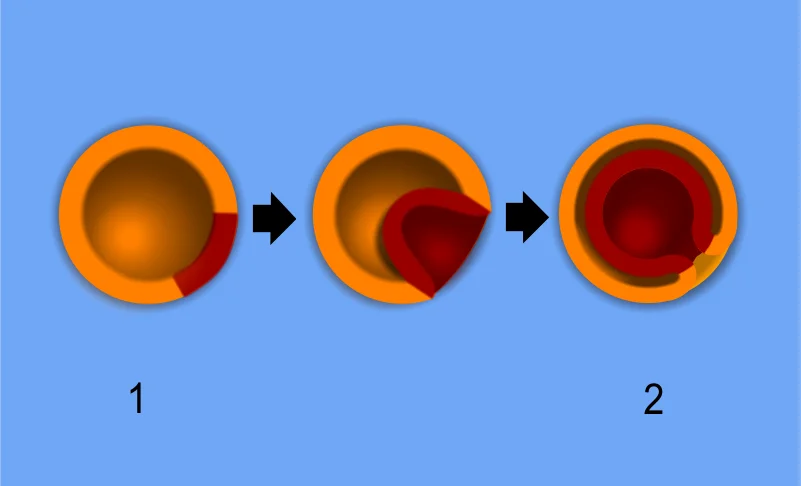
Gastrulation transforms a hollow blastula into a three-layered gastrula through invagination. Focus on: the three germ layers (ectoderm = outer, mesoderm = middle, endoderm = inner) and that the opening formed is the blastopore. Credit: Wikimedia Commons, Public Domain
What Happens During Gastrulation
Gastrulation occurs during the third week of human development. The blastula (a hollow ball of cells) reorganizes into a gastrula - a multi-layered structure with three distinct germ layers:
Ectoderm (outer layer) - “ecto” means outside
Mesoderm (middle layer) - “meso” means middle
Endoderm (inner layer) - “endo” means inside
The process begins with invagination - cells on the surface of the blastula fold inward, pushing into the interior. This inward folding creates a new internal pocket called the archenteron, the primitive gut cavity that will eventually develop into the lining of the digestive tract.
The opening of the archenteron to the outside is called the blastopore. The fate of this opening is one of the most tested embryology facts on the MCAT (more on that below).
Interactive 3D Gastrulation. Rotate to see how the three germ layers form during weeks 3-4 of human embryonic development.Credit: Deepankar.Parmar via Sketchfab, CC BY
Deuterostomes vs. Protostomes
The fate of the blastopore splits the animal kingdom into two major groups - and the MCAT expects you to know which group humans belong to.
In deuterostomes (“second mouth”), the blastopore becomes the anus, and the mouth forms as a second, separate opening. Humans, all vertebrates, and echinoderms (sea stars, sea urchins) are deuterostomes.
In protostomes (“first mouth”), the blastopore becomes the mouth, and the anus forms second. Insects, mollusks, annelids, and nematodes are protostomes.
Feature
Deuterostomes
Protostomes
Blastopore becomes
Anus (mouth forms second)
Mouth (anus forms second)
Cleavage type
Indeterminate (radial)
Determinate (spiral)
Coelom formation
Enterocoely (pouches from gut)
Schizocoely (splitting of mesoderm)
Examples
Humans, vertebrates, echinoderms
Insects, mollusks, worms
The Primitive Streak
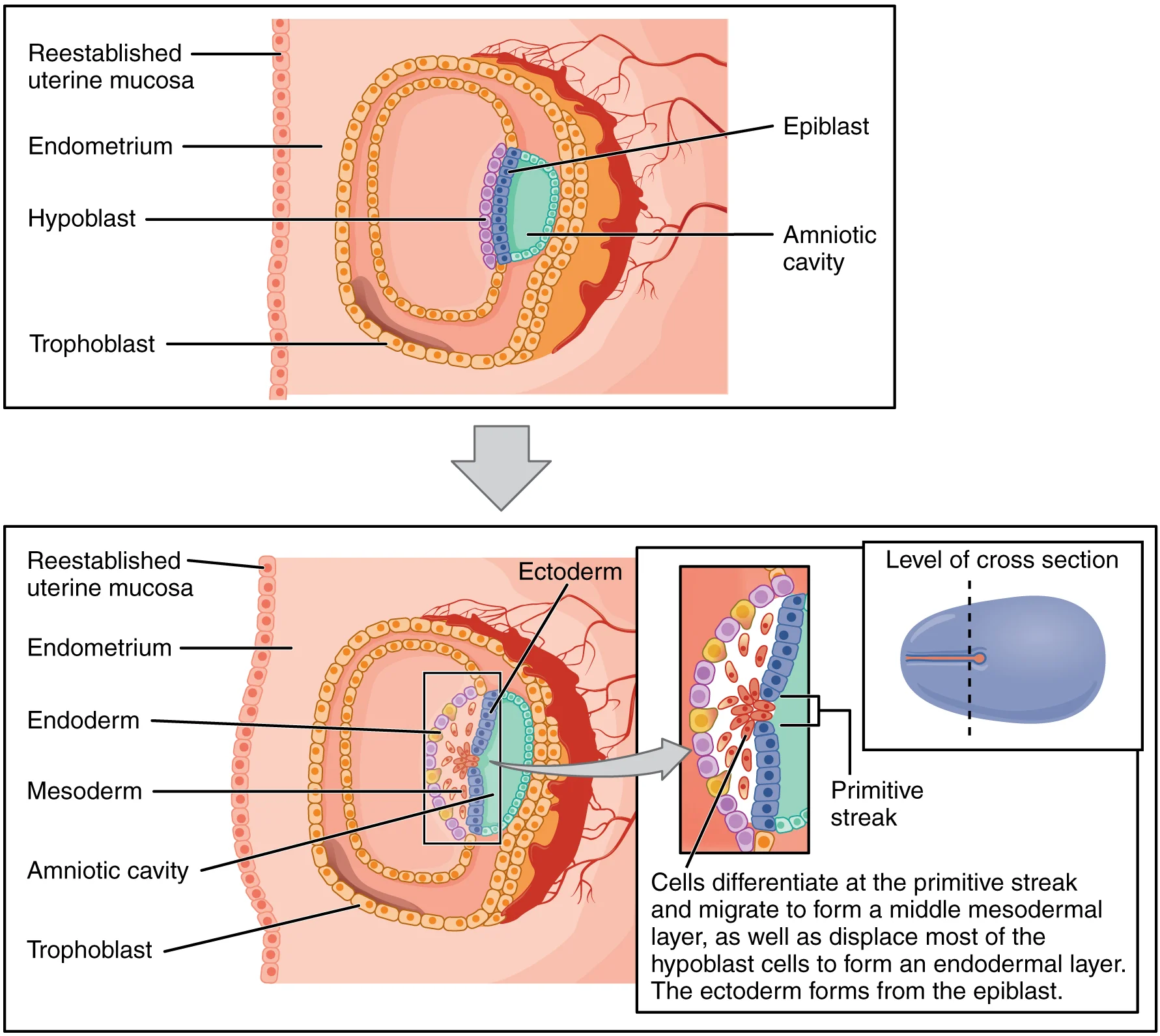
In mammals specifically, gastrulation does not happen by simple invagination of a hollow ball (that model applies to simpler organisms like sea urchins). Instead, the human embryo is a flat disc at this point, and gastrulation is initiated by the formation of the primitive streak - a groove that appears on the surface of the embryonic disc (the epiblast).
Cells on the surface migrate through this groove in a process called ingression:
Some cells move inward and displace the underlying layer to form the endoderm
Other cells migrate between the ectoderm and endoderm to form the mesoderm
The cells remaining on the surface become the ectoderm
The primitive streak also establishes the embryo’s body axes for the first time:
Anterior-posterior (head to tail) - the streak forms at the posterior end
Dorsal-ventral (back to belly)
Left-right symmetry
This is the moment the embryo goes from a featureless disc to having a defined “front,” “back,” “top,” and “bottom.” Before the primitive streak, the embryo has no recognizable orientation.
The node at the anterior tip of the primitive streak acts as the organizer - it secretes signaling molecules that direct surrounding cells to adopt specific fates. This is analogous to the Spemann-Mangold organizer discovered in amphibian embryos (covered in more detail in Section 3.8, Developmental Signaling).
The Three Germ Layers: A Quick Preview
Each germ layer will give rise to specific tissues and organs. The full list is covered in Section 3.5, but here is the overview you need to start connecting the dots:
Germ Layer
Position
Major Derivatives
Ectoderm
Outermost
Skin (epidermis), nervous system (brain, spinal cord, peripheral nerves), lens of the eye, inner ear, tooth enamel, adrenal medulla
Gut lining (epithelium of GI tract), lungs, liver, pancreas, thyroid, parathyroids, bladder lining
Gastrulation Errors
Disruptions during gastrulation can have devastating consequences because this is when the entire body plan is laid down. Teratogens (substances that cause birth defects) are particularly dangerous during gastrulation because they can affect the formation of all three germ layers simultaneously.
Alcohol exposure during the third week of development - when gastrulation is occurring - is one reason why fetal alcohol spectrum disorders can affect so many different organ systems. The damage is not limited to one germ layer; it hits the organizational blueprint itself.
In deuterostomes, what does the blastopore become? What about in protostomes?
Click to reveal answer
In deuterostomes (including humans and all vertebrates), the blastopore develops into the anus, and the mouth forms as a secondary opening. In protostomes (insects, mollusks, worms), the blastopore becomes the mouth, and the anus forms second. "Deutero" = second (mouth is second); "proto" = first (mouth is first).
What are the three primary germ layers, and what is each layer's position in the gastrula?
Click to reveal answer
Ectoderm (outermost layer) - forms skin and nervous system. Mesoderm (middle layer) - forms muscles, bones, blood, heart, kidneys. Endoderm (innermost layer) - forms gut lining, lungs, liver, pancreas. Remember: ecto = outside, meso = middle, endo = inside.
What is the archenteron, and how does it form?
Click to reveal answer
The archenteron is the primitive gut cavity formed during gastrulation when cells on the surface of the blastula invaginate (fold inward). It opens to the outside through the blastopore. The archenteron will eventually develop into the lining of the digestive tract. In deuterostomes, the blastopore (its opening) becomes the anus.
The adrenal cortex and adrenal medulla come from which germ layers, respectively?
Click to reveal answer
The adrenal cortex derives from mesoderm. The adrenal medulla derives from ectoderm (specifically, neural crest cells). This dual origin is a favorite MCAT topic because it tests deep understanding of germ layer derivatives rather than simple memorization.
Imagine you are rolling up a sheet of paper into a tube, then sealing it shut along the top edge. A few stray bits of paper fall away from the seam and scatter across the table. That rolled-up tube becomes your entire central nervous system. Those scattered scraps become an astonishing variety of other structures - from pigment cells in your skin to the core of your adrenal glands. That, in a nutshell, is neurulation.
Neurulation is the process by which the embryo builds the foundation of the nervous system. It begins immediately after gastrulation and is one of the most heavily tested embryology topics on the MCAT.
The Notochord: The Signal That Starts Everything
Before any neural tissue forms, a rod-shaped structure called the notochord must appear. Think of the notochord as a temporary scaffold - like the wooden frame builders erect before pouring concrete for a building. It provides structural support and, more importantly, sends the chemical messages that kick off the entire process.
The notochord is a mesodermal structure. It runs along the midline (head to tail) of the embryo and serves two critical functions:
Structural support - it acts as the embryo’s primitive spine, providing a flexible axis before vertebrae develop
Induction - it releases signaling molecules that instruct the ectoderm directly above it to thicken into neural tissue
In adult humans, the notochord almost entirely degenerates. The only remnant is the nucleus pulposus - the gel-like center of each intervertebral disc. When someone has a “herniated disc,” it is the nucleus pulposus that has bulged out of position.
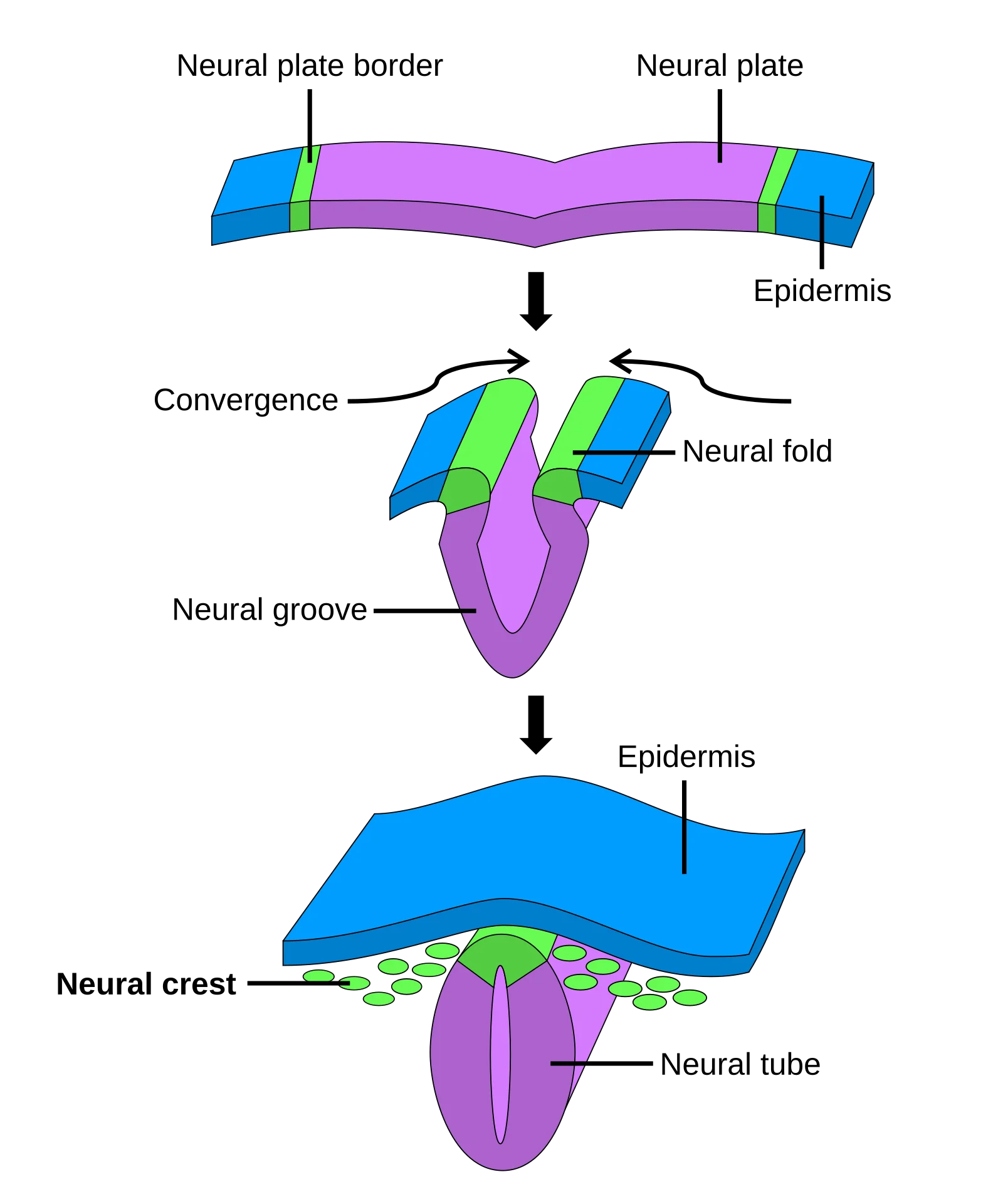
Neurulation: the neural plate folds into a groove, the folds rise and fuse to form the neural tube, and neural crest cells migrate away. Focus on: the notochord inducing the neural plate, and that neural crest cells give rise to PNS neurons, melanocytes, and adrenal medulla. Credit: Wikimedia Commons, Public Domain
Step-by-Step: How the Neural Tube Forms
Neurulation proceeds in an orderly sequence of four steps. Follow along with the “rolling paper” analogy:
Step 1 - Neural plate formation. Chemical signals from the notochord cause the overlying strip of ectoderm to thicken into a flat sheet called the neural plate. This is like laying out your sheet of paper flat on the table. These ectodermal cells are now committed to a neural fate.
Step 2 - Neural groove and neural folds. The center of the neural plate sinks inward along its midline, forming a depression called the neural groove. The raised edges on either side are called the neural folds. This is like pressing your finger along the center of the paper so the edges start to curl upward.
Step 3 - Neural tube closure. The neural folds continue rising, bend toward each other, and eventually meet and fuse at the top, creating a sealed hollow tube - the neural tube. Closure begins in the middle of the embryo and then zips shut toward both the head (anterior) and tail (posterior) ends. This is the moment you seal the top of the rolled paper.
Step 4 - Differentiation of the neural tube. Once closed, the neural tube develops into the entire central nervous system:
The anterior (head) end expands into three bulges that become the brain
The posterior portion becomes the spinal cord
The hollow interior becomes the ventricles of the brain and the central canal of the spinal cord, filled with cerebrospinal fluid
Neural Tube Closure and What Happens When It Fails
Neural tube closure happens remarkably early - around days 22 to 28 of development (weeks 3-4). The tube zips shut from the middle outward, meaning the anterior (head) end and the posterior (tail) end are the last parts to close. If either end fails to seal, devastating birth defects result.
Defect
What Fails to Close
Consequence
Spina bifida
Posterior (caudal) neuropore
Spinal cord remains partially exposed; severity ranges from a small gap in vertebrae (spina bifida occulta) to full exposure of the spinal cord (myelomeningocele)
Anencephaly
Anterior (cranial) neuropore
The brain fails to develop properly; incompatible with life
Folic acid (vitamin B9) supplementation before and during early pregnancy dramatically reduces the risk of neural tube defects. This is why prenatal vitamins contain high doses of folate, and why many countries fortify grain products with folic acid.
Neural Crest Cells: The “Fourth Germ Layer”
Here is where neurulation gets really interesting. As the neural folds rise and fuse to form the neural tube, a special population of cells at the very tips of the folds breaks free. These are neural crest cells, and they are unlike any other cells in the embryo.
Neural crest cells are derived from the ectoderm, but they do something no other ectodermal cells do - they migrate throughout the entire body and differentiate into a staggeringly diverse set of structures. Because of this extraordinary versatility, neural crest cells are sometimes called the “fourth germ layer.”
Here is what neural crest cells become:
Category
Neural Crest Derivatives
Peripheral nervous system
Sensory ganglia (dorsal root ganglia), autonomic ganglia (sympathetic and parasympathetic), Schwann cells (myelinate PNS axons), enteric nervous system (“gut brain”)
Endocrine
Adrenal medulla (chromaffin cells that produce epinephrine and norepinephrine), calcitonin-producing parafollicular (C) cells of the thyroid
Pigment
Melanocytes (pigment-producing cells of the skin, hair, and eyes)
Craniofacial
Bones and cartilage of the face and skull, odontoblasts (cells that make tooth dentin)
Cardiovascular
Smooth muscle of the great vessels (aorta, pulmonary artery), aorticopulmonary septum (divides the outflow tract of the heart)
Why Neural Crest Cells Are MCAT Gold
The MCAT loves neural crest cells because their derivatives are “unexpected.” Here are the traps:
Adrenal medulla comes from ectoderm (via neural crest), NOT mesoderm like the adrenal cortex. Same gland, different germ layers.
Melanocytes come from neural crest, NOT from the surface ectoderm that forms the epidermis. They live in the skin but did not originate there - they migrated in.
Schwann cells come from neural crest, NOT from the CNS. Oligodendrocytes (which myelinate CNS axons) come from the neural tube, but Schwann cells (which myelinate PNS axons) come from neural crest.
Calcitonin-producing C cells of the thyroid come from neural crest. The rest of the thyroid is endoderm. One gland, two origins.
If an MCAT question asks about the embryonic origin of something surprising, neural crest is very often the answer.
What structure signals the overlying ectoderm to form the neural plate, and what germ layer is it derived from?
Click to reveal answer
The notochord, which is derived from mesoderm. It runs along the midline of the embryo and releases chemical signals that induce the overlying ectoderm to thicken into the neural plate. In adults, the notochord's only remnant is the nucleus pulposus of intervertebral discs.
Name five structures derived from neural crest cells.
Click to reveal answer
Any five of: melanocytes, adrenal medulla (chromaffin cells), PNS sensory ganglia, autonomic ganglia, Schwann cells, enteric nervous system, craniofacial bones and cartilage, odontoblasts (tooth dentin), smooth muscle of great vessels, calcitonin-producing C cells of the thyroid. Use the mnemonic MOTEL CAPS.
What is the difference between spina bifida and anencephaly?
Click to reveal answer
Spina bifida results from failure of the posterior (caudal) neuropore to close, leaving the spinal cord partially exposed. Severity varies from mild (occulta) to severe (myelomeningocele). Anencephaly results from failure of the anterior (cranial) neuropore to close, preventing normal brain development - it is fatal. Both are neural tube defects that can be prevented by adequate folic acid intake.
Every cell in your body carries the same complete set of DNA. A liver cell has the genes for making brain proteins, and a skin cell has the genes for making hemoglobin. So why does a liver cell act like a liver cell and not a neuron? The answer is selective transcription - and it is the engine that drives organogenesis.
Think of your genome as a massive cookbook with 20,000+ recipes. Every cell owns a copy of the entire cookbook, but each cell type only opens to a specific chapter. A liver cell reads the “liver chapter,” ignoring the recipes for keratin and neurotransmitters. A neuron reads the “brain chapter,” skipping the recipes for bile and albumin. The cookbook is identical; the bookmark is different.
From Germ Layers to Organs
Gastrulation gave the embryo three germ layers. Neurulation built the beginnings of the nervous system. Now, during organogenesis (weeks 3 through 8 of human development), those three layers transform into every organ and tissue in the body.
By the end of week 8, the embryo has a recognizable human form with limb buds, a beating heart, and the foundations of every major organ system. At this point, the embryo is reclassified as a fetus.
Organogenesis is not a single event. It is a coordinated cascade of cell division, migration, differentiation, and programmed cell death - all controlled by selective gene expression.
Selective Transcription: The Master Switch
Every cell in the developing embryo contains the same genome. What makes a cell become a cardiomyocyte instead of a hepatocyte is which genes are turned on and which are silenced.
This process - called selective transcription or differential gene expression - is controlled by several mechanisms:
Transcription factors - proteins that bind to promoter or enhancer regions and activate (or repress) specific genes
Signaling molecules - neighboring cells release chemical signals (morphogens, growth factors) that tell a cell which transcription factors to produce
Epigenetic modifications - DNA methylation and histone modifications can permanently silence certain genes, locking a cell into its fate
Cytoplasmic determinants - molecules unevenly distributed in the egg cytoplasm during cleavage, giving daughter cells different starting instructions
Cell Specialization Is (Usually) a One-Way Street
As cells become more specialized, they typically lose the ability to become other cell types. This progressive restriction is called a loss of potency:
Potency Level
Can Become
Example
Totipotent
Any cell type + placenta
Zygote, cells up to ~4-cell stage
Pluripotent
Any cell type (not placenta)
Inner cell mass / embryonic stem cells
Multipotent
Several related cell types
Hematopoietic stem cells, neural crest cells
Unipotent
Only one cell type
Mature muscle satellite cells
Once a cell has differentiated (e.g., a mature red blood cell), it generally cannot reverse course. The genes it does not need have been epigenetically silenced.
The Danger Zone: Teratogens and Critical Periods
Because organogenesis involves such precisely timed gene expression and cell signaling, it is extremely vulnerable to disruption. A teratogen is any agent that causes abnormal development (birth defects).
The effect of a teratogen depends entirely on timing:
Period
Weeks
Vulnerability
Pre-embryonic
1-2
All-or-nothing: the embryo either dies or recovers completely
Embryonic (organogenesis)
3-8
Maximum vulnerability - organs are actively forming; disruption causes major structural defects
Fetal
9-38
Organs are formed but still maturing; teratogens cause functional defects and growth restriction, rarely major structural malformations
Each organ has its own critical period - the specific window when it is forming and most susceptible to damage. The heart’s critical period is earlier (weeks 3-6) than the brain’s (which extends through the fetal period because the brain continues developing after birth).
Common teratogens include:
Alcohol - the most common preventable cause of birth defects
Certain medications - isotretinoin (Accutane), valproic acid, warfarin
Beyond specific teratogen exposure, the overall health of the mother profoundly affects organogenesis:
Folic acid deficiency increases the risk of neural tube defects (spina bifida, anencephaly). Folate is essential for DNA synthesis and cell division - processes that occur at breakneck speed during weeks 3-4 when the neural tube is closing. This is why supplementation is recommended before conception.
Maternal diabetes (particularly poorly controlled pre-gestational diabetes) increases the risk of congenital heart defects, neural tube defects, and caudal regression syndrome. High glucose levels disrupt the delicate signaling cascades that guide organ formation.
Maternal infections can cross the placenta and directly damage developing organs. The classic teaching mnemonic is “TORCH” - Toxoplasmosis, Other (syphilis, Zika), Rubella, CMV, Herpes - but for the MCAT, the key concept is that timing of infection determines which organs are affected.
The Big Picture: Organogenesis in One Sentence
Every cell has the same DNA, but selective transcription ensures each cell reads only the genes it needs - and if anything disrupts this process during the critical period of organ formation (weeks 3-8), structural birth defects can result.
The specific organs each germ layer produces are covered in detail in the next section on germ layer derivatives.
All cells in the body have the same DNA. How does a liver cell "know" to act like a liver cell?
Click to reveal answer
Selective transcription (differential gene expression). Each cell type activates only the subset of genes relevant to its function and silences the rest through transcription factor activity, epigenetic modifications (DNA methylation, histone modification), and signaling from neighboring cells. No genes are lost during differentiation - they are only turned on or off.
Why is weeks 3-8 the most dangerous time for teratogen exposure?
Click to reveal answer
Weeks 3-8 is the period of organogenesis - when all major organs are actively forming. Each organ has a critical period during which it is most vulnerable to disruption. Teratogen exposure during this window causes severe structural birth defects. Before week 3, damage is all-or-nothing (embryo dies or recovers). After week 8, organs are formed but still maturing, so defects tend to be functional rather than structural.
What is a teratogen? Give three examples from different categories.
Click to reveal answer
A teratogen is any agent that causes abnormal embryonic or fetal development (birth defects). Examples: (1) Alcohol (drug/substance) - causes fetal alcohol syndrome. (2) Rubella virus (infection) - causes congenital heart defects, deafness, cataracts. (3) Radiation or mercury (environmental agent) - causes growth restriction and CNS damage. The severity depends on timing of exposure relative to each organ's critical period.
This is the section you will return to again and again. Knowing which germ layer produces which structure is one of the single highest-yield topics on the MCAT. The test loves to describe a disease, drug, or mutation affecting a specific tissue and then ask: “From which embryonic germ layer does this tissue derive?”
The good news? Germ layer derivatives follow a logical pattern. If you understand the pattern, you barely need to memorize anything.
The three germ layers established during gastrulation. Ectoderm (outer) becomes skin and nervous system, mesoderm (middle) becomes muscle, bone, and circulatory system, endoderm (inner) becomes gut lining and associated organs. Credit: OpenStax College, CC BY 3.0
The Burrito Model: Understanding the Pattern
Think of the embryo as a burrito.
The tortilla on the outside is the ectoderm. It wraps around everything and covers the exterior surface. Just like the tortilla is the part you see and touch, the ectoderm makes the structures that interact with the outside world - skin, sensory organs, and the nervous system.
The filling in the center is the endoderm. It is tucked deep inside, forming the inner tube. Just like the rice, beans, and meat run through the core of the burrito, the endoderm lines the gut tube and everything that buds off from it - digestive organs, lungs, liver, pancreas.
The cheese, sour cream, and guacamole layered between the tortilla and the filling represent the mesoderm. This middle layer provides structural support and “plumbing” - muscle, bone, blood, heart, kidneys. It fills in all the space between the outer wrapper and the inner core.
Ectoderm Derivatives: The Outer Layer
The ectoderm is the outermost germ layer. Its derivatives fall into three categories: surface structures, neural tube structures, and neural crest structures.
Surface Ectoderm - everything at the body’s surface and openings:
Structure
Notes
Epidermis (outermost skin)
NOT the dermis - that is mesoderm
Hair, nails, sweat glands, sebaceous glands
All skin appendages
Lens of the eye
The retina is neuroectoderm - different origin!
Inner ear
Sensory structures for hearing and balance
Epithelial lining of the mouth
The oral cavity is an ectodermal invagination
Epithelial lining of the anal canal
The distal anus is an ectodermal invagination
Epithelial lining of the nose
Another surface opening
Tooth enamel
Hardest substance in the body - ectoderm; dentin is neural crest
Anterior pituitary
From Rathke’s pouch - an upward ectodermal invagination from the mouth roof
Neuroectoderm (from the neural tube) - the entire central nervous system:
Structure
Notes
Brain
All regions
Spinal cord
Entire CNS
Retina
Outgrowth of the brain (diencephalon)
Posterior pituitary
Downward extension of the brain (infundibulum)
Pineal gland
Outgrowth of the diencephalon
Neural Crest Cells (from the tips of the neural folds) - the “fourth germ layer”:
Structure
Notes
PNS sensory ganglia (dorsal root ganglia)
Peripheral nervous system
Autonomic ganglia (sympathetic + parasympathetic)
Both divisions
Schwann cells
Myelinate PNS axons (NOT oligodendrocytes - those are neural tube)
Pigment cells; they migrate into the epidermis but originate from neural crest
Calcitonin-producing C cells (parafollicular cells)
In the thyroid gland
Craniofacial bones and cartilage
Facial skeleton
Odontoblasts
Make tooth dentin (enamel is surface ectoderm - one tooth, two origins!)
Enteric nervous system
”Gut brain” - neurons in the wall of the GI tract
Smooth muscle of great vessels
Aorta, pulmonary artery
Aorticopulmonary septum
Divides the cardiac outflow tract
Mesoderm Derivatives: The Middle Layer
The mesoderm is the middle germ layer, and it produces more different tissue types than either of the other two layers.
Musculoskeletal:
Structure
Notes
All skeletal muscle
Every voluntary muscle
All smooth muscle
Gut wall, blood vessels, bronchi (except great vessel smooth muscle = neural crest)
Cardiac muscle
The heart’s working tissue
Bones and cartilage
All axial and appendicular skeleton (except craniofacial = neural crest)
Tendons and ligaments
Connective tissue that attaches muscle to bone and bone to bone
Dermis
The deep skin layer; the epidermis above it is ectoderm
Circulatory and Blood:
Structure
Notes
Heart
The first functional organ - beating by day 22
All blood vessels
Arteries, veins, capillaries
All blood cells
Red blood cells, white blood cells, platelets
Spleen
Filters blood; mesodermal origin
Lymphatic system
Lymph nodes, lymph vessels
Bone marrow
Site of hematopoiesis
Urogenital:
Structure
Notes
Kidneys and ureters
Internal organs, but NOT endoderm
Gonads (testes and ovaries)
Mesodermal, even though they are internal
Adrenal cortex
The outer portion of the adrenal gland - NOT the medulla
Connective Tissue and Serous Membranes:
Structure
Notes
All connective tissue
Areolar, adipose, dense regular/irregular
Serous membranes
Pleura, peritoneum, pericardium
Muscular and connective tissue layers of the digestive and respiratory systems
The muscular wall of the gut is mesoderm; only the inner epithelial lining is endoderm
Endoderm Derivatives: The Inner Layer
The endoderm is the innermost germ layer, and it follows one beautifully simple rule: the endoderm lines the gut tube and everything that buds off from it.
Digestive Tract Lining:
Structure
Notes
Epithelial lining of the GI tract
Stomach, small intestine, large intestine
Exception: mouth and anal canal linings
These are ectoderm (surface openings)
Organs That Bud from the Gut Tube:
Structure
Notes
Liver and gallbladder
Bud off the foregut
Pancreas (exocrine and endocrine)
Buds off the foregut-midgut junction
Thyroid gland (follicular cells)
Buds off the floor of the pharynx (but C cells are neural crest!)
Parathyroid glands
Bud from pharyngeal pouches
Thymus
Buds from the third pharyngeal pouch
Respiratory System:
Structure
Notes
Epithelial lining of the trachea, bronchi, and lungs
The lungs bud off the foregut; the surrounding smooth muscle and cartilage are mesoderm
Other Endoderm Derivatives:
Structure
Notes
Urinary bladder lining
Endodermal; the muscular wall is mesoderm
Urethra lining
Distal portions
Epithelial lining of the auditory (Eustachian) tube
Connects middle ear to pharynx
Epithelial lining of the middle ear cavity
Derived from the first pharyngeal pouch
The Complete Comparison Table
Here is the master reference table. Use it for quick review before the exam.
These structures have germ layer origins that surprise students. Expect to see them on test day.
Structure
Germ Layer
Why It Is Tricky
Adrenal cortex
Mesoderm
Same organ as the medulla, but different germ layer
Adrenal medulla
Ectoderm (neural crest)
The inner part of the adrenal gland comes from the outer germ layer
Anterior pituitary
Surface ectoderm (Rathke’s pouch)
It is inside the skull, so it seems like it should be endoderm
Posterior pituitary
Neuroectoderm
Outgrowth of the brain - same gland, two embryonic origins
Kidneys
Mesoderm
You might think “internal organ = endoderm,” but kidneys do NOT bud from the gut tube
Dermis
Mesoderm
The deep skin layer is mesoderm; only the superficial epidermis is ectoderm
Tooth enamel
Surface ectoderm
Enamel is ectoderm; dentin is neural crest (odontoblasts) - one tooth, two germ layers
Melanocytes
Ectoderm (neural crest)
They live in the epidermis but did NOT originate there - they migrated in from the neural crest
Lens of the eye
Surface ectoderm
The retina is neuroectoderm, but the lens is surface ectoderm - same organ, different origins
C cells of the thyroid
Neural crest
The follicular cells that make T3/T4 are endoderm; the C cells that make calcitonin are neural crest
Schwann cells
Neural crest
Oligodendrocytes (CNS myelin) are from the neural tube; Schwann cells (PNS myelin) are from neural crest
One Last Rule of Thumb
When you are stuck on a germ layer question, use this decision tree:
Does it line the gut tube or bud from the gut? —> Endoderm
Does it cover the surface, sense the outside world, or conduct electrical signals? —> Ectoderm
Is it structural, circulatory, or “in between”? —> Mesoderm
Is it surprisingly derived from something that migrated from the neural folds? —> Neural crest (ectoderm)
If you know the burrito model and the tricky exceptions, you can reason through almost any germ layer derivative question.
A researcher studies a tumor of melanocyte origin. From which germ layer do melanocytes derive?
Click to reveal answer
Ectoderm - specifically, neural crest cells. Melanocytes originate from the neural crest during development and migrate into the epidermis, hair follicles, and uvea of the eye. They are NOT derived from the same surface ectoderm that forms the epidermis, even though they reside there. This is a classic "unexpected origin" question.
The adrenal gland's cortex and medulla derive from which germ layers, respectively?
Click to reveal answer
Adrenal cortex = mesoderm.Adrenal medulla = ectoderm (neural crest). The cortex produces steroid hormones (cortisol, aldosterone, androgens). The medulla produces catecholamines (epinephrine, norepinephrine) and is essentially made of modified postganglionic sympathetic neurons. One gland, two germ layers, two hormone classes.
Why does the liver derive from endoderm, even though it is a solid internal organ?
Click to reveal answer
The liver buds off the foregut (the primitive gut tube), which is lined by endoderm. The rule: all organs that develop as outpouchings of the gut tube - liver, pancreas, thyroid, lungs, gallbladder - derive from endoderm, regardless of whether they end up hollow or solid. If it buds from the gut, it is endoderm.
Kidneys are internal organs, but they are NOT endoderm. Why?
Click to reveal answer
Kidneys derive from mesoderm (specifically, intermediate mesoderm). The endoderm rule is "lines the gut and buds from the gut." Kidneys do NOT bud from the gut tube - they develop from mesodermal tissue alongside it. Similarly, the gonads (testes and ovaries) are internal organs that come from mesoderm, not endoderm.
Name three structures in the body that have dual germ layer origins.
Every cell in your body carries the same complete genome - roughly 20,000 genes in every nucleus. A liver cell, a neuron, and a red blood cell all contain identical DNA. So how does each cell “know” what to become?
The answer comes down to two related but distinct processes: first a cell commits to a fate, then it physically transforms to carry out that fate. Understanding the difference between these stages is one of the most commonly tested concepts in MCAT developmental biology.
Determination: The Irreversible Commitment
Determination is the point at which a cell becomes irreversibly committed to a particular developmental lineage. Before determination, a cell’s fate is flexible - transplant it to a new location and it will follow the cues of its new neighbors. After determination, the cell will develop along its committed path no matter where you place it.
Determination is an internal molecular event. You cannot see it by looking at the cell under a microscope. The cell still looks like its undetermined neighbors. What has changed is the pattern of transcription factors and epigenetic marks inside the nucleus.
A cell may pass through a reversible stage called specification before reaching full determination. Specified cells are leaning toward a fate but can still be redirected by new environmental signals. Determined cells cannot.
Stage
Reversible?
Visible Change?
Key Feature
Specification
Yes
No
Cell is influenced by local signals but can switch if moved
Determination
No
No
Internal commitment is locked in; transplantation does not change fate
Differentiation
No
Yes
Cell physically transforms - new proteins, new shape, new function
Differentiation: Becoming Specialized
Differentiation is the process by which a determined cell actually becomes its final, specialized cell type. This involves activating a specific subset of genes and producing the proteins, organelles, and structures that define that cell type.
Examples of differentiation in action:
A neuron precursor grows an axon and dendrites, synthesizes neurotransmitter receptors, and becomes electrically excitable
A red blood cell precursor produces massive amounts of hemoglobin and (in mammals) ejects its nucleus
A muscle cell precursor fuses with neighboring cells to form a multinucleated fiber packed with actin and myosin
The key mechanism behind differentiation is selective gene expression - also called differential gene expression.
Selective Gene Expression: Same Genome, Different Readout
Differentiation does not involve deleting genes. Every differentiated cell retains the full genome. Instead, cells activate different combinations of genes while keeping others silent.
This principle is proven by cloning experiments. Dolly the sheep was produced by transferring the nucleus of a mammary gland cell into an enucleated egg cell. That single nucleus, from a fully differentiated cell, contained all the genetic information needed to build an entire organism.
The molecular tools that control selective expression include:
Transcription factors - proteins that bind DNA regulatory regions and activate or repress gene transcription
Epigenetic modifications - chemical marks on DNA (methylation) or histone proteins (acetylation, methylation) that make genes more or less accessible without changing the DNA sequence
mRNA processing - alternative splicing allows a single gene to produce different protein variants in different cell types
Translational and post-translational control - regulating how much protein is made from each mRNA and how proteins are modified after synthesis
Induction: Cells Telling Other Cells What to Become
Cells do not determine their fate in isolation. They rely on signals from their neighbors. Induction is the process by which one group of cells (the inducer) sends signals that influence the developmental fate of a nearby group (the responder).
For induction to work, the responder must be competent - it must express the right receptors and intracellular signaling machinery to detect and respond to the inducer’s signal. Without competence, the signal goes unnoticed.
The classic demonstration is the Spemann-Mangold organizer experiment. In amphibian embryos, Hans Spemann and Hilde Mangold transplanted the dorsal lip of the blastopore to the opposite side of a host embryo. The transplanted tissue induced the host cells to form a complete second body axis - a second head, neural tube, and set of somites. This proved that one group of cells can redirect the fate of neighboring cells through induction.
Reciprocal Induction
In many cases, induction is not a one-way conversation. Reciprocal induction occurs when two tissues take turns inducing each other, with each signal triggering the next step in development.
The best example is vertebrate eye development:
The optic vesicle (an outgrowth of the developing brain) contacts the overlying surface ectoderm
The optic vesicle induces the surface ectoderm to thicken and form the lens placode
The developing lens placode signals back and induces the optic vesicle to invaginate, forming the optic cup (which becomes the retina)
The optic cup then induces the lens placode to detach and form the lens vesicle
Neither structure can form properly without the other. Remove the optic vesicle before it contacts the ectoderm, and no lens forms. Prevent the lens from forming, and the optic cup fails to develop correctly. This back-and-forth signaling is why reciprocal induction is sometimes called a “developmental dialogue.”
Cell-Cell Communication in Development
Developmental signals travel between cells through several distinct modes. The MCAT expects you to distinguish these based on the distance the signal travels and the mechanism of delivery.
Signaling Type
Distance
Mechanism
Example in Development
Autocrine
Self
Cell secretes a signal that binds its own receptors
Growth factor released by a cell stimulates its own proliferation
Paracrine
Short-range (nearby cells)
Signal diffuses locally through extracellular fluid
Morphogen gradients; most inductive signals during embryogenesis
Juxtacrine
Direct contact
Signal requires physical cell-cell or cell-matrix contact
Delta-Notch signaling between adjacent cells; lateral inhibition
Endocrine
Long-range (distant)
Signal travels through the bloodstream
Hormones from placenta affecting fetal development
Most developmental signaling is paracrine - short-range signals that diffuse through tissue. Morphogen gradients, which you will study in section 8, are the classic example.
Juxtacrine signaling is notable because it requires direct physical contact. The Delta-Notch pathway is the most important juxtacrine mechanism for the MCAT. When a differentiating cell expresses the Delta ligand on its surface, it binds Notch receptors on immediately adjacent cells and suppresses them from adopting the same fate. This creates the regular spacing patterns seen in hair follicles, sensory neurons, and other structures.
Putting It All Together
Development proceeds through a coordinated sequence: inductive signals from neighboring cells guide a responsive cell through specification, then determination, and finally differentiation. At each step, selective gene expression narrows the cell’s options until it is locked into its final identity.
This is not a one-time event. It happens at every stage of embryogenesis - during gastrulation, neurulation, and organogenesis - with increasingly refined signals directing increasingly specialized cell types.
What is the difference between determination and differentiation?
Click to reveal answer
Determination is the irreversible internal commitment of a cell to a specific developmental fate - the cell has decided what it will become, but it has not physically changed yet. Differentiation is the process of actually becoming that specialized cell type, with visible changes in structure, gene expression, and function (e.g., growing axons, producing hemoglobin). Determination = invisible decision. Differentiation = visible transformation.
Explain reciprocal induction using eye development as an example.
Click to reveal answer
In reciprocal induction, two tissues take turns signaling each other. During eye development: (1) the optic vesicle contacts the surface ectoderm and induces it to form the lens placode, (2) the developing lens signals back and induces the optic vesicle to invaginate into the optic cup (future retina). Neither structure forms properly without signals from the other - it is a two-way developmental dialogue.
What are the four types of cell-cell signaling, and how do they differ?
Click to reveal answer
Autocrine - a cell signals itself. Paracrine - signal diffuses locally to nearby cells (most developmental signaling, including morphogen gradients). Juxtacrine - requires direct cell-cell contact (e.g., Delta-Notch). Endocrine - signal travels through the bloodstream to distant targets. The key distinction is the distance over which the signal acts.
A fertilized egg can become literally anything - a brain cell, a bone cell, a placenta cell, or even an entire identical twin. A blood stem cell in your bone marrow can become any blood cell type, but it will never become a neuron. A mature red blood cell cannot become anything else at all.
This spectrum of developmental potential is called potency, and understanding how it narrows during development is essential for the MCAT.
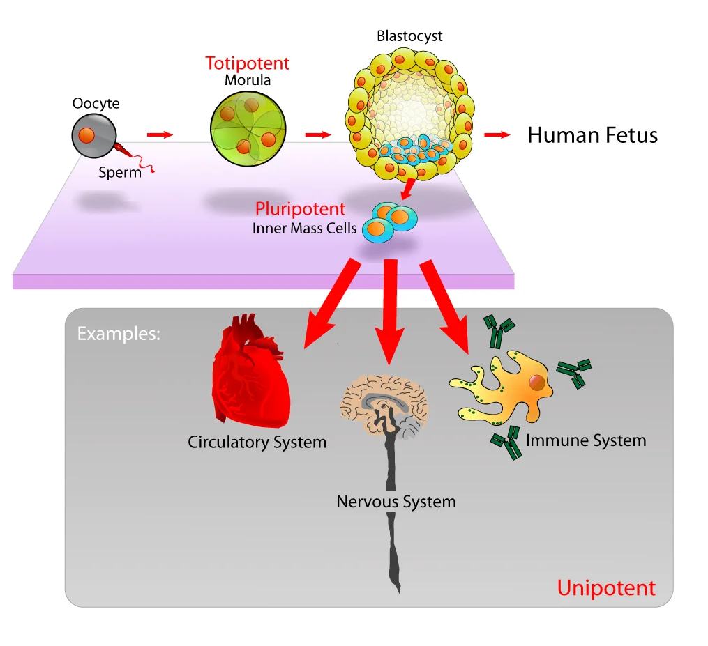
Stem cell potency hierarchy. Totipotent cells (zygote, early blastomeres) can become any cell type plus placenta. Pluripotent cells (inner cell mass) can become any body cell but not placenta. Multipotent cells (e.g., hematopoietic stem cells) are restricted to one lineage. Credit: Wikimedia Commons, CC BY-SA 2.5
The Four Levels of Potency
Level
Definition
Can Become
Example
Totipotent
Can form ANY cell type, including extraembryonic tissues
Embryo + placenta + everything
Zygote; cells up to ~4-cell stage
Pluripotent
Can form any cell type in the body, but NOT extraembryonic tissues
All 200+ body cell types, but not placenta
Inner cell mass of blastocyst; embryonic stem (ES) cells
The zygote and the cells from the first few cleavage divisions (up to about the 4-cell or 8-cell stage) are totipotent. They can form every cell type in the body AND the extraembryonic structures - the placenta, chorion, amnion, and yolk sac.
This is why identical (monozygotic) twins are possible. If the cells of an early embryo separate, each totipotent cell can develop into a complete organism with all its supporting structures.
Totipotency is lost once the blastocyst forms and its cells specialize into two populations: the trophoblast (committed to extraembryonic structures) and the inner cell mass (committed to the embryo proper).
Pluripotent Cells
The inner cell mass (ICM) of the blastocyst contains pluripotent cells. These can differentiate into any of the 200+ cell types derived from all three germ layers (ectoderm, mesoderm, endoderm) but can no longer form the placenta or other extraembryonic tissues.
Embryonic stem (ES) cells are pluripotent cells isolated from the inner cell mass and grown in culture. They are powerful research tools because they can be directed to become virtually any cell type under the right conditions.
Induced pluripotent stem cells (iPSCs) are adult differentiated cells that have been reprogrammed back to a pluripotent state. In 2006, Shinya Yamanaka demonstrated that introducing four specific transcription factors (Oct4, Sox2, Klf4, c-Myc - the “Yamanaka factors”) could convert adult skin cells into cells that behave like embryonic stem cells. This breakthrough proved that differentiation is based on gene silencing, not gene loss.
Multipotent Cells
Multipotent cells can generate several cell types, but only within a single lineage or tissue family. These are the “adult stem cells” that persist in mature tissues and provide ongoing repair and renewal.
Important examples:
Hematopoietic stem cells (in bone marrow) - can become red blood cells, all types of white blood cells, and platelets, but cannot become neurons or muscle cells
Neural stem cells (in the brain) - can become neurons, astrocytes, and oligodendrocytes, but cannot become blood cells
Mesenchymal stem cells - can become bone, cartilage, fat, and muscle cells
Unipotent Cells
Unipotent cells can self-renew (make copies of themselves) but only produce one differentiated cell type. Examples:
Muscle satellite cells - produce only new muscle fibers for repair after injury
Spermatogonial stem cells - produce only sperm precursors
Epidermal basal cells - produce only keratinocytes
Even unipotent cells are still considered stem cells because they retain the ability to self-renew. A fully differentiated cell that has lost the ability to divide (like a mature neuron) is not a stem cell at all.
When Potency Narrows During Development
Potency decreases in a predictable sequence as the embryo develops:
Zygote through ~4-cell stage - totipotent
Blastocyst inner cell mass - pluripotent
Germ layer formation (gastrulation) - cells become restricted to one germ layer’s lineages (multipotent)
Organogenesis and beyond - cells progressively narrow to tissue-specific progenitors and eventually unipotent or terminally differentiated cells
This narrowing is driven by the same mechanisms covered in the previous section: inductive signals trigger selective gene expression, epigenetic modifications lock in committed states, and determination becomes irreversible.
Embryonic vs. Adult Stem Cells
Feature
Embryonic Stem Cells
Adult Stem Cells
Source
Inner cell mass of blastocyst
Various tissues (bone marrow, brain, skin, gut)
Potency
Pluripotent
Usually multipotent or unipotent
Self-renewal
Essentially unlimited in culture
Limited (declines with age)
Differentiation range
Any body cell type
Restricted to their tissue lineage
Ethical considerations
Controversial (requires embryo destruction)
Fewer ethical concerns
Immune rejection risk
High (donor cells may not match recipient)
Low if patient’s own cells are used
Cell Migration During Development
Stem cells and their progeny do not always stay where they are born. Cell migration is essential for proper embryonic development. Cells must travel - sometimes across the entire embryo - to reach their final destination.
Key examples of developmental cell migration:
Neural crest cells migrate extensively from the dorsal neural tube to become melanocytes in the skin, neurons of the peripheral nervous system, adrenal medulla cells, and craniofacial cartilage
Primordial germ cells originate in the yolk sac wall and migrate through the gut mesentery to reach the developing gonads
Immune cell precursors migrate from the bone marrow to the thymus (T cell maturation) or remain in the bone marrow (B cell maturation)
Migration is guided by chemical signals (chemotaxis), adhesion molecules on cell surfaces, and components of the extracellular matrix. Errors in migration can cause developmental disorders - for example, failure of neural crest cell migration can result in Hirschsprung’s disease (absence of enteric ganglia in the colon).
Apoptosis: Programmed Cell Death
Development is not just about building new structures - it is also about strategic destruction. Apoptosis is a carefully regulated process in which cells self-destruct when they are no longer needed or when they pose a danger to the organism.
Think of apoptosis as a controlled demolition. The building (cell) is taken apart piece by piece, the debris is neatly collected, and the site is quietly cleared. Compare this to necrosis - an uncontrolled explosion where rubble flies everywhere and damages the surroundings.
Examples of apoptosis in development:
Digit separation - your fingers and toes begin as a solid paddle of tissue; apoptosis removes the webbing between them to create individual digits
Immune system pruning - T cells that react against self-antigens undergo apoptosis in the thymus (negative selection)
Neural sculpting - the developing brain overproduces neurons, then eliminates those that fail to make functional synaptic connections
Tail resorption - tadpoles lose their tails during metamorphosis via apoptosis
Cell swells; membrane ruptures; contents spill out
Inflammation
No - neighboring cells quietly engulf apoptotic bodies
Yes - released contents trigger an inflammatory response
DNA degradation
Orderly - cut into ~180 bp fragments (nucleosome-sized) by endonucleases
Random, uncontrolled degradation
Role
Normal development, tissue homeostasis, immune regulation
Pathological - response to disease, trauma, or ischemia
Cancer and Potency
Cancer cells often reactivate developmental programs that should be permanently silenced. Some tumors contain cancer stem cells - a subpopulation that can self-renew, resist chemotherapy, and regenerate the tumor even after treatment kills the majority of cells. This connection between stem cell biology and oncology is increasingly tested on the MCAT.
What is the key difference between totipotent and pluripotent cells?
Click to reveal answer
Totipotent cells can form ALL cell types including extraembryonic tissues (placenta, chorion, amnion). Pluripotent cells can form any cell type in the body (all three germ layers) but CANNOT form extraembryonic tissues. The zygote and early cleavage-stage cells are totipotent; the inner cell mass of the blastocyst is pluripotent.
How do apoptosis and necrosis differ in terms of inflammation?
Click to reveal answer
Apoptosis does NOT cause inflammation. The dying cell packages its contents into neat apoptotic bodies that are quietly engulfed by neighboring cells or macrophages. Necrosis DOES cause inflammation because the cell membrane ruptures, spilling intracellular contents into the surrounding tissue and triggering an immune response. This distinction is a key MCAT differentiator.
What are induced pluripotent stem cells (iPSCs), and what do they prove about differentiation?
Click to reveal answer
iPSCs are adult differentiated cells reprogrammed back to a pluripotent state by introducing four Yamanaka factors (Oct4, Sox2, Klf4, c-Myc). They prove that differentiation is based on gene silencing, not gene loss - the full genome remains intact in every differentiated cell. The silenced pluripotency genes can be reactivated, restoring the cell's ability to become any cell type.
How does a cell at the tip of your finger know it is at the tip of your finger and not in your liver? Cells cannot look around and figure out where they are. Instead, they read chemical signals in their environment - and the concentration of those signals tells each cell its position in the embryo and, therefore, what it should become.
This is the molecular GPS system of development.
Morphogens: Positional Information Through Gradients
A morphogen is a signaling molecule that is secreted from a localized source and diffuses outward, forming a concentration gradient across the developing tissue. Cells respond differently depending on how much morphogen they detect.
How Gradients Specify Multiple Cell Types
A morphogen is produced at one location and diffuses outward. Cells close to the source encounter high concentrations; cells far away encounter low concentrations. Different concentration thresholds activate different gene programs:
High concentration - activates gene set A (e.g., ventral motor neurons)
Medium concentration - activates gene set B (e.g., interneurons)
Low concentration - activates gene set C (e.g., dorsal sensory neurons)
This means a single morphogen can pattern an entire tissue with multiple cell types. The information is encoded in the concentration, not just the identity of the molecule. This is an elegant and efficient system - one signal, many outcomes.
Growth Factors
Growth factors are secreted peptides that promote cell proliferation, differentiation, or survival. They bind to receptors on target cells (usually receptor tyrosine kinases) and activate intracellular signaling cascades.
Growth factors play overlapping roles with morphogens, but the MCAT distinction is useful:
Morphogens specify cell fate based on concentration gradients (positional information)
Growth factors promote proliferation and differentiation of cells that are already committed
For the MCAT, you do not need to memorize specific growth factor names. The key concept is that cells secrete peptide signals, those signals bind surface receptors, and the downstream cascade drives target cells to proliferate, differentiate, or migrate. Some molecules (like BMPs) function as both morphogens and growth factors - the categories are not mutually exclusive.
Homeobox (Hox) Genes: Master Regulators of Body Plan
If morphogens tell a cell where it is, homeobox genes tell a body segment what it should become. Homeobox genes (called Hox genes in vertebrates) are a family of master regulatory transcription factors that control segment identity along the anterior-posterior (head-to-tail) axis.
Key features of Hox genes:
1. Colinearity
Hox genes are arranged on the chromosome in the same order as the body segments they control. Genes at the 3’ end of the Hox cluster control anterior (head) structures. Genes at the 5’ end control posterior (tail) structures. The physical order of genes on the DNA mirrors the spatial order of segments in the body.
2. Homeodomain
Each Hox gene encodes a protein containing a homeodomain - a 60-amino-acid DNA-binding motif that functions as a transcription factor. This homeodomain recognizes and binds specific DNA sequences, activating or repressing the downstream genes that actually build the structures of that segment.
3. Extreme conservation
Hox genes are remarkably conserved across the animal kingdom. The same basic set of genes patterns body segments in fruit flies, fish, mice, and humans. A mouse Hox gene can even partially rescue a Hox mutation in a fly - a striking demonstration of evolutionary conservation over hundreds of millions of years.
Homeotic Transformations: When Hox Genes Go Wrong
Mutations in Hox genes cause homeotic transformations - one body segment develops the identity (and structures) of a different segment.
Classic examples:
In Drosophila, a mutation in the Antennapedia gene causes legs to grow where antennae should be
In Drosophila, the Ultrabithorax mutation causes a second pair of wings to develop where halteres (balancing organs) normally form
In mice, Hox gene knockouts can cause vertebrae to adopt the identity of adjacent segments (e.g., a lumbar vertebra developing ribs like a thoracic vertebra)
The key insight is that Hox genes specify segment identity, not structure construction. The building program executes correctly - it just runs the wrong program for that location.
Lateral Inhibition and Delta-Notch Signaling
Not all developmental signaling works through long-range gradients. Lateral inhibition is a mechanism where a cell that commits to a particular fate actively prevents its immediate neighbors from adopting the same fate.
This works through the Delta-Notch pathway:
A cell beginning to differentiate expresses the Delta ligand on its surface
Delta binds to Notch receptors on adjacent cells
Notch activation suppresses the differentiation program in those neighbors
Result: only scattered individual cells differentiate, creating a regular spacing pattern
Lateral inhibition produces the even spacing of hair follicles, sensory bristles in insects, and neurons in developing tissues.
Regeneration
Some organisms and tissues can replace lost structures after injury. The capacity for regeneration varies enormously.
Complete regeneration involves regrowing an entire complex structure with full function:
Salamanders can regenerate entire limbs, including bone, muscle, nerves, and blood vessels
Planaria (flatworms) can regenerate an entire organism from a small body fragment
Starfish can regenerate lost arms
Incomplete regeneration involves wound repair with scar tissue rather than full restoration of the original structure:
The human liver has remarkable regenerative capacity - it can regrow to its original size after up to 70% is removed, though this involves compensatory hyperplasia (existing cells enlarging and dividing) rather than true morphological regeneration
The human heart has very limited regenerative capacity - damaged cardiac muscle is replaced by scar tissue (fibrosis), which is why heart attacks cause permanent loss of function
Peripheral nerves can slowly regenerate if the cell body is intact, but CNS neurons generally cannot
The key MCAT distinction: tissues with high mitotic rates (skin, gut epithelium, blood cells, liver) regenerate well. Tissues with low or no mitotic activity (cardiac muscle, CNS neurons) regenerate poorly or not at all.
Senescence, Telomeres, and Telomerase
Cells do not divide forever. Cellular senescence is the state in which a cell permanently stops dividing but remains alive and metabolically active. Senescent cells accumulate with age and contribute to tissue aging.
A major driver of senescence is telomere shortening:
Telomeres are repetitive DNA sequences (TTAGGG in humans) that cap the ends of chromosomes, protecting them from degradation and fusion
Each round of DNA replication shortens the telomeres slightly because DNA polymerase cannot fully replicate the 3’ end of a linear chromosome (the “end-replication problem”)
When telomeres become critically short, the cell enters senescence or triggers apoptosis
Telomerase is a reverse transcriptase enzyme that extends telomeres by adding TTAGGG repeats. It is active in:
Germ cells (to maintain telomere length across generations)
Stem cells (to support ongoing self-renewal)
Most cancer cells (reactivation of telomerase allows unlimited division - a hallmark of cancer)
Most normal somatic cells have very low or no telomerase activity, which is why they have a finite number of divisions (the Hayflick limit, approximately 50-70 divisions for human cells).
Putting It All Together
Embryonic development is orchestrated by a hierarchy of signals. Morphogen gradients give cells positional information. Growth factors promote proliferation and differentiation of committed cells. Hox genes assign segment identity along the body axis. Lateral inhibition fine-tunes cell fate decisions among neighbors. And throughout all of this, the balance between cell proliferation, differentiation, migration, and apoptosis shapes the final organism.
After development is complete, these same signaling systems remain active at lower levels - maintaining tissue homeostasis, enabling regeneration where possible, and (when dysregulated) driving diseases like cancer.
What is a morphogen, and how does a single morphogen specify multiple cell types?
Click to reveal answer
A morphogen is a signaling molecule secreted from a localized source that diffuses outward, forming a concentration gradient. Cells respond differently depending on the concentration they detect: high concentration activates one gene set, medium activates another, and low activates yet another. This allows a single morphogen to pattern an entire tissue with multiple distinct cell types based on distance from the source.
What is colinearity in the context of Hox genes?
Click to reveal answer
Colinearity means Hox genes are arranged on the chromosome in the same order as the body segments they control along the anterior-posterior axis. Genes at the 3' end of the cluster control anterior (head) structures, while genes at the 5' end control posterior (tail) structures. The physical gene order mirrors the spatial order of body segments.
Why do most cancer cells express telomerase, and why is this significant?
Click to reveal answer
Normal somatic cells have limited telomerase activity, so their telomeres shorten with each division until the cell enters senescence (the Hayflick limit). Cancer cells reactivate telomerase, which extends telomeres and allows unlimited cell division - one of the hallmarks of cancer. This bypasses the normal proliferative countdown that protects against uncontrolled growth.
What is a homeotic transformation, and what type of gene mutation causes it?
Click to reveal answer
A homeotic transformation occurs when one body segment develops the structures of a different segment - for example, legs growing where antennae should be in Drosophila. It is caused by mutations in Hox (homeobox) genes. The key point is that the structure itself forms normally; it is simply the wrong structure for that location because the segment was given the wrong identity.
Think about a building under construction. The HVAC system and water filtration plant are not online yet, so the construction crew runs temporary hoses from the building next door to supply air and clean water. Once the building is finished and its own systems come online, the temporary hoses are disconnected.
That is exactly what fetal circulation does. The fetus’s lungs and liver are not yet functional, so the circulatory system builds temporary bypass routes - shunts - that redirect blood away from those organs. The placenta handles gas exchange and detoxification instead.
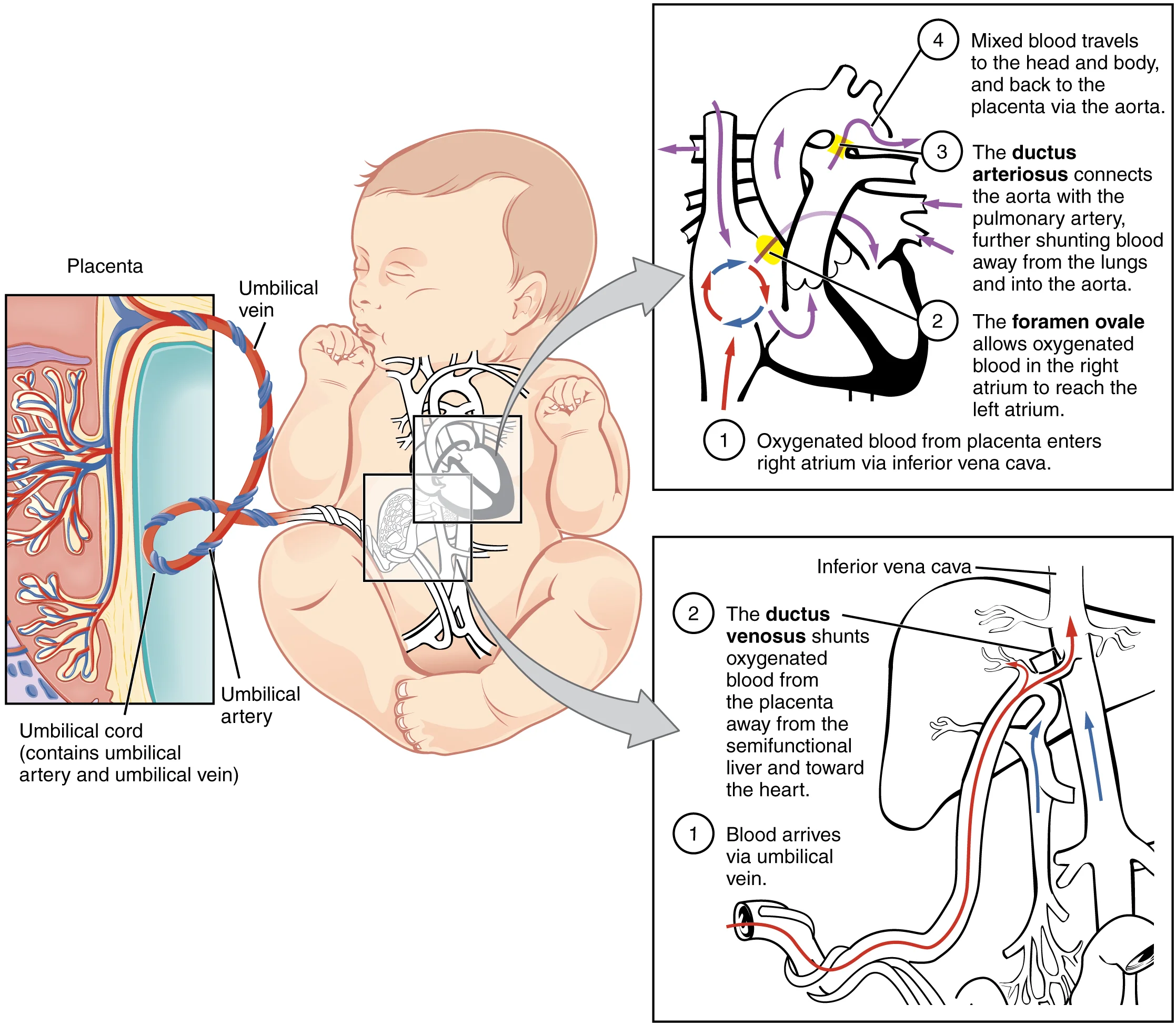
Fetal circulation with its three shunts. Focus on: ductus venosus (bypasses liver), foramen ovale (right atrium → left atrium, bypasses lungs), and ductus arteriosus (pulmonary artery → aorta, bypasses lungs). All three close at birth. Credit: OpenStax College, CC BY 3.0
Why Fetal Circulation Is Different
In adult circulation, the lungs oxygenate blood and the liver filters toxins from digested food. A fetus does neither of these things:
The fetal lungs are collapsed and filled with fluid. They do not perform gas exchange.
The fetal liver does not process nutrients from digestion because the fetus does not eat.
Both of these jobs fall to the placenta, which connects the fetus to the mother’s blood supply. Since the lungs and liver are offline, it would be wasteful to send large volumes of blood through them. The fetal circulatory system solves this problem with three temporary shunts.
The Placenta: Structure and Function
The placenta is the critical interface between mother and fetus. It is a disc-shaped organ embedded in the uterine wall, and it performs four major functions:
1. Gas exchange. Oxygen diffuses from maternal blood into fetal blood; carbon dioxide diffuses in the opposite direction. Maternal and fetal blood never mix directly - they flow in close proximity, separated by thin membranes, allowing diffusion across the placental barrier.
2. Nutrient and waste transfer. Glucose, amino acids, vitamins, and other nutrients cross from mother to fetus by diffusion and active transport. Fetal metabolic waste (urea, CO2) crosses back to the mother for disposal.
3. Immune protection. Maternal IgG antibodies cross the placenta, giving the fetus passive immunity. This is why newborns have temporary protection against diseases the mother has encountered.
4. Endocrine function. The placenta is a hormone-producing powerhouse:
Hormone
Role
hCG (human chorionic gonadotropin)
Maintains the corpus luteum in early pregnancy; basis for pregnancy tests
Progesterone
Maintains the endometrium; prevents uterine contractions; suppresses maternal immune rejection of fetus
Estrogen
Stimulates uterine growth; promotes blood flow to the uterus
Human placental lactogen (hPL)
Adjusts maternal metabolism to increase glucose availability for the fetus
Umbilical Vessels
The umbilical cord connects the fetus to the placenta and contains three vessels:
One umbilical vein - carries oxygenated, nutrient-rich blood FROM the placenta TO the fetus
Two umbilical arteries - carry deoxygenated, waste-laden blood FROM the fetus TO the placenta
Notice something counterintuitive: the umbilical vein carries oxygenated blood, and the umbilical arteries carry deoxygenated blood. This seems backwards compared to the normal rule (arteries = oxygenated, veins = deoxygenated), but it actually follows the true definitions perfectly.
The real definition of arteries and veins has nothing to do with oxygen content:
Arteries carry blood AWAY from the heart
Veins carry blood TOWARD the heart
The umbilical arteries carry blood away from the fetal heart (to the placenta). The umbilical vein carries blood toward the fetal heart (from the placenta). The naming is correct - it is only confusing if you rely on the oxygen-based shortcut instead of the true definitions.
The Three Fetal Shunts
Here is what each shunt does and where it connects:
1. Ductus venosus - bypasses the liver
Oxygenated blood from the placenta enters the fetus through the umbilical vein. Instead of routing all this blood through the liver’s sinusoids, most of it is shunted directly into the inferior vena cava (IVC) via the ductus venosus. This gets oxygen-rich blood to the heart as quickly as possible.
2. Foramen ovale - bypasses the lungs (shunt #1)
Blood enters the right atrium from the IVC. Normally, right atrial blood would travel to the right ventricle and then to the lungs. But fetal lungs are non-functional, so a hole between the right and left atria - the foramen ovale - allows most oxygenated blood to flow directly from the right atrium into the left atrium, bypassing the pulmonary circuit entirely. From the left atrium, blood enters the left ventricle and is pumped through the aorta to the body.
3. Ductus arteriosus - bypasses the lungs (shunt #2)
Some blood still enters the right ventricle and gets pumped into the pulmonary artery. Instead of going to the lungs, most of this blood is diverted from the pulmonary artery directly into the descending aorta through the ductus arteriosus. This is a second bypass around the non-functional lungs.
Shunt
Connects
Organ Bypassed
Adult Remnant
Ductus venosus
Umbilical vein → IVC
Liver
Ligamentum venosum
Foramen ovale
Right atrium → Left atrium
Lungs
Fossa ovalis
Ductus arteriosus
Pulmonary artery → Aorta
Lungs
Ligamentum arteriosum
What Happens at Birth
The moment a baby takes its first breath, a cascade of changes converts the fetal circulatory pattern to the adult pattern:
Lungs inflate - pulmonary vascular resistance drops dramatically as the alveoli open
Blood rushes to the lungs - the pulmonary artery now sends blood to the functional lungs instead of through the ductus arteriosus
Left atrial pressure rises - increased blood return from the lungs raises pressure in the left atrium. This pushes a flap of tissue over the foramen ovale, functionally sealing it shut. It eventually fuses to become the fossa ovalis.
Ductus arteriosus constricts - rising oxygen levels and falling prostaglandin levels trigger constriction. The ductus closes within hours to days, becoming the ligamentum arteriosum.
Umbilical cord is clamped - the umbilical vessels cease function. The ductus venosus closes and becomes the ligamentum venosum.
The key trigger is the pressure reversal: before birth, right heart pressure is higher than left (blood shunts right-to-left through the foramen ovale). After the first breath, left heart pressure exceeds right heart pressure, and the shunt closes.
Fetal Hemoglobin (HbF)
Fetal blood must pick up oxygen from maternal blood at the placenta. For this to work, fetal hemoglobin must bind oxygen more tightly than maternal hemoglobin - otherwise the fetus could never “steal” oxygen from the mother’s blood.
Fetal hemoglobin (HbF) contains two alpha chains and two gamma chains (compared to adult HbA, which has two alpha and two beta chains). The gamma chains are the key: they do not bind 2,3-BPG (2,3-bisphosphoglycerate) as strongly as beta chains do.
Why does this matter? In adult red blood cells, 2,3-BPG binds to hemoglobin and reduces its oxygen affinity (shifts the curve right), promoting oxygen release to tissues. Since HbF is less affected by 2,3-BPG, it retains a higher oxygen affinity than HbA.
On an oxygen-hemoglobin dissociation curve, HbF is shifted to the left compared to HbA:
At any given partial pressure of O2, HbF holds onto more oxygen than HbA
In the placenta, maternal HbA releases O2 (lower affinity), and fetal HbF grabs it (higher affinity)
This ensures efficient oxygen transfer from mother to fetus
After birth, production gradually shifts from gamma chains to beta chains. HbF is replaced by HbA over the first several months of life.
Putting It All Together: Blood Flow Through the Fetus
Follow the path of a red blood cell through the fetal circulatory system:
Oxygenated blood leaves the placenta via the umbilical vein
Most blood bypasses the liver through the ductus venosus and enters the IVC
Blood enters the right atrium
Most blood crosses through the foramen ovale into the left atrium (bypassing the lungs)
Blood flows to the left ventricle and out through the aorta to the body
Some blood from the right atrium enters the right ventricle and is pumped into the pulmonary artery
Most of this blood bypasses the lungs through the ductus arteriosus and enters the descending aorta
Deoxygenated blood returns to the placenta via the two umbilical arteries
At the placenta, CO2 is exchanged for O2, and the cycle repeats
Name the three fetal shunts, what each connects, what organ each bypasses, and what each becomes after birth.
Click to reveal answer
1) Ductus venosus - connects the umbilical vein to the IVC - bypasses the liver - becomes the ligamentum venosum. 2) Foramen ovale - connects right atrium to left atrium - bypasses the lungs - becomes the fossa ovalis. 3) Ductus arteriosus - connects the pulmonary artery to the aorta - bypasses the lungs - becomes the ligamentum arteriosum. Mnemonic: "Forget Da Liver, Forget Da Lungs."
Why does fetal hemoglobin (HbF) have a higher oxygen affinity than adult hemoglobin (HbA)?
Click to reveal answer
HbF has gamma chains instead of beta chains. Gamma chains bind 2,3-BPG less effectively than beta chains. Since 2,3-BPG normally decreases hemoglobin's oxygen affinity, the reduced 2,3-BPG binding in HbF means it retains higher oxygen affinity (left-shifted dissociation curve). This allows the fetus to extract oxygen from maternal blood at the placenta.
The umbilical vein carries oxygenated blood, while the umbilical arteries carry deoxygenated blood. Why is this not a contradiction?
Click to reveal answer
Arteries are defined as vessels carrying blood AWAY from the heart, and veins carry blood TOWARD the heart. The umbilical arteries carry deoxygenated blood away from the fetal heart to the placenta. The umbilical vein carries oxygenated blood from the placenta toward the fetal heart. The naming follows the true directional definitions, not the oxygen-content shortcut that only works for adult systemic circulation.
Imagine a house being built in three phases. In the first phase, the architect lays out every room, hallway, and pipe - the blueprint becomes a real structure. In the second phase, the crew builds upward and outward - the walls rise, the house takes shape. In the third phase, the finishing touches go in - insulation, wiring, paint. That is roughly what the three trimesters of pregnancy accomplish.
Gestation Overview
Human gestation lasts approximately 280 days (40 weeks) from the last menstrual period (LMP), or about 266 days (38 weeks) from fertilization. This period is divided into three trimesters, each roughly 13 weeks long.
First Trimester (Weeks 1-12): The Blueprint
The first trimester is all about organogenesis - laying down the body plan and forming every major organ system. This is the most critical period for development and the most vulnerable to teratogens (substances that cause birth defects).
Key milestones:
Week 3: Gastrulation establishes the three germ layers; neurulation begins
Week 3-4: The heart begins to beat (around day 22 - the first functional organ)
Weeks 4-8: All major organ systems form in rudimentary form (brain, limbs, eyes, ears, digestive tract)
Week 8: The embryo is now called a fetus. This transition marks the shift from organ formation to organ growth and maturation.
Week 12: The fetus is about 6-7 cm long. External genitalia become distinguishable.
Second Trimester (Weeks 13-26): Rapid Growth
With the basic organ blueprint in place, the second trimester focuses on growth and refinement:
The fetus grows rapidly, reaching 30-36 cm by the end of this trimester
Movement becomes noticeable to the mother (“quickening,” typically around weeks 16-20)
Facial features become recognizably human
Bones begin to ossify (replacing cartilage with bone)
Lanugo (fine body hair) and vernix caseosa (waxy coating) cover the skin
The fetus begins to swallow amniotic fluid and produce urine
Third Trimester (Weeks 27-40): Finishing Touches
The third trimester is about maturation and preparation for life outside the womb:
Brain development accelerates - rapid neuronal proliferation, synapse formation, and myelination
Lungs mature - type II alveolar cells begin producing surfactant (around weeks 26-34), which is essential for breathing at birth
Maternal IgG antibodies cross the placenta, providing the fetus with passive immunity
Fat deposition increases - the fetus gains insulating subcutaneous fat for temperature regulation after birth
Growth rate begins to slow as the fetus runs out of room
Trimester
Duration
Key Theme
Major Events
First
Weeks 1-12
Organogenesis
Germ layers form, heart beats (day 22), all organs laid down, embryo becomes fetus at week 8
Second
Weeks 13-26
Growth
Rapid size increase, movement, bone ossification, facial features
Birth - medically called parturition - occurs in three stages, driven by prostaglandins and oxytocin in one of the body’s few examples of positive feedback.
Stage 1 - Dilation (longest stage: 6-12+ hours)
Uterine contractions begin, stimulated by prostaglandins and oxytocin from the posterior pituitary
The cervix thins (effacement) and dilates from 0 to 10 cm
The amniotic sac typically ruptures (“water breaking”)
Positive feedback loop: the baby’s head presses on the cervix, which triggers more oxytocin release, which causes stronger contractions, which pushes the head harder against the cervix
Stage 2 - Expulsion (minutes to a few hours)
The cervix is fully dilated (10 cm)
Strong uterine contractions, aided by voluntary abdominal muscle effort, deliver the fetus through the birth canal
The baby takes its first breath, inflating the lungs
Stage 3 - Placental delivery (15-30 minutes)
Continued uterine contractions expel the placenta and umbilical cord (the “afterbirth”)
Oxytocin continues to drive uterine contraction, which compresses blood vessels at the former placental attachment site and minimizes bleeding
What Happens to the Newborn
The transition from fetal to neonatal life involves several rapid changes:
First breath: Lungs inflate, pulmonary vascular resistance drops, fetal shunts close (see Section 3.9)
Surfactant prevents alveolar collapse after each breath
Fetal hemoglobin (HbF) is gradually replaced by adult hemoglobin (HbA) over the first several months
Umbilical cord is clamped and the remnant becomes the navel
Postnatal Hormonal Shifts
After placental delivery, the sudden drop in progesterone and estrogen (which the placenta had been producing in large quantities) triggers important changes:
Prolactin is no longer inhibited by progesterone, so milk production begins
Oxytocin stimulates the milk let-down reflex during breastfeeding
Cortisol from the fetal adrenal glands helps trigger surfactant production and lung maturation
Growth, Regenerative Capacity, and Senescence
Development does not stop at birth. Tissues differ dramatically in how much they can grow and repair themselves over a lifetime:
High turnover tissues (skin, gut epithelium, blood cells, liver) contain active stem cell populations and regenerate well after injury
Low turnover tissues (cardiac muscle, most CNS neurons) have very limited regenerative capacity - damage is replaced by scar tissue (fibrosis) rather than functional cells
This is why a heart attack causes permanent loss of cardiac function, while a cut on your skin heals completely
Aging reflects the gradual exhaustion of these repair systems. As covered in Section 3.8, somatic cells have a finite number of divisions (the Hayflick limit) because telomeres shorten with each replication. Senescent cells accumulate, stem cell pools decline, and tissue repair slows - all of which contribute to the phenotypes of aging.
What are the three stages of labor, and what hormone drives the positive feedback loop?
Click to reveal answer
The three stages are: 1) Dilation - cervix thins and opens to 10 cm, 2) Expulsion - baby is delivered through the birth canal, 3) Placental delivery - placenta and cord are expelled as afterbirth. Oxytocin drives the positive feedback loop: cervical stretching triggers oxytocin release from the posterior pituitary, which strengthens contractions, which increases cervical stretch, which triggers more oxytocin. The loop ends when the baby is delivered.
At what point during gestation does the embryo become a fetus, and what does this transition signify?
Click to reveal answer
The embryo becomes a fetus at week 8 of development. This transition marks the end of organogenesis (formation of all major organ systems) and the beginning of the growth and maturation phase. After week 8, no new organs form - existing organs grow larger, mature, and become functional.