
Early Embryonic Development
Imagine you are cutting a pizza into smaller and smaller slices, but the pizza never gets bigger. That is cleavage. The fertilized egg divides rapidly - 1 cell becomes 2, then 4, then 8, then 16 - but the total mass stays the same. Each new cell is just a smaller piece of the original pie.
This section covers everything from the moment sperm meets egg to the moment the embryo burrows into the uterine wall and starts building the structures it needs to survive. Every step here is high-yield for the MCAT.
Fertilization
Fertilization typically occurs in the ampulla of the fallopian tube - the widest section, closest to the ovary. Hundreds of millions of sperm begin the journey, but only a few hundred reach the egg.
To reach the egg’s membrane, a sperm must penetrate two barriers:
- Corona radiata - a layer of granulosa (follicle) cells surrounding the egg, providing nutrients and protection
- Zona pellucida - a thick glycoprotein shell encasing the egg itself
The sperm breaks through using the acrosome reaction. The acrosomal apparatus - a specialized, membrane-bound vesicle at the tip of the sperm head - contains hydrolytic enzymes (including hyaluronidase and acrosin). When the sperm contacts the zona pellucida, the acrosomal membrane fuses with the sperm’s plasma membrane, releasing these enzymes. They digest the glycoproteins of the zona pellucida, clearing a path to the egg’s plasma membrane.
Once one sperm fuses with the egg membrane, two things happen immediately to prevent polyspermy (more than one sperm entering the egg). Polyspermy would create a triploid (or higher) embryo that cannot develop normally.
- Fast block to polyspermy: The egg’s membrane rapidly depolarizes (shifts from a negative to a positive resting potential), electrically repelling other sperm within seconds. This is a transient change - it buys time for the permanent block.
- Slow block to polyspermy (cortical reaction): A wave of calcium ions (Ca2+) is released from the endoplasmic reticulum inside the egg. This calcium signal triggers cortical granules - vesicles just below the egg’s membrane - to fuse with the plasma membrane and release their contents into the space between the membrane and the zona pellucida. These enzymes harden the zona pellucida into the fertilization membrane, permanently blocking additional sperm.
The sperm and egg nuclei (pronuclei) then merge, forming the zygote - the first cell of the new organism, containing the full diploid (2n = 46) chromosome count.
Twins: Dizygotic and Monozygotic
Twinning is a natural experiment in embryonic development - and a concept the MCAT uses to test your understanding of early cleavage and implantation.
Dizygotic (fraternal) twins form when two separate eggs are fertilized by two separate sperm during the same cycle. Each twin has its own placenta, its own chorion, and its own amnion. Genetically, they are as similar as any two siblings (sharing roughly 50% of their DNA). They are always dichorionic-diamniotic (di/di).
Monozygotic (identical) twins form when a single fertilized egg splits into two embryos. They are genetically identical (or near-identical). The timing of the split determines the membrane arrangement - and this is what the MCAT really cares about:
| Timing of Split | Membrane Configuration | What Happens |
|---|---|---|
| Days 1-3 (before morula) | Dichorionic-diamniotic (di/di) | Split before trophoblast differentiates; each twin gets its own chorion and amnion |
| Days 4-8 (after trophoblast forms) | Monochorionic-diamniotic (mono/di) | Twins share one chorion (and placenta) but have separate amnions |
| Days 8-12 (after amnion forms) | Monochorionic-monoamniotic (mono/mono) | Twins share both chorion and amnion; higher risk |
| After day 12 | Conjoined twins | Incomplete separation; twins are physically connected |
Cleavage: Dividing Without Growing
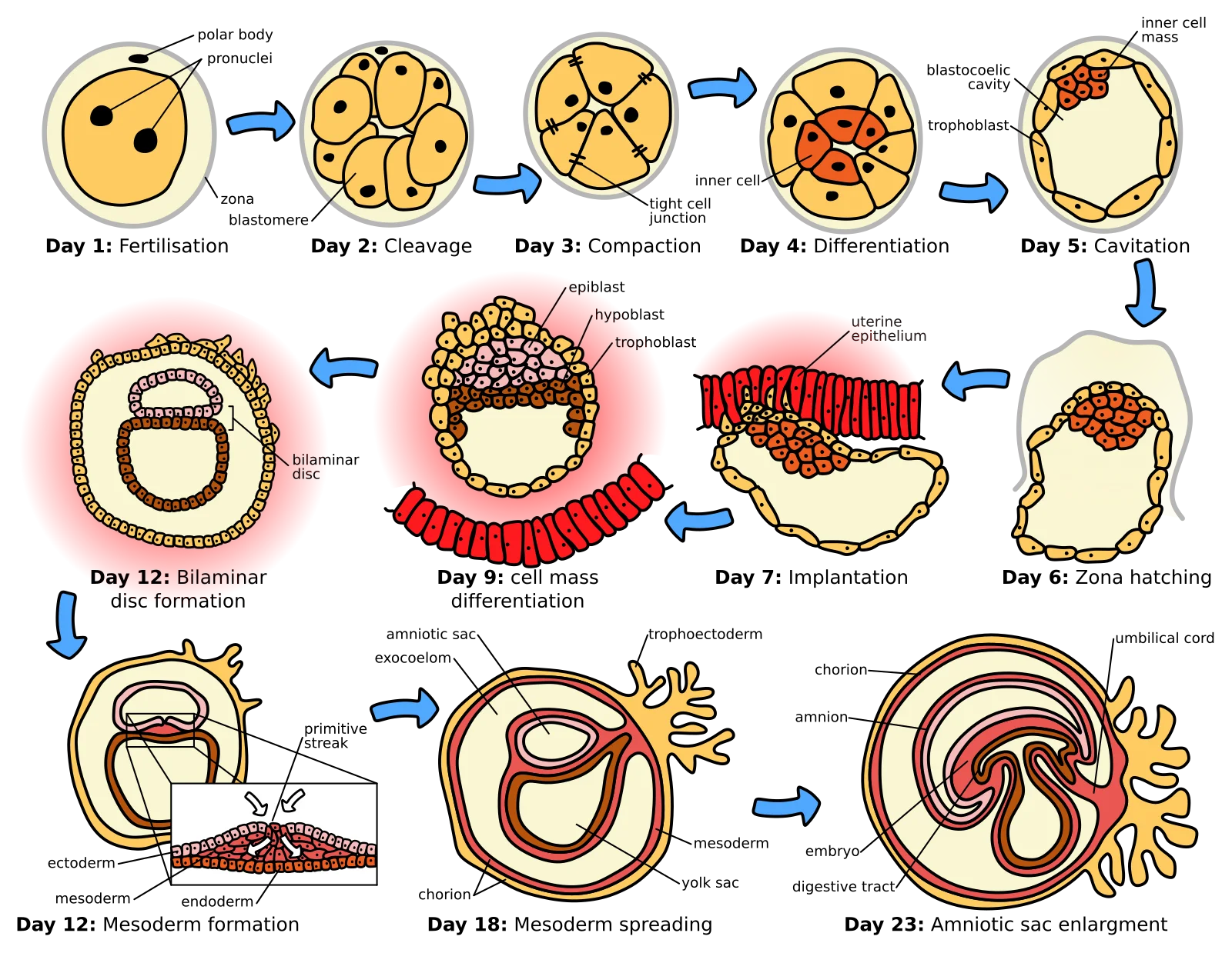
After fertilization, the zygote begins cleavage - a series of rapid mitotic divisions as it travels down the fallopian tube toward the uterus. This journey takes about 5-6 days.
The key feature of cleavage is that cells divide but the embryo does not grow. Each division produces smaller and smaller cells called blastomeres. Two ratios change during cleavage, and both are high-yield:
- Increased nuclear-to-cytoplasmic (N:C) ratio: The zygote starts as one giant cell with a relatively small nucleus. As cleavage produces smaller cells, each cell’s nucleus takes up a larger proportion of the total cell volume. This gives the nucleus better control over cellular activity.
- Increased surface area-to-volume ratio: Smaller cells have more membrane surface relative to their volume. This allows more efficient exchange of gases, nutrients, and waste products - essential for cells that lack a blood supply.
There are two types of cleavage that the MCAT distinguishes:
| Type | Cell Fate | Key Feature | Example |
|---|---|---|---|
| Indeterminate | Each blastomere can become a complete organism | Explains identical twins | Humans, vertebrates |
| Determinate | Each blastomere is committed to a specific fate early | Cells cannot develop independently | Most invertebrates |
Identical twins form when blastomeres from indeterminate cleavage separate early - each cell retains the ability to build an entire person. In organisms with determinate cleavage, losing a single blastomere means the embryo is missing an essential part.
The Morula
At around the 16-cell stage (about 3 days after fertilization), the embryo is a solid ball of cells called the morula (Latin for “little mulberry” - it looks like a mulberry fruit under the microscope).
The morula is still traveling down the fallopian tube at this point. No internal cavity has formed yet - it is a solid cluster of blastomeres compacted tightly together. The process of compaction flattens outer cells, which begin forming tight junctions with one another. This creates a distinction between outer cells and inner cells for the first time.
The Blastocyst (Blastula)
As the morula continues dividing, fluid-filled spaces appear between cells and merge to form a central cavity called the blastocoel. The morula has now become a blastula.
In mammals, we call this stage the blastocyst. It has two distinct regions:
- Trophoblast: The outer ring of cells that will form the placenta and other extraembryonic structures. These cells will never become part of the baby.
- Inner cell mass (ICM): A clump of cells on one side of the blastocoel that will become the embryo itself. These are the cells that contain the potential to form every tissue in the body.
Implantation
Around day 6-7 after fertilization, the blastocyst reaches the uterus and implants into the endometrium (uterine lining). The endometrium must be in the secretory phase (prepared by progesterone from the corpus luteum) to be receptive to implantation. Timing matters - if the blastocyst arrives too early or too late, implantation may fail.
During implantation:
- The trophoblast cells invade the endometrial lining, burrowing into the tissue
- The trophoblast differentiates into two layers: the inner cytotrophoblast and the outer syncytiotrophoblast (a multinucleated layer that directly contacts maternal blood)
- The trophoblast forms the chorion, which develops finger-like projections called chorionic villi that anchor the embryo and vastly increase the surface area for nutrient and gas exchange with maternal blood
- The placenta forms from the chorion (fetal tissue) combined with the decidua basalis (maternal uterine tissue)
Once implantation occurs, the trophoblast begins secreting human chorionic gonadotropin (hCG), which maintains the corpus luteum and its production of progesterone. This is the hormone detected by pregnancy tests.
The Umbilical Cord
The umbilical cord connects the developing fetus to the placenta and contains:
- Two umbilical arteries - carry deoxygenated blood and waste FROM the fetus TO the placenta
- One umbilical vein - carries oxygenated blood and nutrients FROM the placenta TO the fetus
This is the opposite of what you would normally expect: arteries typically carry oxygenated blood. But remember, arteries are defined by direction (away from the heart), not oxygen content. The fetal heart pumps blood to the placenta via the arteries, and the placenta returns oxygenated blood via the vein.
Extraembryonic Membranes
Four membranes develop to support the embryo. These are not part of the embryo’s body but are essential for its survival:
| Membrane | Function |
|---|---|
| Chorion | Outermost membrane; forms the fetal part of the placenta; enables gas and nutrient exchange with maternal blood |
| Amnion | Creates the amniotic sac; filled with amniotic fluid that cushions the fetus, maintains temperature, and allows movement |
| Yolk sac | Produces the earliest blood cells and blood vessels; transfers nutrients to the embryo before the placenta is functional |
| Allantois | Involved in early waste removal; its blood vessels become the umbilical arteries and vein; contributes to bladder development |
Summary: The Timeline
| Time After Fertilization | Stage | Key Event |
|---|---|---|
| 0 hours | Zygote | Sperm and egg nuclei fuse; diploid cell forms |
| Day 1-3 | Cleavage | Rapid mitotic divisions; blastomere number increases |
| Day 3 | Morula | Solid ball of ~16 cells; compaction begins |
| Day 4-5 | Blastocyst | Blastocoel forms; trophoblast and ICM differentiate |
| Day 6-7 | Implantation | Blastocyst burrows into endometrium |
| Day 7+ | Placentation | Chorion and chorionic villi develop; placenta forms |