
Fertilization
Of the 200-300 million sperm released during ejaculation, only a few hundred reach the egg in the fallopian tube. Of those, only one will fertilize it. The journey from vagina to ampulla of the fallopian tube is a brutal obstacle course - acidic pH, cervical mucus, immune cells, wrong turns, and sheer distance all eliminate the vast majority. The sperm that makes it is not the “strongest” - it is the luckiest, with help from some clever molecular biology at the finish line.
Capacitation
Freshly ejaculated sperm cannot fertilize an egg. They must first undergo capacitation - a process that occurs in the female reproductive tract over several hours. During capacitation:
- Cholesterol is removed from the sperm membrane, making it more fluid and reactive
- Intracellular calcium levels increase
- The sperm becomes hyperactive (vigorous, whip-like tail movement)
- The acrosomal membrane becomes primed for the acrosome reaction
Only capacitated sperm can undergo the acrosome reaction and bind to the zona pellucida.
Penetrating the Egg - Two Barriers
Barrier 1: The Corona Radiata
The corona radiata is a layer of granulosa cells surrounding the egg. Sperm must physically push through these cells, aided by hyaluronidase and other enzymes released from the acrosome and from the collective action of many sperm (though only one will ultimately fertilize the egg).
Barrier 2: The Zona Pellucida
The zona pellucida is a thick glycoprotein shell around the oocyte. It contains specific receptor proteins:
- ZP3 - the sperm receptor. It binds species-specific sperm and triggers the acrosome reaction.
- ZP2 - helps maintain sperm binding after the acrosome reaction.
The Acrosome Reaction
When a capacitated sperm binds to ZP3 on the zona pellucida:
- The acrosomal membrane fuses with the sperm’s plasma membrane
- Hydrolytic enzymes (including acrosin) are released
- These enzymes digest a path through the zona pellucida
- The sperm reaches the oocyte’s plasma membrane and fuses with it
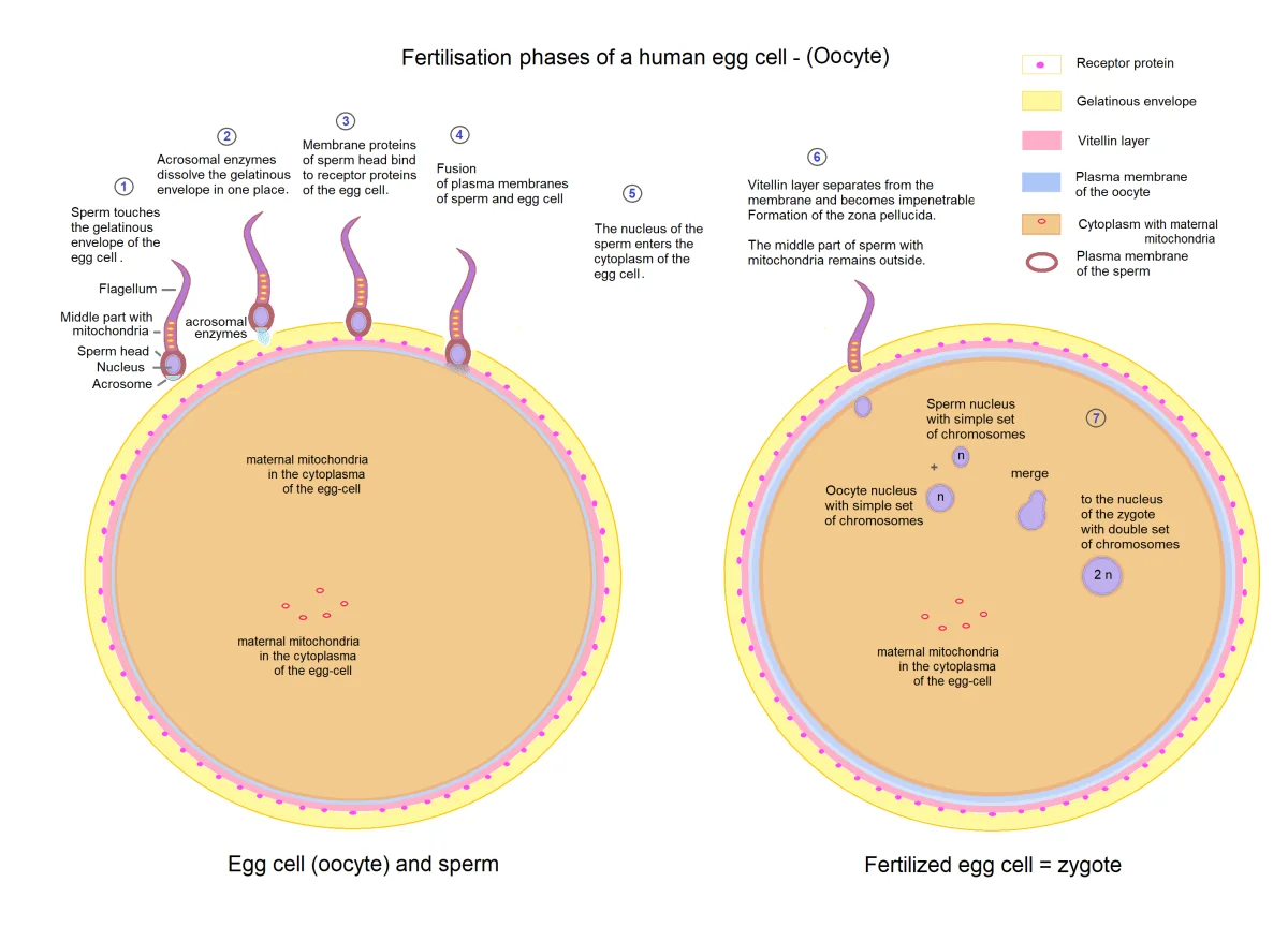
The Cortical Reaction - Blocking Polyspermy
The moment one sperm fuses with the oocyte membrane, two things happen rapidly to prevent additional sperm from entering:
Fast block (immediate):
- The oocyte membrane depolarizes (membrane potential shifts from negative to positive)
- This electrical change prevents other sperm from fusing with the membrane
- Temporary measure, lasting only a few minutes
Slow block (permanent):
- Sperm entry triggers a wave of calcium ions (Ca2+) released from the oocyte’s endoplasmic reticulum
- This calcium wave causes cortical granules (vesicles just beneath the oocyte membrane) to fuse with the membrane and release their contents into the space between the membrane and the zona pellucida
- Cortical granule enzymes harden the zona pellucida into the fertilization membrane, destroying ZP3 receptors and making it impenetrable to additional sperm
Polyspermy (fertilization by more than one sperm) would give the zygote too many chromosomes, which is lethal. The fast and slow blocks ensure this does not happen.
Completing Meiosis and Forming the Zygote
Remember that the “egg” released at ovulation is actually a secondary oocyte arrested at Metaphase II. Sperm entry triggers the completion of Meiosis II:
- The secondary oocyte completes Meiosis II, producing a mature ovum and a second polar body (which degenerates).
- The sperm nucleus (now called the male pronucleus) and the ovum nucleus (the female pronucleus) each form within the cell.
- The two pronuclei migrate toward each other and their membranes dissolve, combining their chromosomes.
- The resulting cell - the zygote - has the full diploid complement of 46 chromosomes (23 from sperm + 23 from egg).
The zygote is the first cell of the new organism.
Early Cleavage
The zygote immediately begins dividing as it travels down the fallopian tube toward the uterus:
Cleavage - rapid mitotic divisions that increase cell number without increasing overall size. Each division produces smaller and smaller cells called blastomeres.
- 2-cell stage → 4-cell → 8-cell → 16-cell
- By the 16-32 cell stage, the embryo is a solid ball of cells called the morula (Latin for “mulberry” - it looks like one)
- The morula then develops a fluid-filled cavity (the blastocoel), becoming the blastocyst (around day 5)
The Blastocyst and Implantation
The blastocyst has two distinct cell populations:
- Inner cell mass (ICM) - a cluster of cells on one side that will become the embryo proper. These cells are pluripotent.
- Trophoblast - the outer layer of cells surrounding the blastocoel. It will form the placenta and other extraembryonic membranes. The trophoblast secretes enzymes that allow the blastocyst to burrow into the endometrium.
Implantation occurs about 6-7 days after fertilization. The trophoblast invades the endometrial lining and establishes connections with the mother’s blood supply. Once implanted, the trophoblast begins secreting hCG to maintain the corpus luteum and its progesterone production. From here, the blastocyst undergoes gastrulation and organogenesis, which we cover in early embryonic development.
Indeterminate vs. Determinate Cleavage
- Indeterminate cleavage (humans and other deuterostomes) - each early blastomere retains the ability to develop into a complete organism. This is why identical twins can form if the embryo splits in the early cleavage stages.
- Determinate cleavage (most protostomes) - the fate of each blastomere is fixed from the first division. Separating cells would not produce complete organisms.
Extraembryonic Membranes
Four membranes develop to support the embryo:
| Membrane | Function |
|---|---|
| Chorion | Outermost membrane; develops into the fetal part of the placenta; chorionic villi establish maternal-fetal exchange |
| Amnion | Encloses the embryo in the amniotic sac filled with amniotic fluid; cushions and protects |
| Allantois | Contributes to umbilical cord and bladder development; waste exchange in non-mammalian vertebrates |
| Yolk sac | Early blood cell formation and nutrient transfer; largely vestigial in humans (no yolk) |
The umbilical cord connects the fetus to the placenta and contains two umbilical arteries (carrying deoxygenated blood from fetus to placenta) and one umbilical vein (carrying oxygenated blood from placenta to fetus).