
Stem Cells
A fertilized egg can become literally anything - a brain cell, a bone cell, a placenta cell, or even an entire identical twin. A blood stem cell in your bone marrow can become any blood cell type, but it will never become a neuron. A mature red blood cell cannot become anything else at all.
This spectrum of developmental potential is called potency, and understanding how it narrows during development is essential for the MCAT.
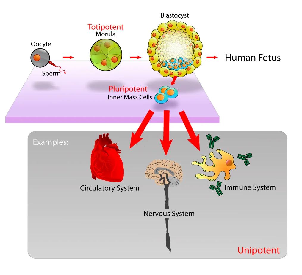
The Four Levels of Potency
| Level | Definition | Can Become | Example |
|---|---|---|---|
| Totipotent | Can form ANY cell type, including extraembryonic tissues | Embryo + placenta + everything | Zygote; cells up to ~4-cell stage |
| Pluripotent | Can form any cell type in the body, but NOT extraembryonic tissues | All 200+ body cell types, but not placenta | Inner cell mass of blastocyst; embryonic stem (ES) cells |
| Multipotent | Can form multiple cell types within one lineage | Limited range within a tissue family | Hematopoietic stem cells; neural stem cells; mesenchymal stem cells |
| Unipotent | Can form only one cell type | One specific cell type | Muscle satellite cells; spermatogonial stem cells |
Totipotent Cells
The zygote and the cells from the first few cleavage divisions (up to about the 4-cell or 8-cell stage) are totipotent. They can form every cell type in the body AND the extraembryonic structures - the placenta, chorion, amnion, and yolk sac.
This is why identical (monozygotic) twins are possible. If the cells of an early embryo separate, each totipotent cell can develop into a complete organism with all its supporting structures.
Totipotency is lost once the blastocyst forms and its cells specialize into two populations: the trophoblast (committed to extraembryonic structures) and the inner cell mass (committed to the embryo proper).
Pluripotent Cells
The inner cell mass (ICM) of the blastocyst contains pluripotent cells. These can differentiate into any of the 200+ cell types derived from all three germ layers (ectoderm, mesoderm, endoderm) but can no longer form the placenta or other extraembryonic tissues.
Embryonic stem (ES) cells are pluripotent cells isolated from the inner cell mass and grown in culture. They are powerful research tools because they can be directed to become virtually any cell type under the right conditions.
Induced pluripotent stem cells (iPSCs) are adult differentiated cells that have been reprogrammed back to a pluripotent state. In 2006, Shinya Yamanaka demonstrated that introducing four specific transcription factors (Oct4, Sox2, Klf4, c-Myc - the “Yamanaka factors”) could convert adult skin cells into cells that behave like embryonic stem cells. This breakthrough proved that differentiation is based on gene silencing, not gene loss.
Multipotent Cells
Multipotent cells can generate several cell types, but only within a single lineage or tissue family. These are the “adult stem cells” that persist in mature tissues and provide ongoing repair and renewal.
Important examples:
- Hematopoietic stem cells (in bone marrow) - can become red blood cells, all types of white blood cells, and platelets, but cannot become neurons or muscle cells
- Neural stem cells (in the brain) - can become neurons, astrocytes, and oligodendrocytes, but cannot become blood cells
- Mesenchymal stem cells - can become bone, cartilage, fat, and muscle cells
Unipotent Cells
Unipotent cells can self-renew (make copies of themselves) but only produce one differentiated cell type. Examples:
- Muscle satellite cells - produce only new muscle fibers for repair after injury
- Spermatogonial stem cells - produce only sperm precursors
- Epidermal basal cells - produce only keratinocytes
Even unipotent cells are still considered stem cells because they retain the ability to self-renew. A fully differentiated cell that has lost the ability to divide (like a mature neuron) is not a stem cell at all.
When Potency Narrows During Development
Potency decreases in a predictable sequence as the embryo develops:
- Zygote through ~4-cell stage - totipotent
- Blastocyst inner cell mass - pluripotent
- Germ layer formation (gastrulation) - cells become restricted to one germ layer’s lineages (multipotent)
- Organogenesis and beyond - cells progressively narrow to tissue-specific progenitors and eventually unipotent or terminally differentiated cells
This narrowing is driven by the same mechanisms covered in the previous section: inductive signals trigger selective gene expression, epigenetic modifications lock in committed states, and determination becomes irreversible.
Embryonic vs. Adult Stem Cells
| Feature | Embryonic Stem Cells | Adult Stem Cells |
|---|---|---|
| Source | Inner cell mass of blastocyst | Various tissues (bone marrow, brain, skin, gut) |
| Potency | Pluripotent | Usually multipotent or unipotent |
| Self-renewal | Essentially unlimited in culture | Limited (declines with age) |
| Differentiation range | Any body cell type | Restricted to their tissue lineage |
| Ethical considerations | Controversial (requires embryo destruction) | Fewer ethical concerns |
| Immune rejection risk | High (donor cells may not match recipient) | Low if patient’s own cells are used |
Cell Migration During Development
Stem cells and their progeny do not always stay where they are born. Cell migration is essential for proper embryonic development. Cells must travel - sometimes across the entire embryo - to reach their final destination.
Key examples of developmental cell migration:
- Neural crest cells migrate extensively from the dorsal neural tube to become melanocytes in the skin, neurons of the peripheral nervous system, adrenal medulla cells, and craniofacial cartilage
- Primordial germ cells originate in the yolk sac wall and migrate through the gut mesentery to reach the developing gonads
- Immune cell precursors migrate from the bone marrow to the thymus (T cell maturation) or remain in the bone marrow (B cell maturation)
Migration is guided by chemical signals (chemotaxis), adhesion molecules on cell surfaces, and components of the extracellular matrix. Errors in migration can cause developmental disorders - for example, failure of neural crest cell migration can result in Hirschsprung’s disease (absence of enteric ganglia in the colon).
Apoptosis: Programmed Cell Death
Development is not just about building new structures - it is also about strategic destruction. Apoptosis is a carefully regulated process in which cells self-destruct when they are no longer needed or when they pose a danger to the organism.
Think of apoptosis as a controlled demolition. The building (cell) is taken apart piece by piece, the debris is neatly collected, and the site is quietly cleared. Compare this to necrosis - an uncontrolled explosion where rubble flies everywhere and damages the surroundings.
Examples of apoptosis in development:
- Digit separation - your fingers and toes begin as a solid paddle of tissue; apoptosis removes the webbing between them to create individual digits
- Immune system pruning - T cells that react against self-antigens undergo apoptosis in the thymus (negative selection)
- Neural sculpting - the developing brain overproduces neurons, then eliminates those that fail to make functional synaptic connections
- Tail resorption - tadpoles lose their tails during metamorphosis via apoptosis
Apoptosis vs. Necrosis
| Feature | Apoptosis | Necrosis |
|---|---|---|
| Trigger | Internal signals (programmed, regulated) | External injury, toxins, oxygen deprivation |
| Cell changes | Cell shrinks; chromatin condenses; membrane blebs form; cell breaks into apoptotic bodies | Cell swells; membrane ruptures; contents spill out |
| Inflammation | No - neighboring cells quietly engulf apoptotic bodies | Yes - released contents trigger an inflammatory response |
| DNA degradation | Orderly - cut into ~180 bp fragments (nucleosome-sized) by endonucleases | Random, uncontrolled degradation |
| Role | Normal development, tissue homeostasis, immune regulation | Pathological - response to disease, trauma, or ischemia |
Cancer and Potency
Cancer cells often reactivate developmental programs that should be permanently silenced. Some tumors contain cancer stem cells - a subpopulation that can self-renew, resist chemotherapy, and regenerate the tumor even after treatment kills the majority of cells. This connection between stem cell biology and oncology is increasingly tested on the MCAT.