
Neurulation
Imagine you are rolling up a sheet of paper into a tube, then sealing it shut along the top edge. A few stray bits of paper fall away from the seam and scatter across the table. That rolled-up tube becomes your entire central nervous system. Those scattered scraps become an astonishing variety of other structures - from pigment cells in your skin to the core of your adrenal glands. That, in a nutshell, is neurulation.
Neurulation is the process by which the embryo builds the foundation of the nervous system. It begins immediately after gastrulation and is one of the most heavily tested embryology topics on the MCAT.
The Notochord: The Signal That Starts Everything
Before any neural tissue forms, a rod-shaped structure called the notochord must appear. Think of the notochord as a temporary scaffold - like the wooden frame builders erect before pouring concrete for a building. It provides structural support and, more importantly, sends the chemical messages that kick off the entire process.
The notochord is a mesodermal structure. It runs along the midline (head to tail) of the embryo and serves two critical functions:
- Structural support - it acts as the embryo’s primitive spine, providing a flexible axis before vertebrae develop
- Induction - it releases signaling molecules that instruct the ectoderm directly above it to thicken into neural tissue
In adult humans, the notochord almost entirely degenerates. The only remnant is the nucleus pulposus - the gel-like center of each intervertebral disc. When someone has a “herniated disc,” it is the nucleus pulposus that has bulged out of position.
Step-by-Step: How the Neural Tube Forms
Neurulation proceeds in an orderly sequence of four steps. Follow along with the “rolling paper” analogy:
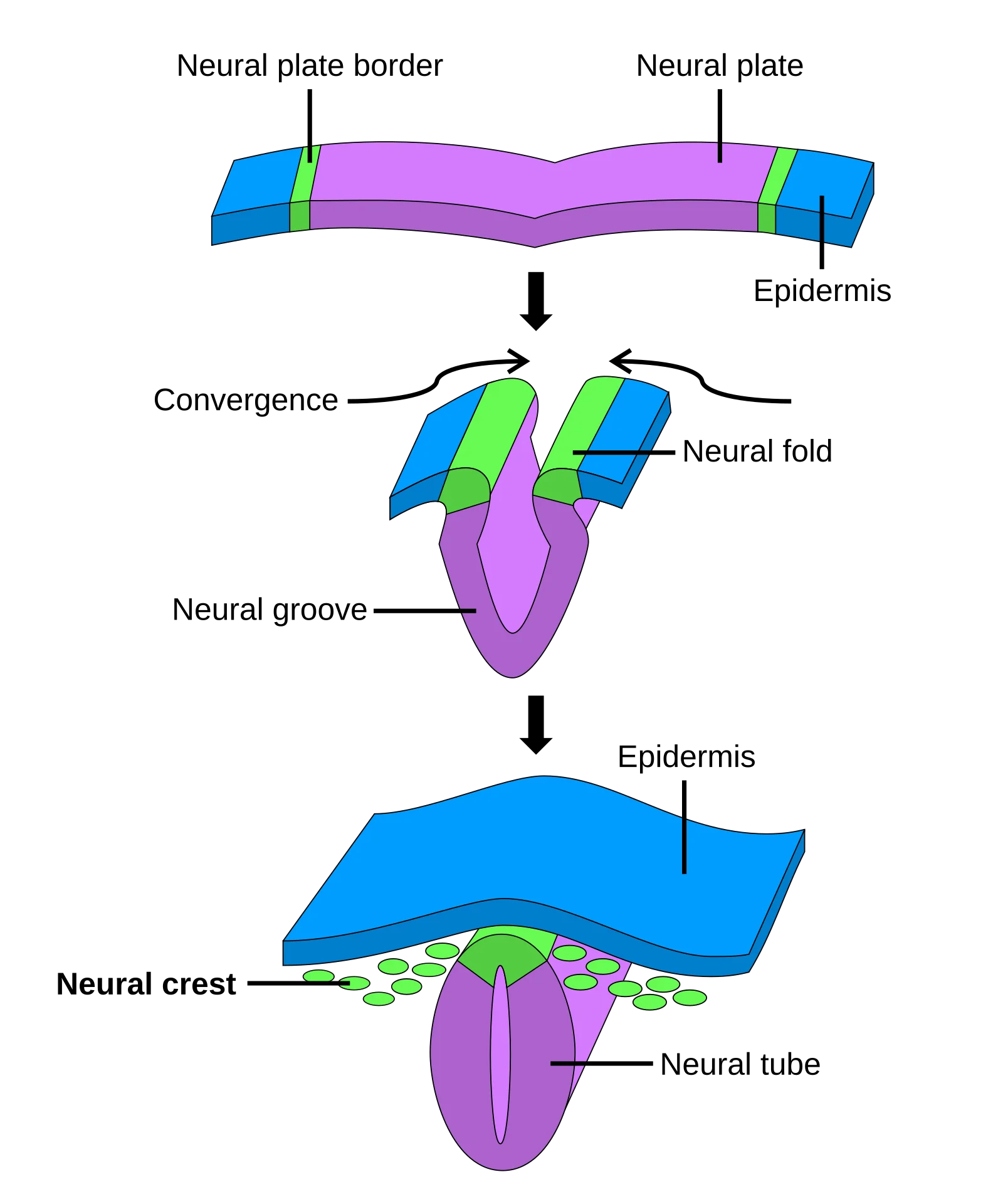
Step 1 - Neural plate formation. Chemical signals from the notochord cause the overlying strip of ectoderm to thicken into a flat sheet called the neural plate. This is like laying out your sheet of paper flat on the table. These ectodermal cells are now committed to a neural fate.
Step 2 - Neural groove and neural folds. The center of the neural plate sinks inward along its midline, forming a depression called the neural groove. The raised edges on either side are called the neural folds. This is like pressing your finger along the center of the paper so the edges start to curl upward.
Step 3 - Neural tube closure. The neural folds continue rising, bend toward each other, and eventually meet and fuse at the top, creating a sealed hollow tube - the neural tube. Closure begins in the middle of the embryo and then zips shut toward both the head (anterior) and tail (posterior) ends. This is the moment you seal the top of the rolled paper.
Step 4 - Differentiation of the neural tube. Once closed, the neural tube develops into the entire central nervous system:
- The anterior (head) end expands into three bulges that become the brain
- The posterior portion becomes the spinal cord
- The hollow interior becomes the ventricles of the brain and the central canal of the spinal cord, filled with cerebrospinal fluid
Neural Tube Closure and What Happens When It Fails
Neural tube closure happens remarkably early - around days 22 to 28 of development (weeks 3-4). The tube zips shut from the middle outward, meaning the anterior (head) end and the posterior (tail) end are the last parts to close. If either end fails to seal, devastating birth defects result.
| Defect | What Fails to Close | Consequence |
|---|---|---|
| Spina bifida | Posterior (caudal) neuropore | Spinal cord remains partially exposed; severity ranges from a small gap in vertebrae (spina bifida occulta) to full exposure of the spinal cord (myelomeningocele) |
| Anencephaly | Anterior (cranial) neuropore | The brain fails to develop properly; incompatible with life |
Folic acid (vitamin B9) supplementation before and during early pregnancy dramatically reduces the risk of neural tube defects. This is why prenatal vitamins contain high doses of folate, and why many countries fortify grain products with folic acid.
Neural Crest Cells: The “Fourth Germ Layer”
Here is where neurulation gets really interesting. As the neural folds rise and fuse to form the neural tube, a special population of cells at the very tips of the folds breaks free. These are neural crest cells, and they are unlike any other cells in the embryo.
Neural crest cells are derived from the ectoderm, but they do something no other ectodermal cells do - they migrate throughout the entire body and differentiate into a staggeringly diverse set of structures. Because of this extraordinary versatility, neural crest cells are sometimes called the “fourth germ layer.”
Here is what neural crest cells become:
| Category | Neural Crest Derivatives |
|---|---|
| Peripheral nervous system | Sensory ganglia (dorsal root ganglia), autonomic ganglia (sympathetic and parasympathetic), Schwann cells (myelinate PNS axons), enteric nervous system (“gut brain”) |
| Endocrine | Adrenal medulla (chromaffin cells that produce epinephrine and norepinephrine), calcitonin-producing parafollicular (C) cells of the thyroid |
| Pigment | Melanocytes (pigment-producing cells of the skin, hair, and eyes) |
| Craniofacial | Bones and cartilage of the face and skull, odontoblasts (cells that make tooth dentin) |
| Cardiovascular | Smooth muscle of the great vessels (aorta, pulmonary artery), aorticopulmonary septum (divides the outflow tract of the heart) |
Why Neural Crest Cells Are MCAT Gold
The MCAT loves neural crest cells because their derivatives are “unexpected.” Here are the traps:
- Adrenal medulla comes from ectoderm (via neural crest), NOT mesoderm like the adrenal cortex. Same gland, different germ layers.
- Melanocytes come from neural crest, NOT from the surface ectoderm that forms the epidermis. They live in the skin but did not originate there - they migrated in.
- Schwann cells come from neural crest, NOT from the CNS. Oligodendrocytes (which myelinate CNS axons) come from the neural tube, but Schwann cells (which myelinate PNS axons) come from neural crest.
- Calcitonin-producing C cells of the thyroid come from neural crest. The rest of the thyroid is endoderm. One gland, two origins.
If an MCAT question asks about the embryonic origin of something surprising, neural crest is very often the answer.
Putting It All Together
Here is the complete neurulation timeline:
| Timing | Event |
|---|---|
| Week 3 | Notochord (mesoderm) signals overlying ectoderm |
| ~Day 18 | Neural plate forms from thickened ectoderm |
| ~Day 20 | Neural plate folds inward, creating neural groove flanked by neural folds |
| ~Days 22-28 | Neural folds fuse to form the neural tube; neural crest cells migrate away from the fold tips |
| Week 4+ | Anterior neural tube expands into brain vesicles; posterior tube becomes spinal cord |