
Fetal Circulation
Think about a building under construction. The HVAC system and water filtration plant are not online yet, so the construction crew runs temporary hoses from the building next door to supply air and clean water. Once the building is finished and its own systems come online, the temporary hoses are disconnected.
That is exactly what fetal circulation does. The fetus’s lungs and liver are not yet functional, so the circulatory system builds temporary bypass routes - shunts - that redirect blood away from those organs. The placenta handles gas exchange and detoxification instead.
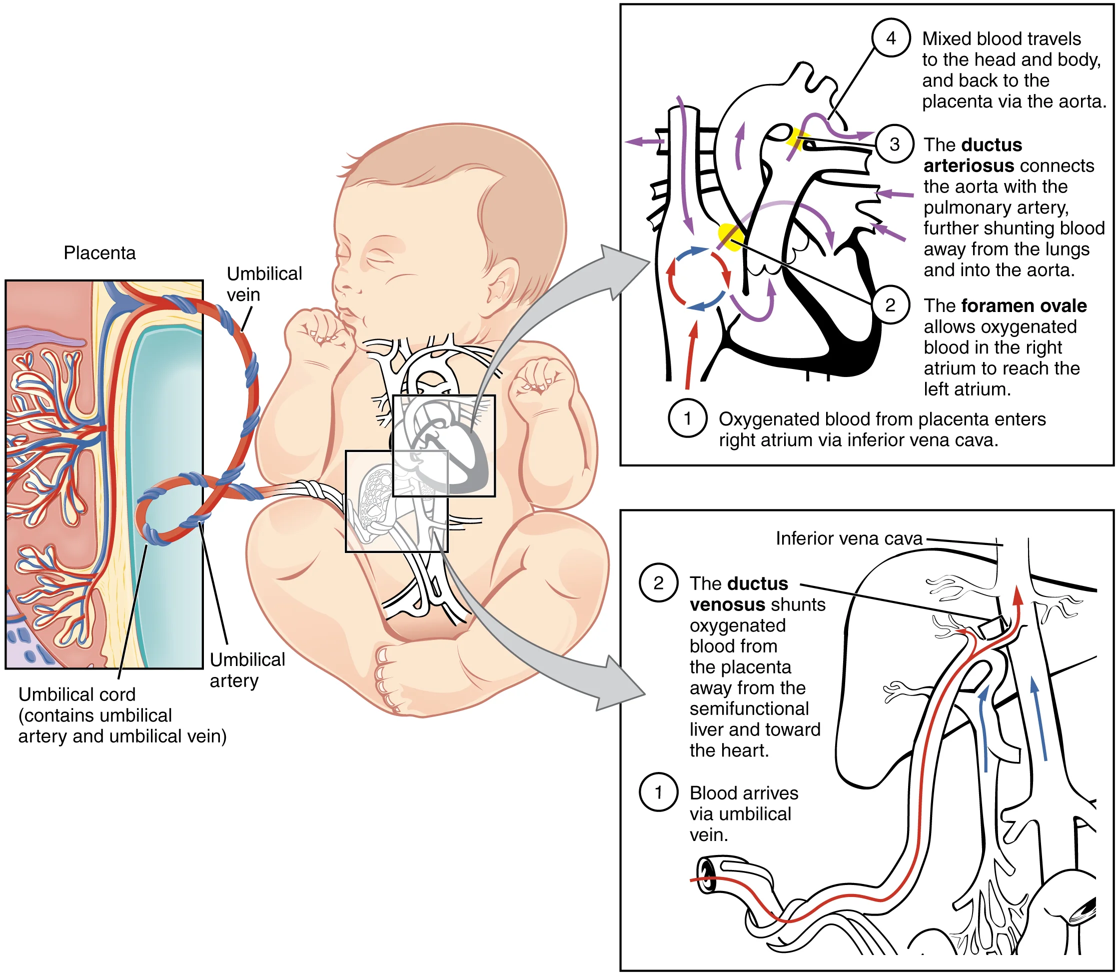
Why Fetal Circulation Is Different
In adult circulation, the lungs oxygenate blood and the liver filters toxins from digested food. A fetus does neither of these things:
- The fetal lungs are collapsed and filled with fluid. They do not perform gas exchange.
- The fetal liver does not process nutrients from digestion because the fetus does not eat.
Both of these jobs fall to the placenta, which connects the fetus to the mother’s blood supply. Since the lungs and liver are offline, it would be wasteful to send large volumes of blood through them. The fetal circulatory system solves this problem with three temporary shunts.
The Placenta: Structure and Function
The placenta is the critical interface between mother and fetus. It is a disc-shaped organ embedded in the uterine wall, and it performs four major functions:
1. Gas exchange. Oxygen diffuses from maternal blood into fetal blood; carbon dioxide diffuses in the opposite direction. Maternal and fetal blood never mix directly - they flow in close proximity, separated by thin membranes, allowing diffusion across the placental barrier.
2. Nutrient and waste transfer. Glucose, amino acids, vitamins, and other nutrients cross from mother to fetus by diffusion and active transport. Fetal metabolic waste (urea, CO2) crosses back to the mother for disposal.
3. Immune protection. Maternal IgG antibodies cross the placenta, giving the fetus passive immunity. This is why newborns have temporary protection against diseases the mother has encountered.
4. Endocrine function. The placenta is a hormone-producing powerhouse:
| Hormone | Role |
|---|---|
| hCG (human chorionic gonadotropin) | Maintains the corpus luteum in early pregnancy; basis for pregnancy tests |
| Progesterone | Maintains the endometrium; prevents uterine contractions; suppresses maternal immune rejection of fetus |
| Estrogen | Stimulates uterine growth; promotes blood flow to the uterus |
| Human placental lactogen (hPL) | Adjusts maternal metabolism to increase glucose availability for the fetus |
Umbilical Vessels
The umbilical cord connects the fetus to the placenta and contains three vessels:
- One umbilical vein - carries oxygenated, nutrient-rich blood FROM the placenta TO the fetus
- Two umbilical arteries - carry deoxygenated, waste-laden blood FROM the fetus TO the placenta
Notice something counterintuitive: the umbilical vein carries oxygenated blood, and the umbilical arteries carry deoxygenated blood. This seems backwards compared to the normal rule (arteries = oxygenated, veins = deoxygenated), but it actually follows the true definitions perfectly.
The real definition of arteries and veins has nothing to do with oxygen content:
- Arteries carry blood AWAY from the heart
- Veins carry blood TOWARD the heart
The umbilical arteries carry blood away from the fetal heart (to the placenta). The umbilical vein carries blood toward the fetal heart (from the placenta). The naming is correct - it is only confusing if you rely on the oxygen-based shortcut instead of the true definitions.
The Three Fetal Shunts
Here is what each shunt does and where it connects:
1. Ductus venosus - bypasses the liver
Oxygenated blood from the placenta enters the fetus through the umbilical vein. Instead of routing all this blood through the liver’s sinusoids, most of it is shunted directly into the inferior vena cava (IVC) via the ductus venosus. This gets oxygen-rich blood to the heart as quickly as possible.
2. Foramen ovale - bypasses the lungs (shunt #1)
Blood enters the right atrium from the IVC. Normally, right atrial blood would travel to the right ventricle and then to the lungs. But fetal lungs are non-functional, so a hole between the right and left atria - the foramen ovale - allows most oxygenated blood to flow directly from the right atrium into the left atrium, bypassing the pulmonary circuit entirely. From the left atrium, blood enters the left ventricle and is pumped through the aorta to the body.
3. Ductus arteriosus - bypasses the lungs (shunt #2)
Some blood still enters the right ventricle and gets pumped into the pulmonary artery. Instead of going to the lungs, most of this blood is diverted from the pulmonary artery directly into the descending aorta through the ductus arteriosus. This is a second bypass around the non-functional lungs.
| Shunt | Connects | Organ Bypassed | Adult Remnant |
|---|---|---|---|
| Ductus venosus | Umbilical vein → IVC | Liver | Ligamentum venosum |
| Foramen ovale | Right atrium → Left atrium | Lungs | Fossa ovalis |
| Ductus arteriosus | Pulmonary artery → Aorta | Lungs | Ligamentum arteriosum |
What Happens at Birth
The moment a baby takes its first breath, a cascade of changes converts the fetal circulatory pattern to the adult pattern:
- Lungs inflate - pulmonary vascular resistance drops dramatically as the alveoli open
- Blood rushes to the lungs - the pulmonary artery now sends blood to the functional lungs instead of through the ductus arteriosus
- Left atrial pressure rises - increased blood return from the lungs raises pressure in the left atrium. This pushes a flap of tissue over the foramen ovale, functionally sealing it shut. It eventually fuses to become the fossa ovalis.
- Ductus arteriosus constricts - rising oxygen levels and falling prostaglandin levels trigger constriction. The ductus closes within hours to days, becoming the ligamentum arteriosum.
- Umbilical cord is clamped - the umbilical vessels cease function. The ductus venosus closes and becomes the ligamentum venosum.
The key trigger is the pressure reversal: before birth, right heart pressure is higher than left (blood shunts right-to-left through the foramen ovale). After the first breath, left heart pressure exceeds right heart pressure, and the shunt closes.
Fetal Hemoglobin (HbF)
Fetal blood must pick up oxygen from maternal blood at the placenta. For this to work, fetal hemoglobin must bind oxygen more tightly than maternal hemoglobin - otherwise the fetus could never “steal” oxygen from the mother’s blood.
Fetal hemoglobin (HbF) contains two alpha chains and two gamma chains (compared to adult HbA, which has two alpha and two beta chains). The gamma chains are the key: they do not bind 2,3-BPG (2,3-bisphosphoglycerate) as strongly as beta chains do.
Why does this matter? In adult red blood cells, 2,3-BPG binds to hemoglobin and reduces its oxygen affinity (shifts the curve right), promoting oxygen release to tissues. Since HbF is less affected by 2,3-BPG, it retains a higher oxygen affinity than HbA.
On an oxygen-hemoglobin dissociation curve, HbF is shifted to the left compared to HbA:
- At any given partial pressure of O2, HbF holds onto more oxygen than HbA
- In the placenta, maternal HbA releases O2 (lower affinity), and fetal HbF grabs it (higher affinity)
- This ensures efficient oxygen transfer from mother to fetus
After birth, production gradually shifts from gamma chains to beta chains. HbF is replaced by HbA over the first several months of life.
Putting It All Together: Blood Flow Through the Fetus
Follow the path of a red blood cell through the fetal circulatory system:
- Oxygenated blood leaves the placenta via the umbilical vein
- Most blood bypasses the liver through the ductus venosus and enters the IVC
- Blood enters the right atrium
- Most blood crosses through the foramen ovale into the left atrium (bypassing the lungs)
- Blood flows to the left ventricle and out through the aorta to the body
- Some blood from the right atrium enters the right ventricle and is pumped into the pulmonary artery
- Most of this blood bypasses the lungs through the ductus arteriosus and enters the descending aorta
- Deoxygenated blood returns to the placenta via the two umbilical arteries
- At the placenta, CO2 is exchanged for O2, and the cycle repeats