The nervous system is a hierarchy: sensory input flows in, motor output flows out, and everything in between is processing. This section lays out the major divisions you need to memorize for any MCAT neuroscience passage.
CNS and PNS
Central Nervous System (CNS) = brain + spinal cord. Where most processing happens.
Peripheral Nervous System (PNS) = everything else. All 12 cranial nerves and 31 pairs of spinal nerves, plus associated ganglia.
Brain Divisions
The brain forms from three primary vesicles in the embryo:
Midbrain → midbrain (sensory relay, motor coordination)
Hindbrain → pons, medulla, cerebellum (automatic and motor control)
The 12 Cranial Nerves
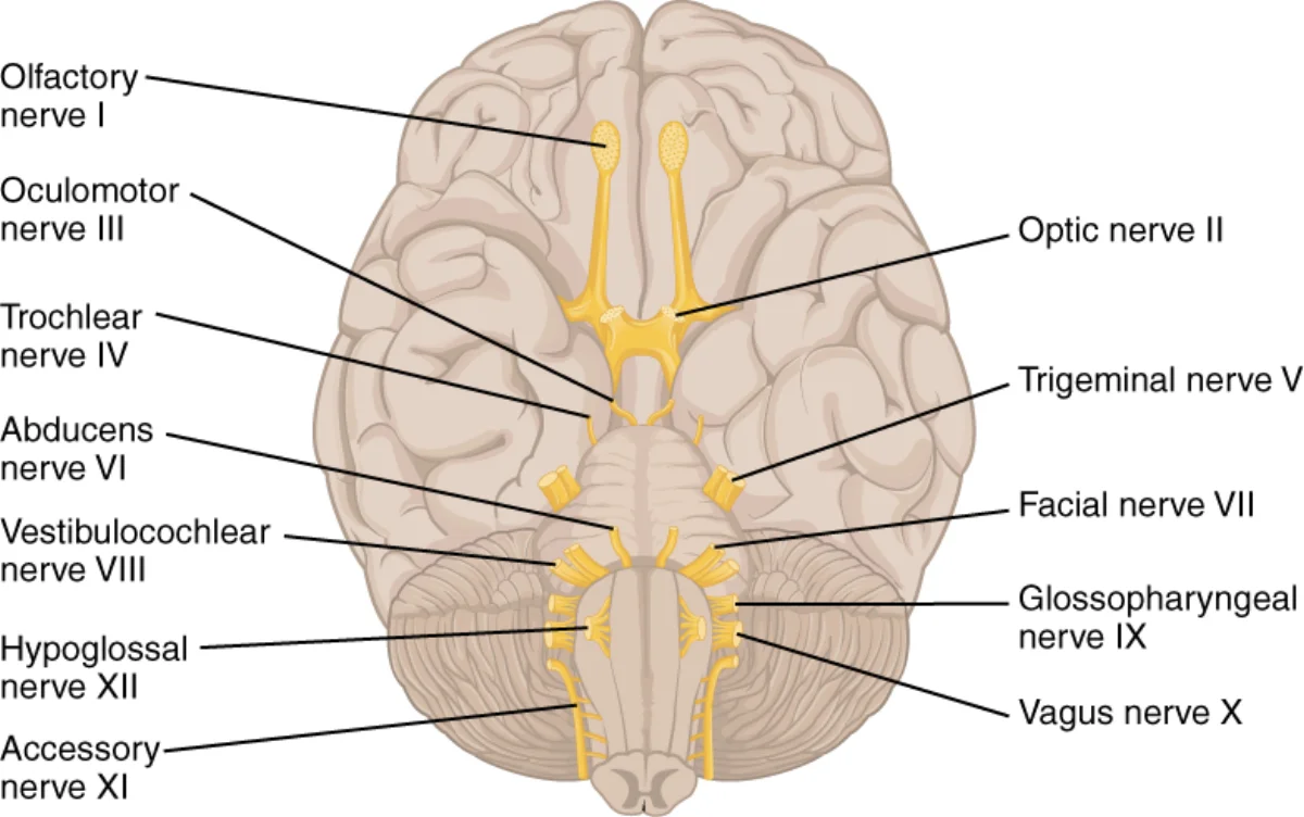
The 12 cranial nerves emerge directly from the brain or brainstem (not the spinal cord). You should know each nerve’s number, name, function class (sensory, motor, or both), and what it does.
The twelve cranial nerves arising from the inferior surface of the brain. Note their anterior-to-posterior emergence order — the same order as their Roman numerals — which is why "On Old Olympus' Towering Tops…" works as a mnemonic. Credit: OpenStax College via Wikimedia Commons (CC BY 4.0).
#
Name
Function
What It Does
I
Olfactory
Sensory
Smell
II
Optic
Sensory
Vision
III
Oculomotor
Motor
Most eye movements, pupil constriction, lens shape
Parasympathetic to heart, lungs, gut; voice (larynx); swallowing
XI
(Spinal) Accessory
Motor
Sternocleidomastoid and trapezius (head turning, shoulder shrug)
XII
Hypoglossal
Motor
Tongue movement
Mnemonic 1: Name Order
“On Old Olympus’ Towering Tops A Finn And German Viewed Some Hops”
Each first letter maps to a nerve in order: Olfactory, Optic, Oculomotor, Trochlear, Trigeminal, Abducens, Facial, (Vestibulo-)Acoustic, Glossopharyngeal, Vagus, Spinal accessory, Hypoglossal.
Mnemonic 2: Function (Sensory / Motor / Both)
“Some Say Marry Money But My Brother Says Big Brains Matter More”
In order I → XII: Sensory, Sensory, Motor, Motor, Both, Motor, Both, Sensory, Both, Both, Motor, Motor.
Clinically High-Yield Nerves
CN II (optic). Tested with visual acuity and visual fields. Afferent limb of the pupillary light reflex.
CN III (oculomotor). Efferent limb of the pupillary light reflex - damage produces a “blown pupil” (dilated, unreactive) and a “down-and-out” eye because only IV and VI still pull.
CN V (trigeminal). Three divisions: V1 ophthalmic, V2 maxillary, V3 mandibular. Corneal reflex afferent.
CN VII (facial). Bell’s palsy affects this nerve and produces unilateral facial droop including the forehead (an upper motor neuron stroke spares the forehead because of bilateral cortical input).
CN X (vagus). The main parasympathetic output to the body. Carries “rest and digest” signals to the heart and gut. Vagal stimulation slows the heart.
The Two Branches of the PNS
Somatic Nervous System
Carries voluntary motor commands to skeletal muscles and brings in sensory information from the skin, joints, and muscles. Under conscious control: you decide to lift your arm; the somatic nervous system makes it happen.
Autonomic Nervous System (ANS)
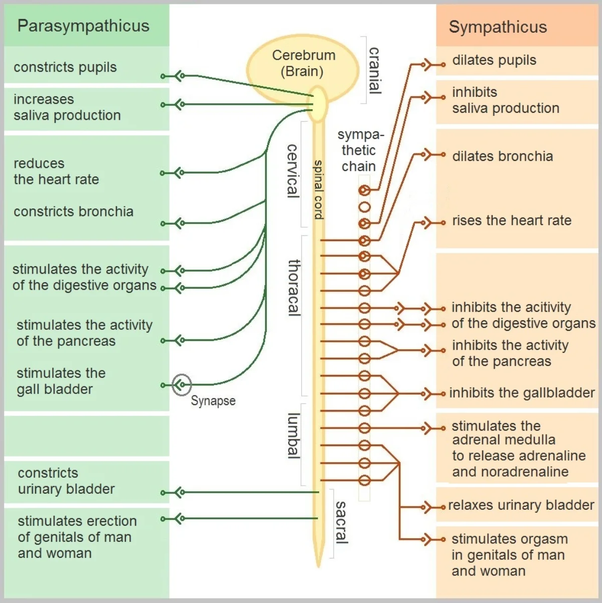
Controls involuntary functions: heart rate, digestion, pupil size, glandular secretion. Not under conscious control. Two opposing branches:
Sympathetic - “fight or flight.” Prepares the body for action.
Digestion resumes, blood flow to the viscera increases.
Long pre-ganglionic axons from brainstem or sacral spinal cord; short post-ganglionic axons.
Both branches usually act on the same organs with opposite effects - the balance between them tunes physiology moment-to-moment.
The two branches of the autonomic nervous system, with their target organs and opposite effects. Parasympathetic ("rest and digest") fibers exit at the cranial and sacral levels; sympathetic ("fight or flight") fibers exit thoracolumbarly via the sympathetic chain. Credit: Geo-Science-International via Wikimedia Commons (CC0).
Afferent vs Efferent Neurons
Remember the A/E distinction: Afferent = Arrival (sensory, into CNS). Efferent = Exit (motor, out of CNS). Receptors send info through afferent axons; motor commands travel out through efferent axons.
Upper and Lower Motor Neurons
Voluntary muscle control is a two-step chain:
Upper motor neurons (UMNs) originate in the cerebral cortex and travel down to synapse on lower motor neurons in the brainstem or spinal cord. Their axons form the corticospinal tract (to spinal cord) or corticobulbar tract (to brainstem, for head/neck).
Lower motor neurons (LMNs) exit the CNS and synapse directly on skeletal muscle at the neuromuscular junction.
UMNs control LMNs. LMNs actually move muscles.
Motor Neuron Signs
Damage to UMNs and LMNs produces characteristically different clinical pictures:
UMN signs. Weakness, hyperreflexia (exaggerated stretch reflexes), clonus (rhythmic contraction), hypertonia (increased muscle tone), extensor plantar response (Babinski sign - toes fan upward instead of downward when the sole is stroked).
The logic: without UMN input, the spinal reflex circuits lose their “moderator” and become hyperactive. Without LMN input, the muscle gets no drive at all - it withers and twitches.
The Muscle Stretch Reflex
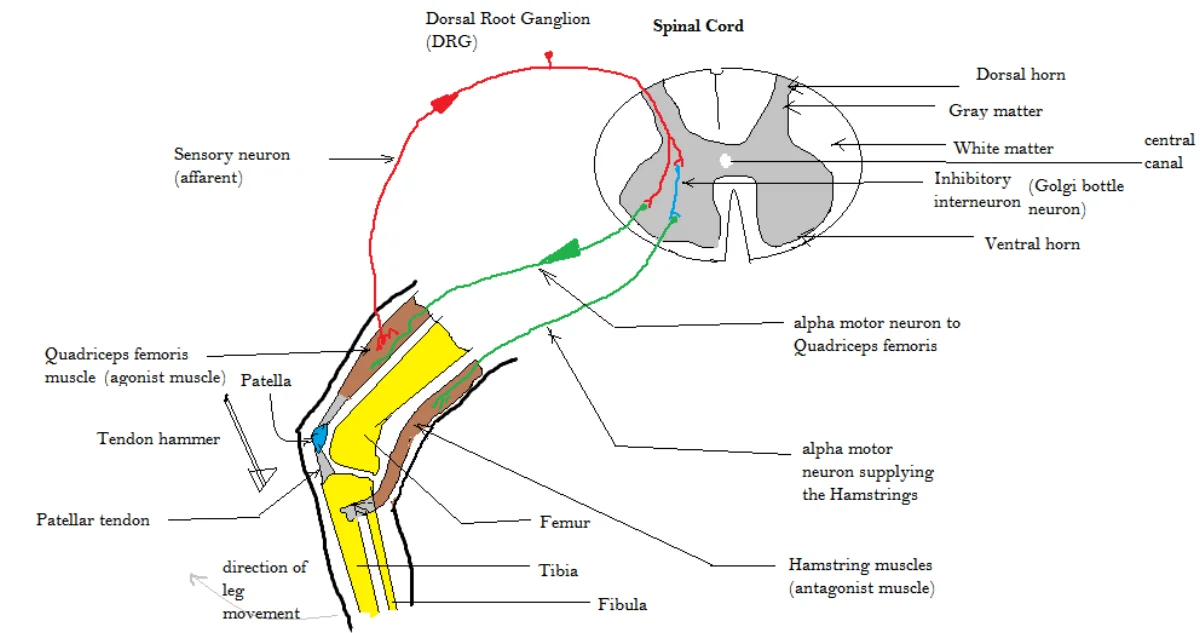
A reflex is a quick, involuntary response to a stimulus that doesn’t require the brain. The classic knee-jerk is a monosynaptic stretch reflex:
The hammer hits the patellar tendon, stretching the quadriceps.
Stretch receptors in the muscle (muscle spindles) fire afferent signals up to the spinal cord.
The afferent neuron synapses directly onto an LMN that innervates the same muscle.
The LMN fires, contracting the quadriceps - the leg kicks out.
At the same time, a separate inhibitory interneuron silences the antagonist muscle (hamstring) so it doesn’t fight the kick. This is reciprocal inhibition.
Reflexes demonstrate that the spinal cord does meaningful processing on its own - you don’t think “leg, kick.” The body protects itself faster than the brain can decide.
The monosynaptic patellar (knee-jerk) reflex. The afferent neuron synapses directly on the LMN to the quadriceps (one synapse → muscle contracts), while a separate inhibitory interneuron silences the hamstring — reciprocal inhibition. The whole circuit runs through the spinal cord without involving the brain. Credit: Amiya Sarkar via Wikimedia Commons (CC BY-SA 4.0).
Gray and White Matter
Gray matter - cell bodies (somas) of neurons, as well as dendrites and synapses.
White matter - myelinated axons. Looks white because of the fatty myelin.
Distributions differ between brain and spinal cord:
Brain. Gray matter on outside (cortex), white matter inside.
Spinal cord. White matter on outside (tracts going up and down), gray matter on inside (where the synapses happen).
What's the difference between the somatic and autonomic nervous systems?
Click to reveal answer
Somatic: voluntary control of skeletal muscle and conscious sensation. Autonomic: involuntary control of smooth muscle, cardiac muscle, and glands. ANS has sympathetic and parasympathetic branches.
Distinguish upper motor neuron signs from lower motor neuron signs.
Click to reveal answer
UMN: hyperreflexia, clonus, hypertonia, Babinski (up-going toes). LMN: atrophy, fasciculations, hypotonia, hyporeflexia. Weakness is common to both but the other signs distinguish them.
Give the mnemonic for the 12 cranial nerves in order.
Click to reveal answer
"On Old Olympus' Towering Tops A Finn And German Viewed Some Hops." Olfactory, Optic, Oculomotor, Trochlear, Trigeminal, Abducens, Facial, (Vestibulo)Acoustic, Glossopharyngeal, Vagus, Spinal accessory, Hypoglossal.
Which cranial nerves are purely sensory, which are purely motor, and which are both?
Click to reveal answer
"Some Say Marry Money But My Brother Says Big Brains Matter More." Sensory: I, II, VIII. Motor: III, IV, VI, XI, XII. Both: V, VII, IX, X.
Which cranial nerves move the eye, and what does "LR6 SO4 rest3" mean?
Click to reveal answer
Eye movement nerves: CN III (oculomotor), IV (trochlear), VI (abducens). LR6 = Lateral Rectus by CN VI. SO4 = Superior Oblique by CN IV. Rest3 = all remaining eye muscles by CN III.
Where are gray and white matter located in the brain vs. the spinal cord?
Click to reveal answer
Brain: gray outside (cortex), white inside. Spinal cord: white outside (tracts), gray inside. The layout is reversed between the two.
The cerebral cortex is the folded, wrinkled outer layer of your brain - the “gray matter” that does the slow, flexible work of perception, planning, and language. Its folds (gyri and sulci) pack more surface area into the skull; most of the cortex sits tucked inside those folds.
The cortex divides into four lobes (per hemisphere). Memorize each lobe’s name, location, and primary functions.
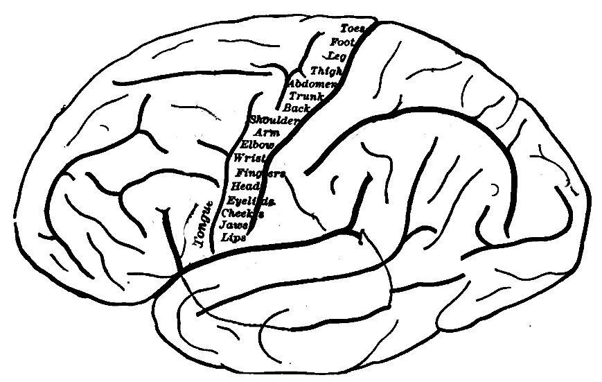
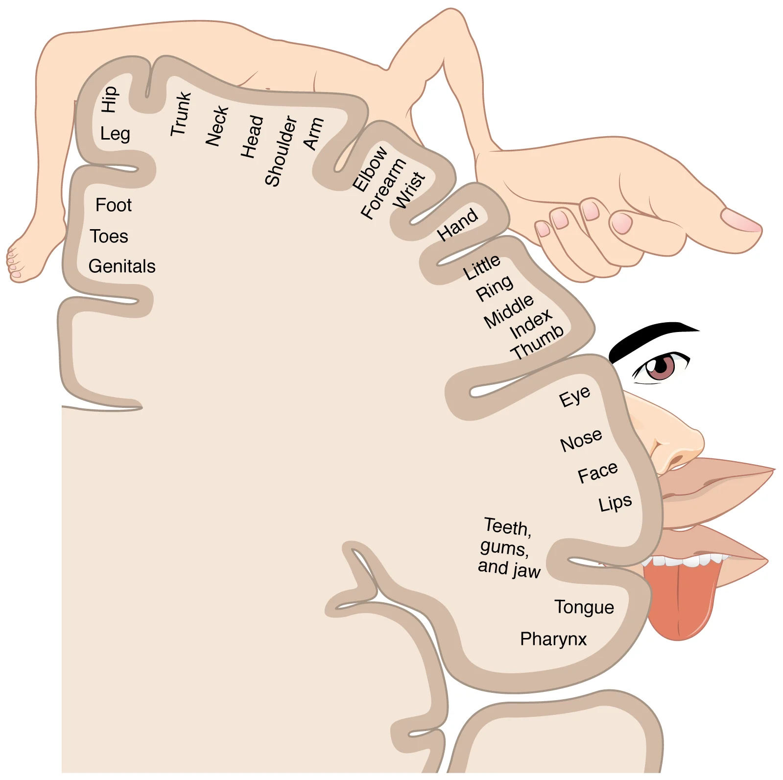
Primary motor cortex - voluntary movement. Organized as a motor homunculus - a distorted body map where fine-control areas (face, hands) get more cortex than coarse-control areas (torso, legs).
Prefrontal cortex - higher executive function: planning, decision-making, working memory, judgment, inhibition of impulses, personality. The last brain region to fully mature (mid-20s).
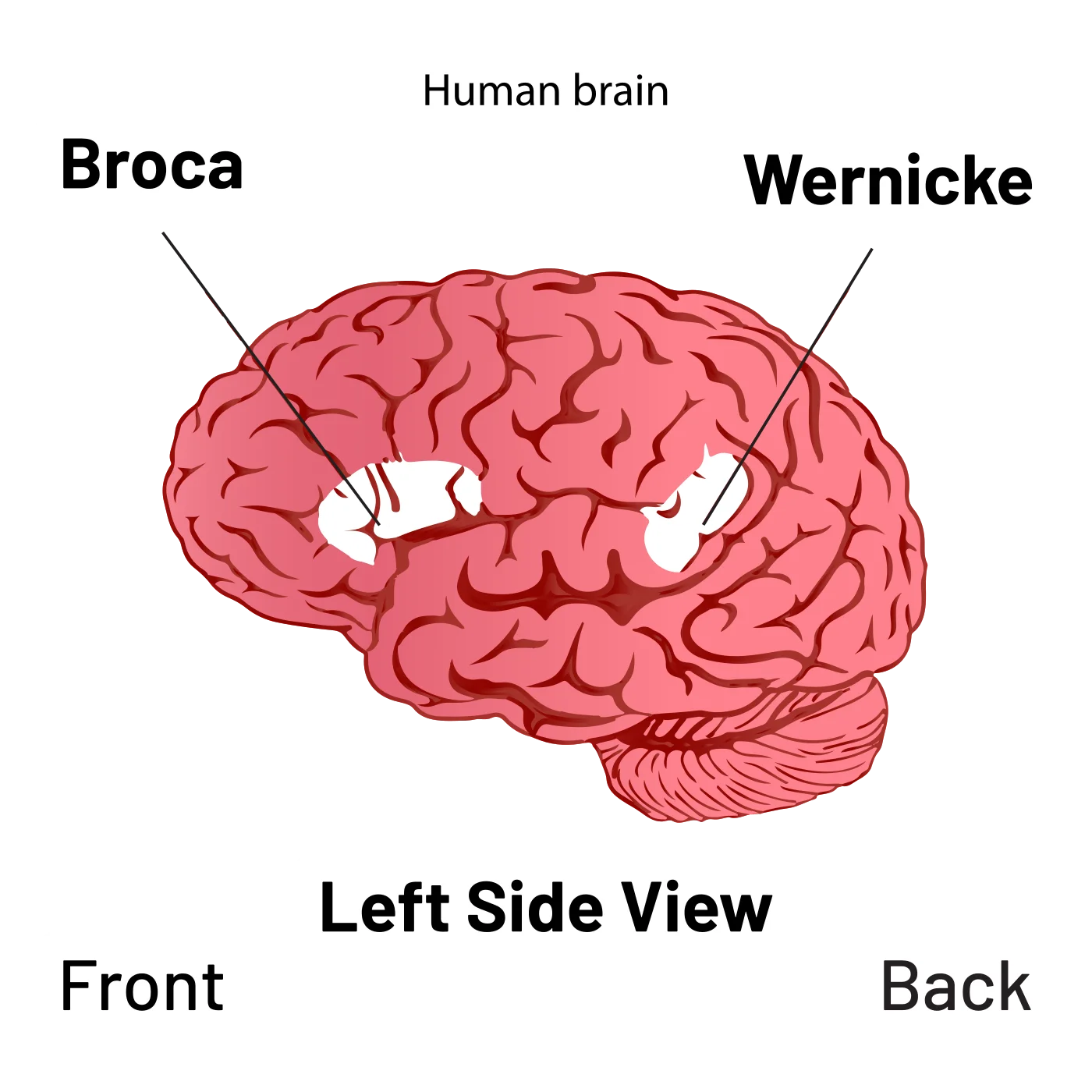
Broca’s area (dominant hemisphere, usually left) - speech production. Damage → Broca’s aphasia (non-fluent, effortful speech with preserved comprehension). We covered this in Chapter 4.
The motor homunculus mapped along the precentral gyrus. Body parts controlled by the largest cortical areas (hands, face, tongue) sit at the bottom of the strip; feet and legs sit at the top. Credit: Ranson, 1920 (Public Domain) via Wikimedia Commons.
Parietal Lobe
Top of the head. Responsible for:
Primary somatosensory cortex - receives touch, pressure, pain, temperature, and proprioception. Has its own sensory homunculus with enormous lips, hands, and face.
Attention - the parietal cortex is critical in directing the attentional spotlight.
The sensory homunculus. Cortical area devoted to each body region scales with receptor density — fingers, lips, and tongue dominate; torso and legs get far less cortex. Credit: OpenStax Anatomy & Physiology 2e, CC BY 3.0.
Damage to the right parietal lobe can produce hemispatial neglect - patients fail to attend to the left side of space, ignoring food on the left side of their plate or shaving only the right side of their face.
Occipital Lobe
Back of the head. Almost entirely devoted to vision.
Primary visual cortex (V1) - receives input from the lateral geniculate nucleus (LGN) of the thalamus. Contains “striate cortex” (named for its striped appearance under a microscope).
Processes form, color, and motion via the parvocellular and magnocellular pathways we covered in Chapter 1.
Damage to V1 causes cortical blindness - the eyes work but the brain cannot process vision.
Temporal Lobe
Side of the head, above the ears. Responsible for:
Primary auditory cortex - receives sound information from the cochlea via the thalamus. Organized tonotopically (frequency-mapped).
Wernicke’s area (dominant hemisphere) - language comprehension. Damage → Wernicke’s aphasia (fluent but meaningless speech, impaired comprehension).
Hippocampus and medial temporal lobe - memory formation.
Fusiform face area - specialized for recognizing faces. Damage produces prosopagnosia (face blindness).
The two classical language areas on the left hemisphere. Broca's area (frontal) handles speech production; Wernicke's area (posterior temporal) handles language comprehension. Credit: UX Stalin via Wikimedia Commons, CC BY-SA 4.0.
Contralateral Control
The brain controls the opposite side of the body. Left hemisphere runs the right side; right hemisphere runs the left side. Holds for motor, sensory, and visual modalities. The exception: smell is ipsilateral - the right nostril projects to the right hemisphere and vice versa, because olfactory signals bypass the typical thalamic crossing.
This is why a right-hemisphere stroke causes left-sided weakness and sensory loss, and why split-brain research (severed corpus callosum) reveals such striking asymmetries.
Hemispheric Dominance
In ~90% of people (including most left-handers), the left hemisphere is the language-dominant hemisphere. It handles:
Language comprehension and production (Wernicke’s, Broca’s)
Logical, sequential, analytical reasoning
Math and detail orientation
The right hemisphere (non-dominant for language) specializes in:
Prosody - emotional tone, rhythm, inflection of speech
Creativity and intuition
Spatial/holistic processing
Face recognition
Big-picture concepts
Despite popular myths, there is no strict “left-brain” or “right-brain” person. Normal cognition uses both hemispheres, communicating across the corpus callosum (the thick band of white matter that connects the two sides).
Left-handed individuals show some differences in lateralization (some have language on the right, or bilateral language), but the differences are subtle and incompletely understood.
Which lobe contains the primary motor cortex and Broca's area?
Click to reveal answer
Frontal lobe. The primary motor cortex controls voluntary movement and is organized as a motor homunculus. Broca's area (in the dominant hemisphere) handles speech production.
Why does damage to the right parietal lobe cause patients to ignore the left side of space?
Click to reveal answer
The right parietal cortex is critical for directing attention - particularly to the left side of space due to contralateral organization. Damage produces hemispatial neglect, where patients fail to attend to stimuli on the left.
Which sense does NOT follow contralateral organization?
Click to reveal answer
Smell (olfaction). Smell is ipsilateral - the right nostril sends to the right hemisphere, the left to the left. Olfactory signals bypass the thalamus and the typical crossover.
Underneath the cerebral cortex sits the “old brain” - a set of evolutionarily ancient structures that handle the functions you don’t want to think about: breathing, balance, arousal, coordination, and emotional tagging. They work below conscious awareness, and damage to any of them can be devastating.
The Brainstem
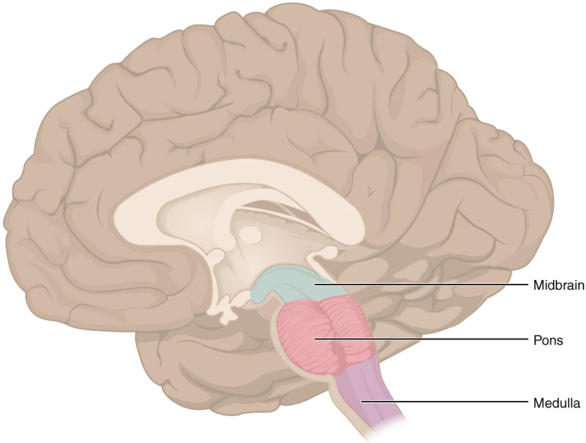
The brainstem connects the cerebrum to the spinal cord. Three segments, bottom to top:
The three segments of the brainstem in mid-sagittal view. The midbrain houses the substantia nigra and the colliculi; the pons relays cortex-cerebellum traffic and contains REM-sleep centers; the medulla controls heart rate, breathing, and other autonomic vital functions. Credit: OpenStax via Wikimedia Commons (CC BY 4.0).
Contains the cardiovascular center and respiratory center.
Damage is often fatal - this is where lethal brainstem strokes live.
Pons
“Bridge” between the cerebrum and the cerebellum.
Regulates sleep and arousal - contains centers for REM sleep onset.
Relay for sensory and motor signals between cortex and spinal cord.
Midbrain
Contains the substantia nigra (source of dopamine for the basal ganglia) and the tectum (superior and inferior colliculi for visual and auditory reflexes).
Substantia nigra damage is the hallmark of Parkinson’s disease - loss of dopamine neurons produces tremor, rigidity, and bradykinesia.
Reticular Formation
A network of neurons running through the brainstem. Critical for:
Arousal and consciousness. The reticular activating system (RAS) sends diffuse glutamate projections to the cortex; its activity level corresponds to your wakefulness.
Sleep-wake cycle. Damage can produce coma.
Attention and autonomic reflexes.
Mnemonic for the brainstem: Pavlov’s Really Frickin’ Mad (Pons, Reticular formation, + Medulla inside, Formation).
Cerebellum (“Little Brain”)
Tucked under the back of the brain. Two roles:
Coordinates voluntary movement. Plans timing and fluidity of movement. Essential for balance, posture, and fine motor control.
Motor learning and proprioception - integrates sensory feedback to refine movements.
Alcohol impairs the cerebellum, which is why intoxicated people slur, stumble, and fail coordination tests. Cerebellar damage produces ataxia (uncoordinated movement), intention tremor (shakiness that worsens as you approach a target), and speech problems.
Subcortical Structures
Thalamus
The relay station of the brain. Virtually all sensory information (except olfaction) passes through the thalamus on its way to the cortex. Different nuclei of the thalamus are specialized for different modalities (LGN for vision, MGN for hearing).
Also contributes to higher cognition and emotion regulation.
Hypothalamus
Just below the thalamus (“hypo” = below). The master regulator of:
Autonomic nervous system - orchestrates sympathetic vs. parasympathetic tone.
Endocrine system - controls the pituitary gland via releasing/inhibiting hormones.
Circadian rhythm - the SCN lives here.
Tiny but mighty. The hypothalamus links the nervous and endocrine systems - a nervous-system command center with hormonal outputs.
Basal Ganglia
A set of subcortical nuclei (caudate, putamen, globus pallidus, substantia nigra, subthalamic nucleus) that:
Help coordinate movement - though they don’t contain UMNs themselves, they modulate motor cortex activity.
Contribute to habit learning and procedural memory.
Participate in cognition and emotion.
Dopamine from the substantia nigra to the striatum (caudate + putamen) is essential for normal basal ganglia function. Loss of this dopamine is what kills motor control in Parkinson’s disease.
Corpus Callosum
A thick band of white matter connecting the two cerebral hemispheres. Allows them to share information. Cutting it (for severe epilepsy) produces split-brain patients - the classic experiments demonstrating hemispheric specialization.
Mnemonic: “Corpus Call Someone.”
Which brainstem structure regulates heart rate and breathing?
Click to reveal answer
The medulla (oblongata). It contains cardiovascular and respiratory centers. Brainstem strokes targeting the medulla are often fatal.
Why does alcohol cause slurred speech and loss of balance?
Click to reveal answer
Alcohol impairs the cerebellum, which coordinates voluntary movement. Cerebellar dysfunction causes ataxia, intention tremor, and slurred speech - the classic drunken motor signs.
Parkinson's disease results from loss of dopamine in which pathway?
Click to reveal answer
The nigrostriatal pathway - from the substantia nigra in the midbrain to the striatum (caudate + putamen) of the basal ganglia. Loss of this dopamine causes tremor, rigidity, and bradykinesia.
What is the function of the corpus callosum?
Click to reveal answer
It connects the left and right cerebral hemispheres, allowing them to communicate. Severed corpus callosum produces split-brain patients, the classic evidence for hemispheric specialization.
Neurons talk to each other with chemicals. Each neurotransmitter has a distinct personality: some excite, some inhibit, some modulate mood or attention. Then we’ll turn to how we see the brain in action - the imaging methods you need to keep straight for the MCAT.
The Major Neurotransmitters
Glutamate
Main excitatory neurotransmitter in the CNS.
Diffuse projections (reticular activating system) keep the cortex aroused.
Too much glutamate → excitotoxicity; kainic acid experiments exploit this to kill neurons.
Involved in learning, memory (long-term potentiation), consciousness.
Mnemonic: GLU = eGLUcites.
GABA and Glycine
Main inhibitory neurotransmitters. GABA in the brain, glycine in the spinal cord.
Alcohol, benzodiazepines, barbiturates all enhance GABA-A receptor activity → sedation.
Loss of inhibition produces seizures - why anticonvulsants often boost GABA.
Acetylcholine (ACh)
Released by all motor neurons at the neuromuscular junction - essential for muscle contraction. Mnemonic: ACE flexes his muscles.
Used by the autonomic nervous system (both sympathetic pre-ganglionic and all parasympathetic).
In the CNS: attention and arousal. Basal forebrain (nucleus basalis, septal nuclei) projects ACh widely to cortex - degeneration is a feature of Alzheimer’s disease.
Dopamine
Reward, motivation, motor control.
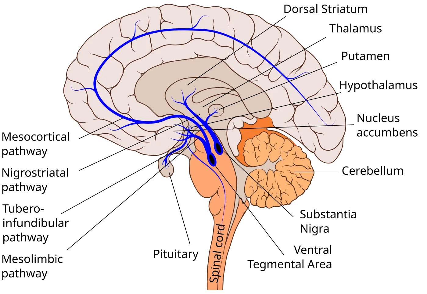
Substantia nigra → striatum (nigrostriatal pathway): motor planning. Degeneration = Parkinson’s.
VTA → nucleus accumbens (mesolimbic pathway): reward and reinforcement. Hyperactivity associated with positive symptoms of schizophrenia (hallucinations, delusions).
VTA → prefrontal cortex (mesocortical pathway): cognition, affect. Hypoactivity associated with negative symptoms of schizophrenia.
Mnemonic: Low dopamine → Parkinson’s; high dopamine → schizophrenia (“ski-zophrenia”).
The four major dopaminergic pathways. Nigrostriatal loss drives Parkinson's disease. Mesolimbic overactivity is linked to the positive symptoms of schizophrenia; mesocortical underactivity is linked to the negative symptoms. Credit: Slashme, Patrick J. Lynch, and Fvasconcellos via Wikimedia Commons, CC BY-SA 4.0.
Serotonin (5-HT)
Released from raphe nuclei in the brainstem. Projects widely.
Regulates mood, appetite, sleep, arousal.
Low serotonin linked to depression (SSRIs boost serotonin). Mnemonic: “Sir Rotten” is depressed.
Also implicated in anxiety and aggression.
Norepinephrine (Noradrenaline)
Released from the locus coeruleus in the pons.
Promotes alertness, attention, arousal.
Low levels → depression; high levels → anxiety/mania.
Also the main transmitter for sympathetic post-ganglionic synapses (with epinephrine).
Histamine
From the hypothalamus to the cortex.
Wakefulness, attention. Antihistamines cause drowsiness by blocking this pathway.
Endorphins (Endogenous Opioids)
Peptide neurotransmitters.
Block pain sensations; produce “runner’s high.”
Mimicked by opiates (morphine, heroin, oxycodone).
Mnemonic: “A running door with fins” (happy because endorphins are happy).
Monoamines (biogenic amines). Serotonin, histamine, dopamine, norepinephrine, epinephrine. Slower-acting, G-protein-coupled receptors. Involved in attention, cognition, emotion. Dopamine, norepinephrine, and epinephrine are also catecholamines (share a catechol group).
Peptide neurotransmitters. Endorphins, enkephalins. Small peptides.
Acetylcholine. In its own chemistry class. ANS and motor neurons.
Brain-Imaging Techniques
Two broad aims: structure (what does the brain look like?) and function (what is it doing?).
Structural Imaging
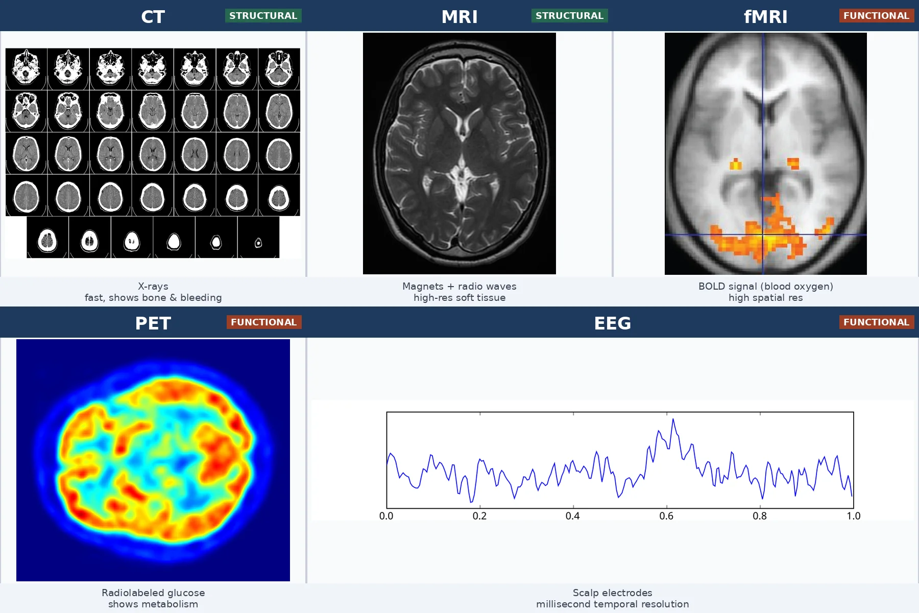
CT (Computerized Axial Tomography). Uses X-rays to build a 3D image. Fast; good for tumors, bleeding, and skull fractures. Lower resolution than MRI for soft tissue. Mnemonic: CATs use X-rays.
MRI (Magnetic Resonance Imaging). Uses powerful magnets and radio waves. Higher resolution for soft tissue than CT. Slower. Good for detailed brain anatomy. Does NOT show function.
Functional Imaging
EEG (Electroencephalogram). Scalp electrodes measure summed electrical activity of cortical neurons. High temporal resolution (milliseconds); poor spatial resolution. Non-invasive, inexpensive. Used for sleep studies, seizures, cognitive tasks. Mnemonic: Electro = EEG = electricity.
MEG (Magnetoencephalogram). Measures magnetic fields produced by brain currents using SQUIDs (Superconducting Quantum Interference Devices). Better spatial resolution than EEG, similar temporal resolution. Rare and expensive.
PET (Positron Emission Tomography). Inject radioactive glucose; active brain regions take up more. Shows metabolic activity. Invasive (radiation exposure); low spatial and temporal resolution. Mnemonic: PETs like glucose.
fMRI (Functional MRI). Measures changes in blood oxygenation (BOLD signal) - active regions use more oxygen. High spatial resolution; moderate temporal resolution (seconds). Non-invasive. Most popular research tool in cognitive neuroscience. Mnemonic: f = function.
The major brain-imaging techniques side by side. Structural methods (CT, MRI) show anatomy; functional methods (fMRI, PET, EEG) show activity. fMRI has the best spatial resolution of the functional group; EEG has the best temporal resolution. Composite credit: CT — Mikael Häggström / Uppsala University Hospital (CC0); MRI — Novaksean (CC BY-SA 4.0); fMRI — OpenStax (CC BY 4.0); PET — Jens Maus (Public Domain); EEG — Hugo Gamboa (CC BY-SA 3.0). Composition released under CC BY-SA 4.0.
Which neurotransmitters are abnormal in Parkinson's disease and in schizophrenia?
Click to reveal answer
Both involve dopamine. Parkinson's: too little dopamine in the nigrostriatal pathway. Schizophrenia: dysregulation of dopamine - typically hyperactivity in mesolimbic pathway (positive symptoms) and hypoactivity in mesocortical pathway (negative symptoms).
What is the most common excitatory neurotransmitter in the CNS, and the most common inhibitory?
Click to reveal answer
Glutamate is the main excitatory neurotransmitter. GABA is the main inhibitory neurotransmitter (glycine is the main inhibitory in the spinal cord).
Which imaging method provides the best TEMPORAL resolution, and which the best SPATIAL resolution of brain function?
Click to reveal answer
Best temporal resolution: EEG (milliseconds). Best spatial resolution for function: fMRI (millimeters). Each involves a trade-off between where and when.
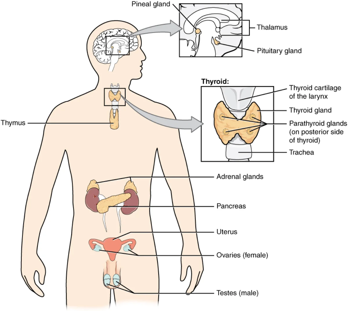
The endocrine system works alongside the nervous system to control behavior, but on a slower timescale. Nervous system = fast, targeted (neurotransmitters across a synapse). Endocrine system = slow, body-wide (hormones through the blood). Together they coordinate physiology.
The major endocrine glands and their anatomical positions. Each gland releases hormones into the bloodstream that travel body-wide to act on tissues bearing the matching receptor. Credit: OpenStax College via Wikimedia Commons (CC BY 3.0).
Steroid hormones. Testosterone, estrogen, progesterone, cortisol. Lipid-soluble, pass directly through the plasma membrane, bind intracellular receptors that act as transcription factors.
Tyrosine derivatives. Thyroid hormones (T3, T4) and catecholamines (epinephrine, norepinephrine). Made from the amino acid tyrosine.
The Hypothalamus-Pituitary Axis
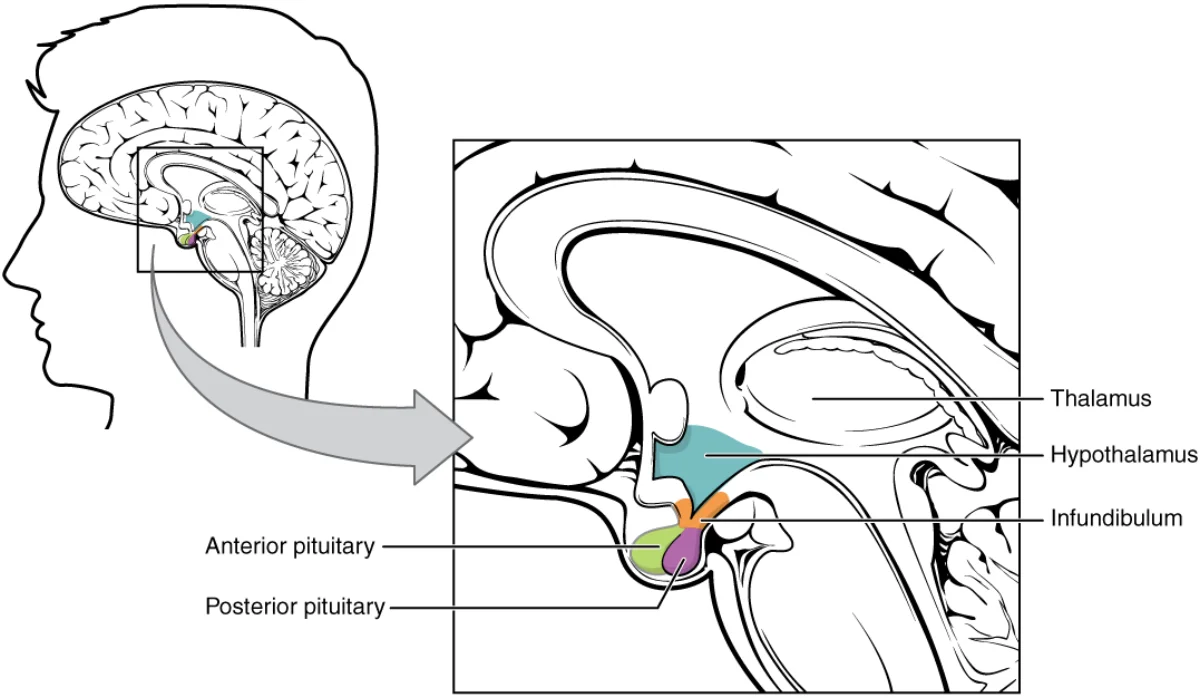
The hypothalamus is the bridge between the nervous and endocrine systems. It controls the pituitary gland (“master gland”), which sits just below it.
The hypothalamus-pituitary complex. The hypothalamus releases hormones (releasing/inhibiting factors) that travel down the infundibulum to the anterior pituitary; the posterior pituitary stores and releases hormones synthesized in the hypothalamus itself. Credit: OpenStax College via Wikimedia Commons (CC BY 3.0).
Anterior Pituitary
Releases hormones in response to releasing factors from the hypothalamus. Mnemonic: FLAT PEG:
Release parathyroid hormone (PTH) to regulate calcium levels.
Adrenal Glands
Each sits on top of a kidney. Two layers, different hormones:
Adrenal medulla (inner) - releases catecholamines: epinephrine and norepinephrine. Fast stress response.
Adrenal cortex (outer) - releases steroids including cortisol (glucocorticoid). Slower, sustained stress response. Also releases aldosterone (mineralocorticoid, salt/water balance) and sex steroids.
Mnemonic: “Cortisol from the CORTex.”
Gonads
Ovaries: estrogen and progesterone; also produce eggs.
Testes: testosterone; also produce sperm.
Stimulated by LH and FSH from the anterior pituitary.
Pancreas
Insulin (lowers blood glucose) and glucagon (raises blood glucose).
Unusually, the pancreas is NOT controlled by the pituitary. Its hormone release is triggered directly by blood glucose.
Negative Feedback Loops
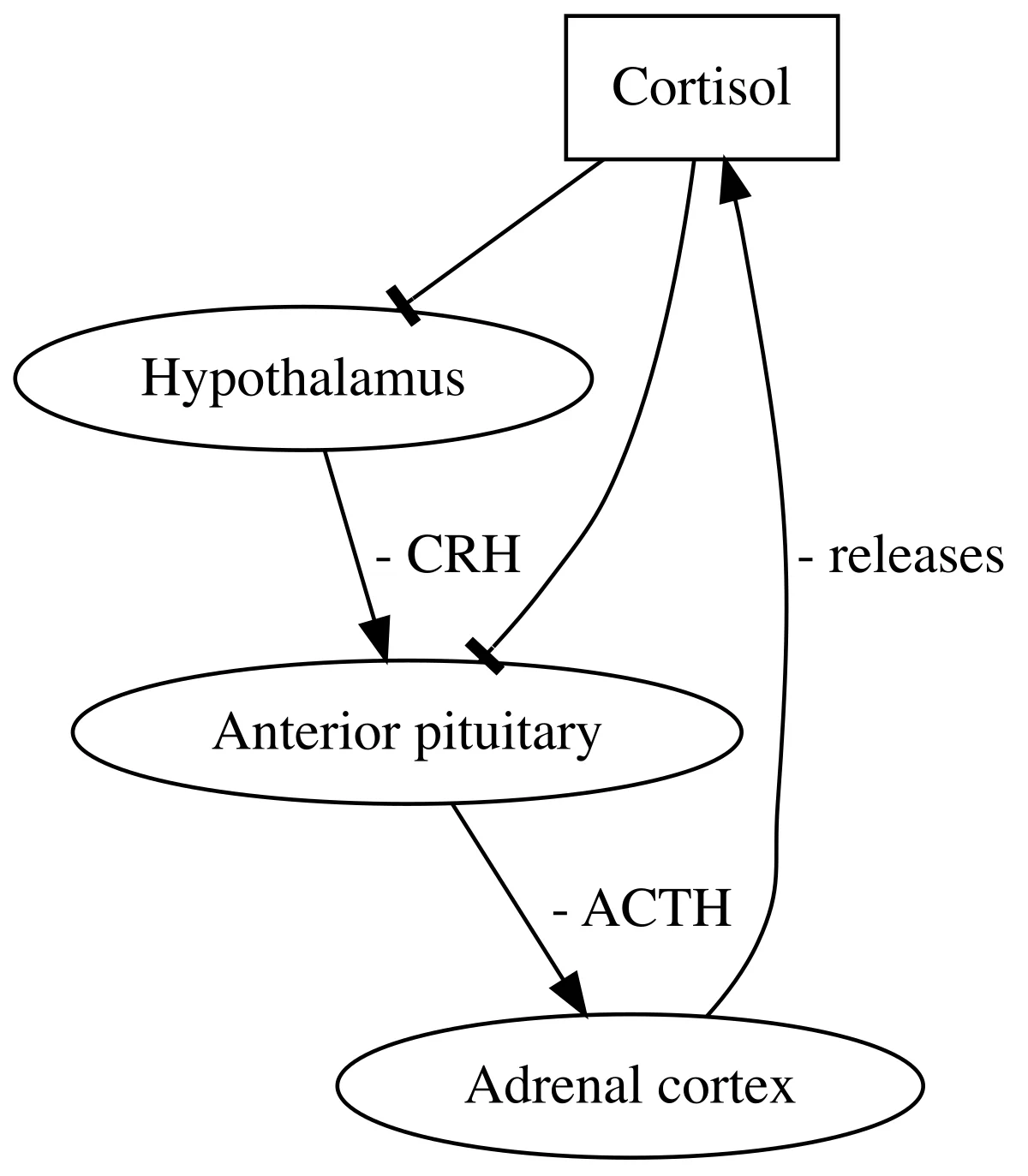
Most endocrine regulation works by negative feedback - the end product of a pathway inhibits its own production. Classic example, thyroid regulation:
Hypothalamus → TRH → anterior pituitary
Anterior pituitary → TSH → thyroid
Thyroid → T3, T4 → tissues
T3 and T4 inhibit TRH and TSH → loop turns itself off
This stable feedback keeps hormone levels within a narrow range. When the loop breaks (e.g., autoimmune destruction of the thyroid), the feedback fails, and levels of TSH skyrocket trying to rescue T3/T4 output.
The HPA axis as the prototypical negative-feedback endocrine loop. Cortisol from the adrenal cortex feeds back to inhibit both the hypothalamus (CRH) and the anterior pituitary (ACTH), keeping its own levels within a narrow range. The same logic governs the thyroid axis (TRH → TSH → T3/T4) and the gonadal axis. Credit: Biolinkage Project via Wikimedia Commons (CC BY-SA 4.0).
Positive feedback loops do exist but are rare. Example: oxytocin during labor - uterine stretching triggers oxytocin release, which triggers more contractions, which stretch more, until delivery. Then the loop stops.
Name the seven hormones released by the anterior pituitary (FLAT PEG).
Click to reveal answer
FSH, LH, ACTH, TSH, Prolactin, Endorphins, Growth hormone. Posterior pituitary releases ADH and Oxytocin (made in the hypothalamus).
Which adrenal region releases cortisol, and which releases epinephrine?
Click to reveal answer
Adrenal CORTEX (outer) = cortisol (and other steroids). Adrenal MEDULLA (inner) = catecholamines (epinephrine, norepinephrine). Cortisol from the CORTex.
How does negative feedback keep thyroid hormones stable?
Click to reveal answer
Hypothalamus TRH → anterior pituitary TSH → thyroid T3/T4. Circulating T3 and T4 inhibit TRH and TSH, turning off their own production. Keeps hormones within a narrow range.
Human development begins with one fertilized cell and ends, roughly nine months later, with a walking-talking toddler not many years after that. This section hits the MCAT-relevant highlights of how that journey unfolds, from fertilization through early childhood.
Fertilization and Embryogenesis
Fertilization (Week 2)
Sperm travels through the female reproductive tract, binds the zona pellucida of the egg.
Acrosome reaction - enzymes on the sperm head digest the zona pellucida.
Sperm plasma membrane fuses with the egg’s, injecting genetic material.
Cortical reaction - enzymes in egg cortical granules rapidly harden the zona pellucida, blocking other sperm (polyspermy block).
Result: zygote with a full diploid genome.
Cleavage and the Morula
The zygote divides (cleavage, no growth) → 2 cells → 4 → 8 → 16 → morula (~32 cells). The outer cells begin to differ from the inner cells: trophoblast outside (will form placenta) and embryoblast inside (will form the embryo).
Blastulation
Blastocyst forms: inner cell mass (embryoblast) + hollow cavity (blastocoel). Zona pellucida breaks down.
Inner cell mass develops an amniotic cavity; cells split into epiblast (above) and hypoblast (below) - the bilaminar disc.
Gastrulation
Formation of the three germ layers from which all organs will develop:
Endoderm - inner layer. Becomes the GI tract lining, lungs, liver, pancreas. Mnemonic: “Endo = insides.”
Neurulation
Special attention to the nervous system’s origin. A rod-like structure called the notochord forms in the mesoderm. It induces the overlying ectoderm to thicken into the neural plate. The neural plate folds up, forming the neural tube. The neural tube becomes the brain and spinal cord.
Failure to close the neural tube causes birth defects (spina bifida at the bottom, anencephaly at the top). Folic acid supplementation during early pregnancy reduces these risks - part of why prenatal vitamins matter.
Implantation and Placenta
The blastocyst (days 6-10 post-fertilization) burrows into the uterine wall (implantation). Trophoblasts differentiate into:
Cytotrophoblast - inner layer.
Syncytiotrophoblast - outer, forms villi that invade maternal blood vessels, eventually forming the placenta. The placenta provides oxygen and nutrients and removes waste.
Gestation Timeline
Total gestation is roughly 40 weeks, measured from the last menstrual period (LMP). Divided into trimesters or weeks.
Week 0: LMP.
Week 2: Fertilization.
Weeks 2-10 (Embryonic Period): Major organ systems form. Most vulnerable to teratogens.
Weeks 10-40 (Fetal Period): Growth and maturation.
Before week 37: Preterm (complications increase).
Week 24: ~50% survival if born.
Weeks 37-42: Full term.
After week 42: Post-term (complications).
Motor Milestones
Approximate ages (with wide individual variation - 50% of children meet each milestone before the listed age):
2-4 months: holds head up, chest up.
2-5 months: rolls over.
5-8 months: sits up (with support, then alone).
5-10 months: stands with support.
6-11 months: pulls up to standing.
7-12 months: crawls.
10-14 months: stands alone.
11-15 months: walks alone.
The sequence is highly conserved across cultures. Infants in every society roll before they crawl, crawl before they walk. The exact timing varies with individual genetics and environment (e.g., infants who sleep on their backs - safer for SIDS prevention - may crawl slightly later).
Head-to-toe (cephalocaudal) development: babies lift their heads before they move their arms, arms before legs. Proximal-to-distal: control of shoulders precedes control of fingers.
Neonatal Reflexes
Babies come pre-loaded with automatic, involuntary motor responses. Some persist (breathing, blinking, pupillary, swallowing). Others are neonatal reflexes that disappear with brain maturation:
Rooting reflex. Stroke the cheek → baby turns head toward the stroke and opens mouth. Helps find the nipple. Disappears by 4 months.
Moro (startle) reflex. Sudden loud noise or loss of support → baby throws arms out, then brings them back. Disappears by 4-6 months.
Babinski reflex. Stroke the sole → toes fan out and up. Disappears by ~12 months (normal response then flips to toes curling down). Re-emergence in adults is a sign of upper motor neuron damage.
Palmar grasp. Object touches palm → fingers close around it. Disappears by 3-4 months.
Tonic neck reflex (fencing posture). Head turned to one side → arm on that side extends, other arm bends. Disappears by 6 months.
Stepping reflex. Hold infant upright with feet on a surface → baby makes stepping motions. Disappears by 2 months.
Sucking reflex. Object in mouth → baby sucks. Disappears by 4 months.
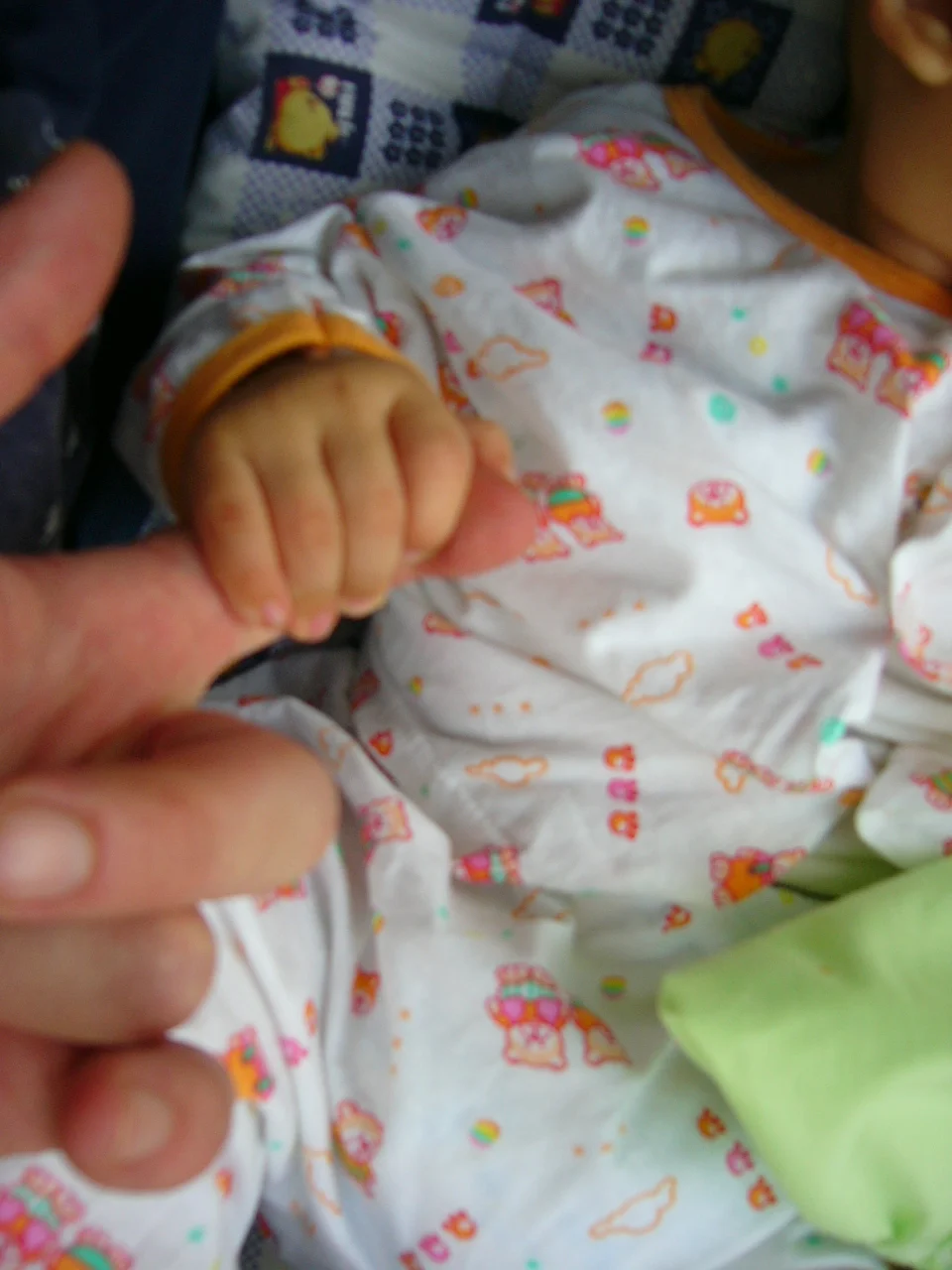
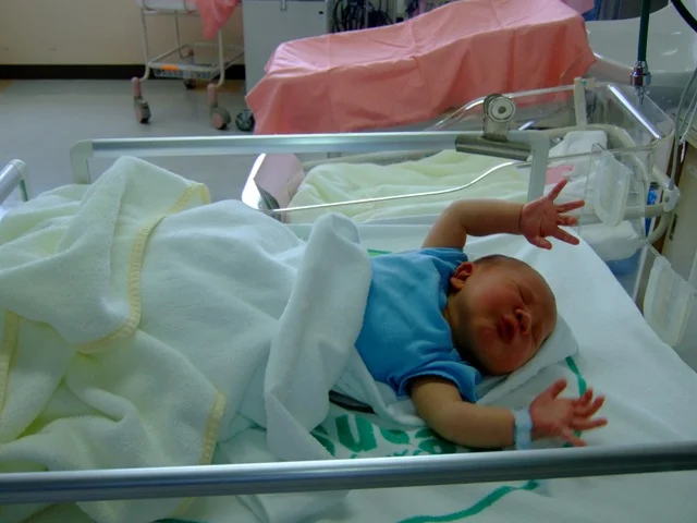
Palmar grasp reflex. Pressure on the infant's palm triggers reflexive finger flexion around the object. Normally disappears by 3-4 months. Credit: Wikimedia Commons, CC BY-SA 3.0.Moro (startle) reflex. A sudden noise or loss of support causes the infant to throw both arms out to the sides before bringing them back in. Disappears by 4-6 months as cortical inhibition matures. Credit: Tawamie via Wikimedia Commons, CC BY-SA 3.0.
Persistence or re-emergence of these reflexes in older children or adults signals neurological problems (lack of cortical control of spinal reflexes).
Which germ layer gives rise to the nervous system?
Click to reveal answer
Ectoderm. Neurulation: the notochord (mesoderm) induces overlying ectoderm to form the neural plate, which folds into the neural tube → brain and spinal cord. Ectoderm also forms skin, hair, nails, sensory organs.
What is the Moro reflex, and when does it typically disappear?
Click to reveal answer
Startle response to sudden loud noise or loss of support - baby throws arms out and then retracts them. Disappears by 4-6 months as cortical inhibition matures.
An adult patient shows a Babinski reflex (toes fan upward when sole is stroked). What does this suggest?
Click to reveal answer
Upper motor neuron damage (e.g., stroke, spinal cord injury). Babinski is normal in infants but should disappear around 12 months. Its reappearance in adults is a classic UMN sign.
The last stretch of this chapter covers two big transitions: the physical and neural remodeling of adolescence, and the genetic and environmental factors that shape behavior across the lifespan. Each is a favorite MCAT topic.
Adolescence
Adolescence begins at sexual maturity (puberty) and ends at adult independence. The exact age markers are cultural, but the biology is universal.
Puberty
About 2 years of sexual maturation. Typically starts around age 11 in girls, 13 in boys (with wide individual variation).
Primary sex characteristics - the reproductive organs themselves (testes, ovaries, genitalia).
Secondary sex characteristics - related to sexual development but not required for reproduction.
Boys: voice deepens, body hair, growth spurt.
Girls: breasts, widening hips.
Both: pubic and underarm hair.
Social Timing Effects
Boys. Early puberty has mixed effects - stronger and taller (advantages in sports, popularity), but also increased risk of delinquency and alcohol use.
Girls. Early puberty is largely negative - increased risk of teasing, harassment, and being out of sync with peers.
Brain Changes
Adolescence is a period of dramatic neural remodeling:
Prefrontal cortex (planning, judgment, impulse control) is the LAST region to mature, continuing into the mid-20s. This lag explains characteristic teenage risk-taking and poor judgment: the emotional (limbic) system is mature, but the prefrontal brake is still developing.
Limbic system (amygdala) is fully developed, driving emotional intensity.
Corpus callosum thickens, improving inter-hemispheric communication (related to language skill development).
Synaptic pruning - unused synapses are eliminated while heavily used ones are strengthened. “Use it or lose it.” What teenagers spend their time doing shapes their adult brains. Total brain volume actually decreases slightly in adolescence due to pruning.
Temperament, Heredity, and Genes
Temperament - innate emotional reactivity and disposition (shy, easy, difficult), relatively stable across life. Observable from infancy, before environmental effects have accumulated.
Heredity - transmission of traits from parents to offspring via genes. Humans have about 20,000-25,000 genes.
Traits cluster into:
Simple (Mendelian) traits. Controlled by one or few genes. Eye color, hair color.
Complex traits. Controlled by many genes interacting with each other and the environment. Intelligence, personality, disease susceptibility. Most behavioral traits are complex.
Twin Studies
Designed to tease apart nature vs nurture.
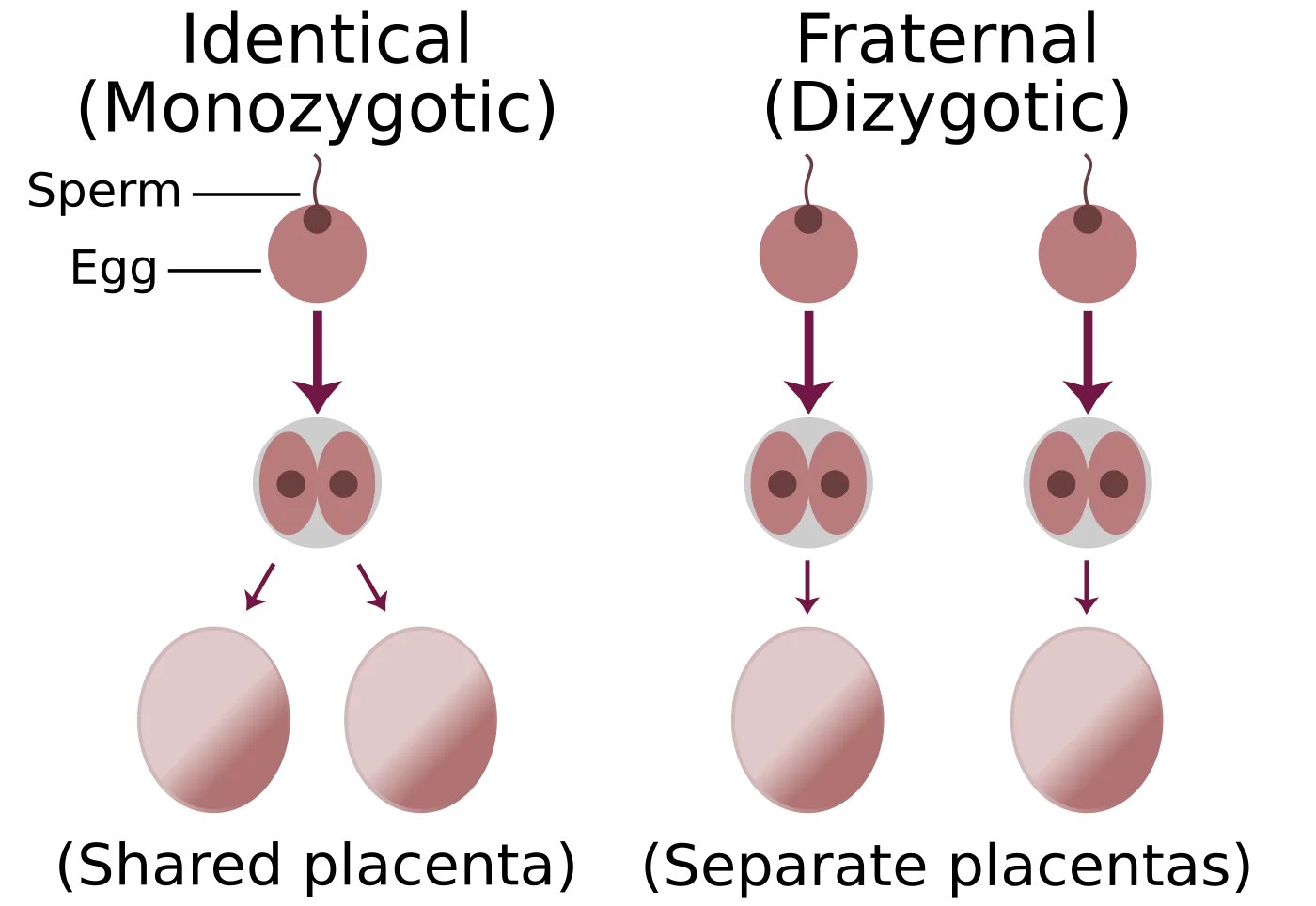
Monozygotic (identical) twins. One fertilized egg splits in two. Share 100% of DNA.
Dizygotic (fraternal) twins. Two separately fertilized eggs. Share 50% of DNA on average - like ordinary siblings, just same womb and age.
Both share the prenatal environment and household. If a trait shows up more similarly in monozygotic twins than dizygotic twins, it suggests a genetic contribution. If both twin types show the same similarity, the trait looks environmentally driven.
Identical twins reared apart are the gold standard: same genes, different environments. The Minnesota Twin Study famously found that identical twins raised apart still converge on many traits (personality, intelligence, religiosity), supporting strong genetic influence.
Monozygotic twins develop when a single fertilized egg splits (100% shared DNA, often one placenta). Dizygotic twins develop from two separate fertilization events (50% shared DNA on average, separate placentas). Credit: Trlkly / ChristinaT3 via Wikimedia Commons, CC BY-SA 3.0.
Adoption Studies
Adopted children share genes with biological parents but environment with adoptive parents. If a trait resembles biological parents more, it’s genetic; if it resembles adoptive parents, it’s environmental.
Problems with twin and adoption studies:
Identical twins often treated more similarly than fraternal twins (confound).
Adoptive families are not randomly distributed (tend to be similar to biological families in SES).
Information about biological parents is often incomplete.
Heritability
Heritability (h²) - the proportion of variance in a trait within a population that is attributable to genetic variance.
Critical nuances the MCAT loves to test:
Heritability is a population statistic, not an individual one. You cannot say “John’s intelligence is 70% genetic.”
Heritability depends on the environment being studied. If everyone lives in identical environments, heritability rises (no environmental variation left to explain differences). In wildly different environments, heritability often falls.
High heritability does NOT mean unchangeable. PKU is highly heritable but managed entirely through diet.
Typical heritability estimates: IQ ~50-70% in adults, personality ~40-50%, schizophrenia ~80% liability.
Regulatory Genes and Epigenetics
Only ~5% of the genome codes for proteins. The other ~95% regulates when and where genes are expressed. Regulatory genes control gene expression in development and behavior.
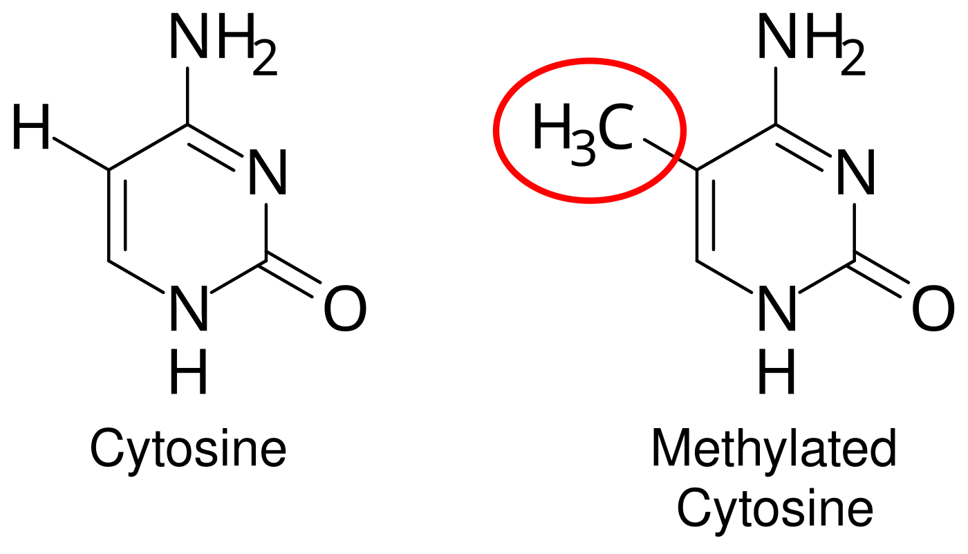
Epigenetics - heritable changes in gene expression that do NOT change the DNA sequence itself. Main mechanism: methylation - attaching methyl groups to DNA, silencing genes. Methylation patterns can be influenced by environment (diet, stress, drugs) and are sometimes passed to offspring.
DNA methylation at the molecular level. A methyl group is added to the 5-carbon of cytosine, producing 5-methylcytosine. This covalent modification typically silences the gene without changing the underlying DNA sequence. Credit: Mariuswalter via Wikimedia Commons, CC BY-SA 4.0.
Epigenetics explains how identical twins can diverge physiologically over time and how maternal experience (nutrition, stress) can affect offspring gene expression without changing the DNA.
Gene-Environment Interaction
Behavior emerges from a constant interplay of genes and environments, not either alone.
Classic Examples
PKU (phenylketonuria). A heritable single-gene disorder: the enzyme phenylalanine hydroxylase is defective, so phenylalanine builds up and causes brain damage. Treatment: a phenylalanine-free diet. A classic case of a “genetic disease” whose effects are entirely determined by environment. If caught on newborn screening, kids with PKU develop normally.
Depression vulnerability. Some genetic variants (e.g., 5-HTT short allele) increase depression risk ONLY in individuals who also experience severe stress. Genes set a vulnerability; environment determines whether it’s expressed.
Older nature-vs-nurture debates have been replaced by the more accurate framing: nature through nurture. Genes and environments are coupled, not competing.
Adaptive Value of Behavioral Traits
Behaviors are shaped by evolution to maintain homeostasis and promote survival and reproduction.
Innate Behaviors
Genetically programmed, present at birth, require no learning.
Fixed-action patterns. Complex innate behaviors triggered by a specific cue (a praying mantis’s strike sequence).
Orientation behaviors.
Kinesis - undirected change in movement speed in response to a stimulus.
Taxis - directed movement toward (positive) or away from (negative) a stimulus. Moths to light = positive phototaxis.
Learned Behaviors
Acquired through experience - covered extensively in Chapter 3 (learning).
Most behaviors are complex, reflecting a mix of innate predisposition and learned refinement. Insects flying is partly innate, partly refined through trial and error.
Which brain region is the LAST to mature during adolescence, and what function does it serve?
Click to reveal answer
The prefrontal cortex, which continues maturing into the mid-20s. It governs planning, decision-making, impulse control, and judgment - hence characteristic teenage risk-taking and poor long-term planning.
What is synaptic pruning, and when does it occur most dramatically?
Click to reveal answer
Elimination of unused synapses while heavily used ones are strengthened. Occurs particularly in adolescence. 'Use it or lose it' - what teenagers spend time doing literally shapes the adult brain.
What common misunderstanding does the phrase 'heritability of intelligence is 50%' produce? Clarify.
Click to reveal answer
It does NOT mean 'your intelligence is 50% genetic.' It means that in the studied population, 50% of the variation between individuals is attributable to genetic variation. It is a population statistic dependent on the environment studied - not a claim about any single person.
What is epigenetics, and how does methylation fit in?
Click to reveal answer
Heritable changes in gene EXPRESSION that don't alter DNA sequence. Methylation - the addition of methyl groups to DNA - typically silences genes. Environmental factors can alter methylation patterns, which sometimes pass to offspring. Explains why identical twins diverge over time.