
The Cerebral Cortex
The cerebral cortex is the folded, wrinkled outer layer of your brain - the “gray matter” that does the slow, flexible work of perception, planning, and language. Its folds (gyri and sulci) pack more surface area into the skull; most of the cortex sits tucked inside those folds.
The cortex divides into four lobes (per hemisphere). Memorize each lobe’s name, location, and primary functions.
The Four Lobes
Mnemonic: F-POT (Frontal, Parietal, Occipital, Temporal).
Frontal Lobe
Front of the head. Responsible for:
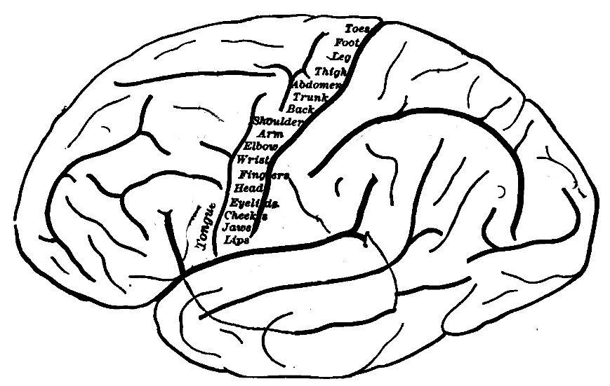
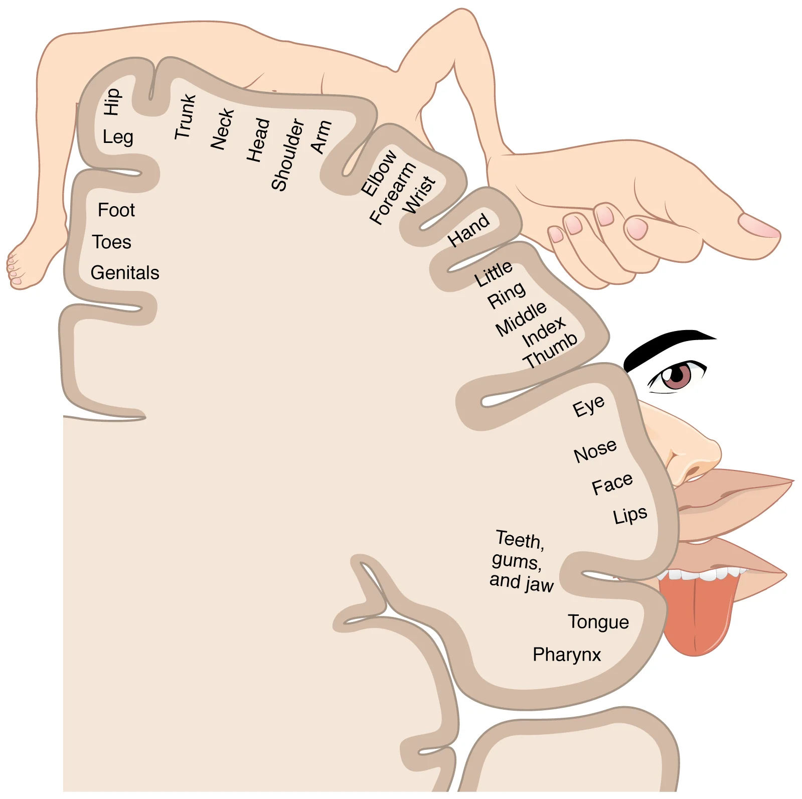
- Primary motor cortex - voluntary movement. Organized as a motor homunculus - a distorted body map where fine-control areas (face, hands) get more cortex than coarse-control areas (torso, legs).
- Prefrontal cortex - higher executive function: planning, decision-making, working memory, judgment, inhibition of impulses, personality. The last brain region to fully mature (mid-20s).
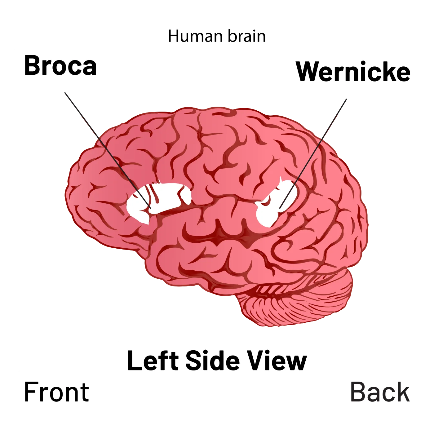
- Broca’s area (dominant hemisphere, usually left) - speech production. Damage → Broca’s aphasia (non-fluent, effortful speech with preserved comprehension). We covered this in Chapter 4.
Parietal Lobe
Top of the head. Responsible for:
- Primary somatosensory cortex - receives touch, pressure, pain, temperature, and proprioception. Has its own sensory homunculus with enormous lips, hands, and face.
- Spatial manipulation - mentally rotating objects, navigating space.
- Attention - the parietal cortex is critical in directing the attentional spotlight.
Damage to the right parietal lobe can produce hemispatial neglect - patients fail to attend to the left side of space, ignoring food on the left side of their plate or shaving only the right side of their face.
Occipital Lobe
Back of the head. Almost entirely devoted to vision.
- Primary visual cortex (V1) - receives input from the lateral geniculate nucleus (LGN) of the thalamus. Contains “striate cortex” (named for its striped appearance under a microscope).
- Processes form, color, and motion via the parvocellular and magnocellular pathways we covered in Chapter 1.
Damage to V1 causes cortical blindness - the eyes work but the brain cannot process vision.
Temporal Lobe
Side of the head, above the ears. Responsible for:
- Primary auditory cortex - receives sound information from the cochlea via the thalamus. Organized tonotopically (frequency-mapped).
- Wernicke’s area (dominant hemisphere) - language comprehension. Damage → Wernicke’s aphasia (fluent but meaningless speech, impaired comprehension).
- Hippocampus and medial temporal lobe - memory formation.
- Fusiform face area - specialized for recognizing faces. Damage produces prosopagnosia (face blindness).
Contralateral Control
The brain controls the opposite side of the body. Left hemisphere runs the right side; right hemisphere runs the left side. Holds for motor, sensory, and visual modalities. The exception: smell is ipsilateral - the right nostril projects to the right hemisphere and vice versa, because olfactory signals bypass the typical thalamic crossing.
This is why a right-hemisphere stroke causes left-sided weakness and sensory loss, and why split-brain research (severed corpus callosum) reveals such striking asymmetries.
Hemispheric Dominance
In ~90% of people (including most left-handers), the left hemisphere is the language-dominant hemisphere. It handles:
- Language comprehension and production (Wernicke’s, Broca’s)
- Logical, sequential, analytical reasoning
- Math and detail orientation
The right hemisphere (non-dominant for language) specializes in:
- Prosody - emotional tone, rhythm, inflection of speech
- Creativity and intuition
- Spatial/holistic processing
- Face recognition
- Big-picture concepts
Despite popular myths, there is no strict “left-brain” or “right-brain” person. Normal cognition uses both hemispheres, communicating across the corpus callosum (the thick band of white matter that connects the two sides).
Left-handed individuals show some differences in lateralization (some have language on the right, or bilateral language), but the differences are subtle and incompletely understood.