
Organization of the Nervous System
The nervous system is a hierarchy: sensory input flows in, motor output flows out, and everything in between is processing. This section lays out the major divisions you need to memorize for any MCAT neuroscience passage.
CNS and PNS
- Central Nervous System (CNS) = brain + spinal cord. Where most processing happens.
- Peripheral Nervous System (PNS) = everything else. All 12 cranial nerves and 31 pairs of spinal nerves, plus associated ganglia.
Brain Divisions
The brain forms from three primary vesicles in the embryo:
- Forebrain → cerebrum, thalamus, hypothalamus (higher functions)
- Midbrain → midbrain (sensory relay, motor coordination)
- Hindbrain → pons, medulla, cerebellum (automatic and motor control)
The 12 Cranial Nerves
The 12 cranial nerves emerge directly from the brain or brainstem (not the spinal cord). You should know each nerve’s number, name, function class (sensory, motor, or both), and what it does.
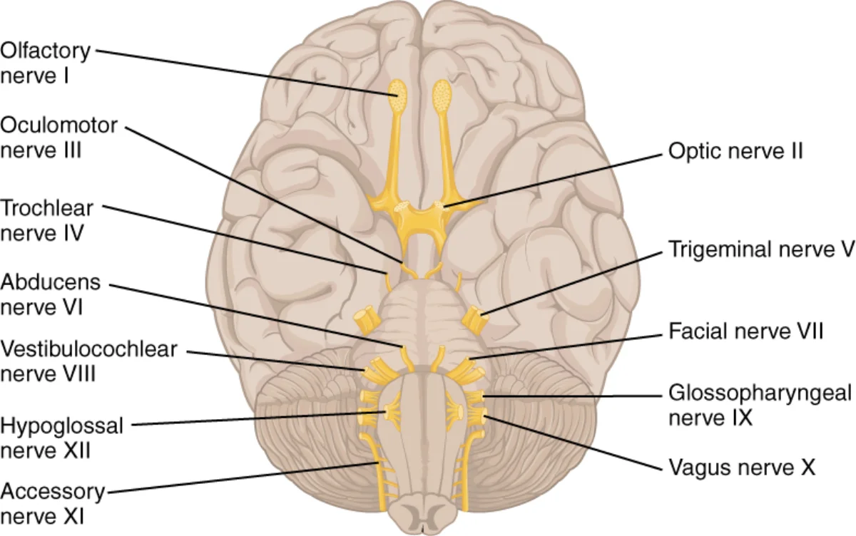
| # | Name | Function | What It Does |
|---|---|---|---|
| I | Olfactory | Sensory | Smell |
| II | Optic | Sensory | Vision |
| III | Oculomotor | Motor | Most eye movements, pupil constriction, lens shape |
| IV | Trochlear | Motor | Superior oblique (eye moves down-and-in) |
| V | Trigeminal | Both | Facial sensation + muscles of chewing |
| VI | Abducens | Motor | Lateral rectus (eye abducts, looks outward) |
| VII | Facial | Both | Muscles of facial expression + taste (anterior tongue) + lacrimation, salivation |
| VIII | Vestibulocochlear | Sensory | Hearing and balance |
| IX | Glossopharyngeal | Both | Taste (posterior tongue), swallowing, parotid salivation, carotid baroreceptors |
| X | Vagus | Both | Parasympathetic to heart, lungs, gut; voice (larynx); swallowing |
| XI | (Spinal) Accessory | Motor | Sternocleidomastoid and trapezius (head turning, shoulder shrug) |
| XII | Hypoglossal | Motor | Tongue movement |
Mnemonic 1: Name Order
“On Old Olympus’ Towering Tops A Finn And German Viewed Some Hops”
Each first letter maps to a nerve in order: Olfactory, Optic, Oculomotor, Trochlear, Trigeminal, Abducens, Facial, (Vestibulo-)Acoustic, Glossopharyngeal, Vagus, Spinal accessory, Hypoglossal.
Mnemonic 2: Function (Sensory / Motor / Both)
“Some Say Marry Money But My Brother Says Big Brains Matter More”
In order I → XII: Sensory, Sensory, Motor, Motor, Both, Motor, Both, Sensory, Both, Both, Motor, Motor.
Clinically High-Yield Nerves
- CN II (optic). Tested with visual acuity and visual fields. Afferent limb of the pupillary light reflex.
- CN III (oculomotor). Efferent limb of the pupillary light reflex - damage produces a “blown pupil” (dilated, unreactive) and a “down-and-out” eye because only IV and VI still pull.
- CN V (trigeminal). Three divisions: V1 ophthalmic, V2 maxillary, V3 mandibular. Corneal reflex afferent.
- CN VII (facial). Bell’s palsy affects this nerve and produces unilateral facial droop including the forehead (an upper motor neuron stroke spares the forehead because of bilateral cortical input).
- CN X (vagus). The main parasympathetic output to the body. Carries “rest and digest” signals to the heart and gut. Vagal stimulation slows the heart.
The Two Branches of the PNS
Somatic Nervous System
Carries voluntary motor commands to skeletal muscles and brings in sensory information from the skin, joints, and muscles. Under conscious control: you decide to lift your arm; the somatic nervous system makes it happen.
Autonomic Nervous System (ANS)
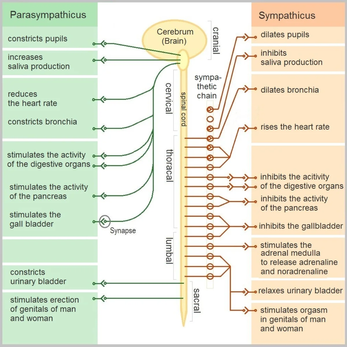
Controls involuntary functions: heart rate, digestion, pupil size, glandular secretion. Not under conscious control. Two opposing branches:
Sympathetic - “fight or flight.” Prepares the body for action.
- Pupils dilate, heart rate increases, respiration deepens.
- Digestion slows; blood diverts to skeletal muscle and vital organs.
- Sweat glands activate, adrenal medulla releases epinephrine.
- Short pre-ganglionic axons near the spinal cord, long post-ganglionic axons to target organs.
Parasympathetic - “rest and digest.” Restores baseline.
- Pupils constrict, heart rate slows, salivation increases.
- Digestion resumes, blood flow to the viscera increases.
- Long pre-ganglionic axons from brainstem or sacral spinal cord; short post-ganglionic axons.
Both branches usually act on the same organs with opposite effects - the balance between them tunes physiology moment-to-moment.
Afferent vs Efferent Neurons
Remember the A/E distinction: Afferent = Arrival (sensory, into CNS). Efferent = Exit (motor, out of CNS). Receptors send info through afferent axons; motor commands travel out through efferent axons.
Upper and Lower Motor Neurons
Voluntary muscle control is a two-step chain:
- Upper motor neurons (UMNs) originate in the cerebral cortex and travel down to synapse on lower motor neurons in the brainstem or spinal cord. Their axons form the corticospinal tract (to spinal cord) or corticobulbar tract (to brainstem, for head/neck).
- Lower motor neurons (LMNs) exit the CNS and synapse directly on skeletal muscle at the neuromuscular junction.
UMNs control LMNs. LMNs actually move muscles.
Motor Neuron Signs
Damage to UMNs and LMNs produces characteristically different clinical pictures:
- LMN signs. Weakness, atrophy, fasciculations (involuntary twitches), hypotonia (low muscle tone), hyporeflexia.
- UMN signs. Weakness, hyperreflexia (exaggerated stretch reflexes), clonus (rhythmic contraction), hypertonia (increased muscle tone), extensor plantar response (Babinski sign - toes fan upward instead of downward when the sole is stroked).
The logic: without UMN input, the spinal reflex circuits lose their “moderator” and become hyperactive. Without LMN input, the muscle gets no drive at all - it withers and twitches.
The Muscle Stretch Reflex
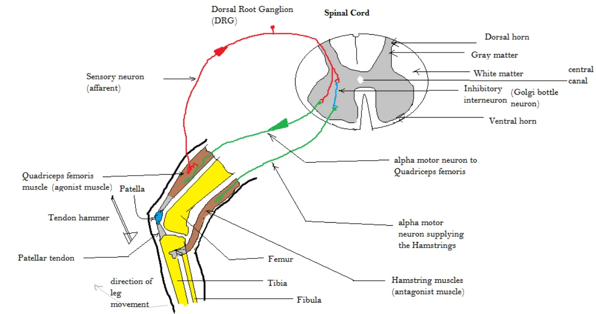
A reflex is a quick, involuntary response to a stimulus that doesn’t require the brain. The classic knee-jerk is a monosynaptic stretch reflex:
- The hammer hits the patellar tendon, stretching the quadriceps.
- Stretch receptors in the muscle (muscle spindles) fire afferent signals up to the spinal cord.
- The afferent neuron synapses directly onto an LMN that innervates the same muscle.
- The LMN fires, contracting the quadriceps - the leg kicks out.
At the same time, a separate inhibitory interneuron silences the antagonist muscle (hamstring) so it doesn’t fight the kick. This is reciprocal inhibition.
Reflexes demonstrate that the spinal cord does meaningful processing on its own - you don’t think “leg, kick.” The body protects itself faster than the brain can decide.
Gray and White Matter
- Gray matter - cell bodies (somas) of neurons, as well as dendrites and synapses.
- White matter - myelinated axons. Looks white because of the fatty myelin.
Distributions differ between brain and spinal cord:
- Brain. Gray matter on outside (cortex), white matter inside.
- Spinal cord. White matter on outside (tracts going up and down), gray matter on inside (where the synapses happen).