
Old Brain and Subcortical Structures
Underneath the cerebral cortex sits the “old brain” - a set of evolutionarily ancient structures that handle the functions you don’t want to think about: breathing, balance, arousal, coordination, and emotional tagging. They work below conscious awareness, and damage to any of them can be devastating.
The Brainstem
The brainstem connects the cerebrum to the spinal cord. Three segments, bottom to top:
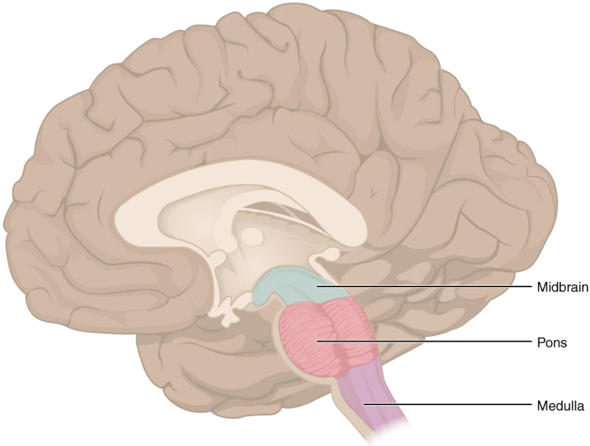
Medulla (Medulla Oblongata)
- Autonomic vital functions. Heart rate, respiration, blood pressure, swallowing, coughing, vomiting.
- Contains the cardiovascular center and respiratory center.
- Damage is often fatal - this is where lethal brainstem strokes live.
Pons
- “Bridge” between the cerebrum and the cerebellum.
- Regulates sleep and arousal - contains centers for REM sleep onset.
- Relay for sensory and motor signals between cortex and spinal cord.
Midbrain
- Contains the substantia nigra (source of dopamine for the basal ganglia) and the tectum (superior and inferior colliculi for visual and auditory reflexes).
- Substantia nigra damage is the hallmark of Parkinson’s disease - loss of dopamine neurons produces tremor, rigidity, and bradykinesia.
Reticular Formation
A network of neurons running through the brainstem. Critical for:
- Arousal and consciousness. The reticular activating system (RAS) sends diffuse glutamate projections to the cortex; its activity level corresponds to your wakefulness.
- Sleep-wake cycle. Damage can produce coma.
- Attention and autonomic reflexes.
Mnemonic for the brainstem: Pavlov’s Really Frickin’ Mad (Pons, Reticular formation, + Medulla inside, Formation).
Cerebellum (“Little Brain”)
Tucked under the back of the brain. Two roles:
- Coordinates voluntary movement. Plans timing and fluidity of movement. Essential for balance, posture, and fine motor control.
- Motor learning and proprioception - integrates sensory feedback to refine movements.
Alcohol impairs the cerebellum, which is why intoxicated people slur, stumble, and fail coordination tests. Cerebellar damage produces ataxia (uncoordinated movement), intention tremor (shakiness that worsens as you approach a target), and speech problems.
Subcortical Structures
Thalamus
The relay station of the brain. Virtually all sensory information (except olfaction) passes through the thalamus on its way to the cortex. Different nuclei of the thalamus are specialized for different modalities (LGN for vision, MGN for hearing).
Also contributes to higher cognition and emotion regulation.
Hypothalamus
Just below the thalamus (“hypo” = below). The master regulator of:
- Homeostasis - hunger, thirst, temperature, fatigue, arousal.
- Autonomic nervous system - orchestrates sympathetic vs. parasympathetic tone.
- Endocrine system - controls the pituitary gland via releasing/inhibiting hormones.
- Circadian rhythm - the SCN lives here.
Tiny but mighty. The hypothalamus links the nervous and endocrine systems - a nervous-system command center with hormonal outputs.
Basal Ganglia
A set of subcortical nuclei (caudate, putamen, globus pallidus, substantia nigra, subthalamic nucleus) that:
- Help coordinate movement - though they don’t contain UMNs themselves, they modulate motor cortex activity.
- Contribute to habit learning and procedural memory.
- Participate in cognition and emotion.
Dopamine from the substantia nigra to the striatum (caudate + putamen) is essential for normal basal ganglia function. Loss of this dopamine is what kills motor control in Parkinson’s disease.
Corpus Callosum
A thick band of white matter connecting the two cerebral hemispheres. Allows them to share information. Cutting it (for severe epilepsy) produces split-brain patients - the classic experiments demonstrating hemispheric specialization.
Mnemonic: “Corpus Call Someone.”