
Neurotransmitters and Brain Imaging
Neurons talk to each other with chemicals. Each neurotransmitter has a distinct personality: some excite, some inhibit, some modulate mood or attention. Then we’ll turn to how we see the brain in action - the imaging methods you need to keep straight for the MCAT.
The Major Neurotransmitters
Glutamate
- Main excitatory neurotransmitter in the CNS.
- Diffuse projections (reticular activating system) keep the cortex aroused.
- Too much glutamate → excitotoxicity; kainic acid experiments exploit this to kill neurons.
- Involved in learning, memory (long-term potentiation), consciousness.
Mnemonic: GLU = eGLUcites.
GABA and Glycine
- Main inhibitory neurotransmitters. GABA in the brain, glycine in the spinal cord.
- Alcohol, benzodiazepines, barbiturates all enhance GABA-A receptor activity → sedation.
- Loss of inhibition produces seizures - why anticonvulsants often boost GABA.
Acetylcholine (ACh)
- Released by all motor neurons at the neuromuscular junction - essential for muscle contraction. Mnemonic: ACE flexes his muscles.
- Used by the autonomic nervous system (both sympathetic pre-ganglionic and all parasympathetic).
- In the CNS: attention and arousal. Basal forebrain (nucleus basalis, septal nuclei) projects ACh widely to cortex - degeneration is a feature of Alzheimer’s disease.
Dopamine
- Reward, motivation, motor control.
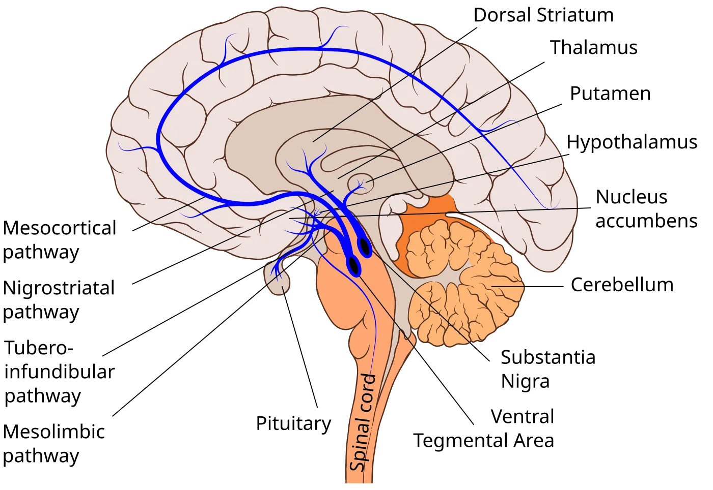
- Substantia nigra → striatum (nigrostriatal pathway): motor planning. Degeneration = Parkinson’s.
- VTA → nucleus accumbens (mesolimbic pathway): reward and reinforcement. Hyperactivity associated with positive symptoms of schizophrenia (hallucinations, delusions).
- VTA → prefrontal cortex (mesocortical pathway): cognition, affect. Hypoactivity associated with negative symptoms of schizophrenia.
- Mnemonic: Low dopamine → Parkinson’s; high dopamine → schizophrenia (“ski-zophrenia”).
Serotonin (5-HT)
- Released from raphe nuclei in the brainstem. Projects widely.
- Regulates mood, appetite, sleep, arousal.
- Low serotonin linked to depression (SSRIs boost serotonin). Mnemonic: “Sir Rotten” is depressed.
- Also implicated in anxiety and aggression.
Norepinephrine (Noradrenaline)
- Released from the locus coeruleus in the pons.
- Promotes alertness, attention, arousal.
- Low levels → depression; high levels → anxiety/mania.
- Also the main transmitter for sympathetic post-ganglionic synapses (with epinephrine).
Histamine
- From the hypothalamus to the cortex.
- Wakefulness, attention. Antihistamines cause drowsiness by blocking this pathway.
Endorphins (Endogenous Opioids)
- Peptide neurotransmitters.
- Block pain sensations; produce “runner’s high.”
- Mimicked by opiates (morphine, heroin, oxycodone).
- Mnemonic: “A running door with fins” (happy because endorphins are happy).
Types of Neurotransmitters by Chemistry
- Amino acid neurotransmitters. Glutamate, GABA, glycine. Fast-acting, ligand-gated channels.
- Monoamines (biogenic amines). Serotonin, histamine, dopamine, norepinephrine, epinephrine. Slower-acting, G-protein-coupled receptors. Involved in attention, cognition, emotion. Dopamine, norepinephrine, and epinephrine are also catecholamines (share a catechol group).
- Peptide neurotransmitters. Endorphins, enkephalins. Small peptides.
- Acetylcholine. In its own chemistry class. ANS and motor neurons.
Brain-Imaging Techniques
Two broad aims: structure (what does the brain look like?) and function (what is it doing?).
Structural Imaging
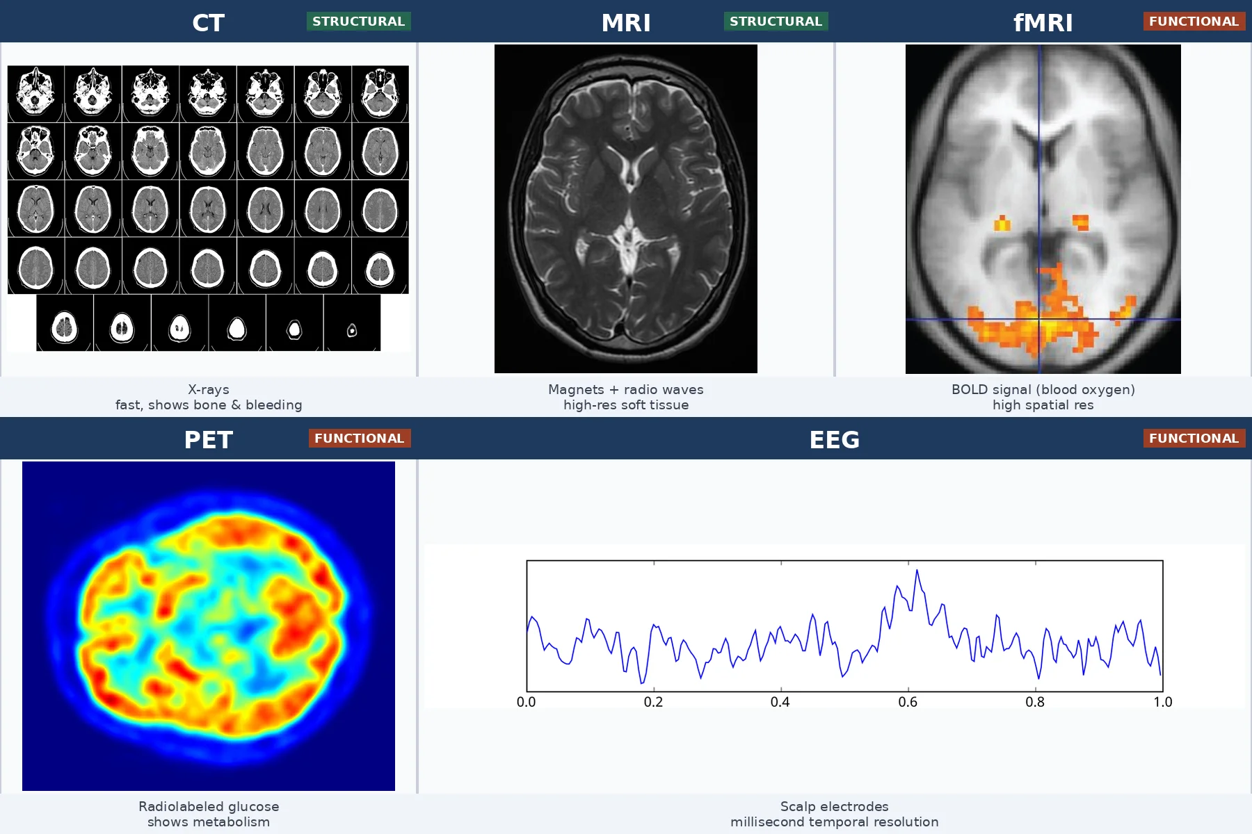
- CT (Computerized Axial Tomography). Uses X-rays to build a 3D image. Fast; good for tumors, bleeding, and skull fractures. Lower resolution than MRI for soft tissue. Mnemonic: CATs use X-rays.
- MRI (Magnetic Resonance Imaging). Uses powerful magnets and radio waves. Higher resolution for soft tissue than CT. Slower. Good for detailed brain anatomy. Does NOT show function.
Functional Imaging
- EEG (Electroencephalogram). Scalp electrodes measure summed electrical activity of cortical neurons. High temporal resolution (milliseconds); poor spatial resolution. Non-invasive, inexpensive. Used for sleep studies, seizures, cognitive tasks. Mnemonic: Electro = EEG = electricity.
- MEG (Magnetoencephalogram). Measures magnetic fields produced by brain currents using SQUIDs (Superconducting Quantum Interference Devices). Better spatial resolution than EEG, similar temporal resolution. Rare and expensive.
- PET (Positron Emission Tomography). Inject radioactive glucose; active brain regions take up more. Shows metabolic activity. Invasive (radiation exposure); low spatial and temporal resolution. Mnemonic: PETs like glucose.
- fMRI (Functional MRI). Measures changes in blood oxygenation (BOLD signal) - active regions use more oxygen. High spatial resolution; moderate temporal resolution (seconds). Non-invasive. Most popular research tool in cognitive neuroscience. Mnemonic: f = function.
Which neurotransmitters are abnormal in Parkinson's disease and in schizophrenia?
Click to reveal answer
Both involve dopamine. Parkinson's: too little dopamine in the nigrostriatal pathway. Schizophrenia: dysregulation of dopamine - typically hyperactivity in mesolimbic pathway (positive symptoms) and hypoactivity in mesocortical pathway (negative symptoms).
What is the most common excitatory neurotransmitter in the CNS, and the most common inhibitory?
Click to reveal answer
Glutamate is the main excitatory neurotransmitter. GABA is the main inhibitory neurotransmitter (glycine is the main inhibitory in the spinal cord).
Which imaging method provides the best TEMPORAL resolution, and which the best SPATIAL resolution of brain function?
Click to reveal answer
Best temporal resolution: EEG (milliseconds). Best spatial resolution for function: fMRI (millimeters). Each involves a trade-off between where and when.