
Blood Composition
Blood is not just a red liquid. It is a complex tissue made up of cells suspended in a protein-rich fluid. An average adult has about 5 liters of blood, and every component has a specific job. Understanding blood composition is essential for MCAT questions on gas transport, immunity, clotting, and transfusion compatibility.
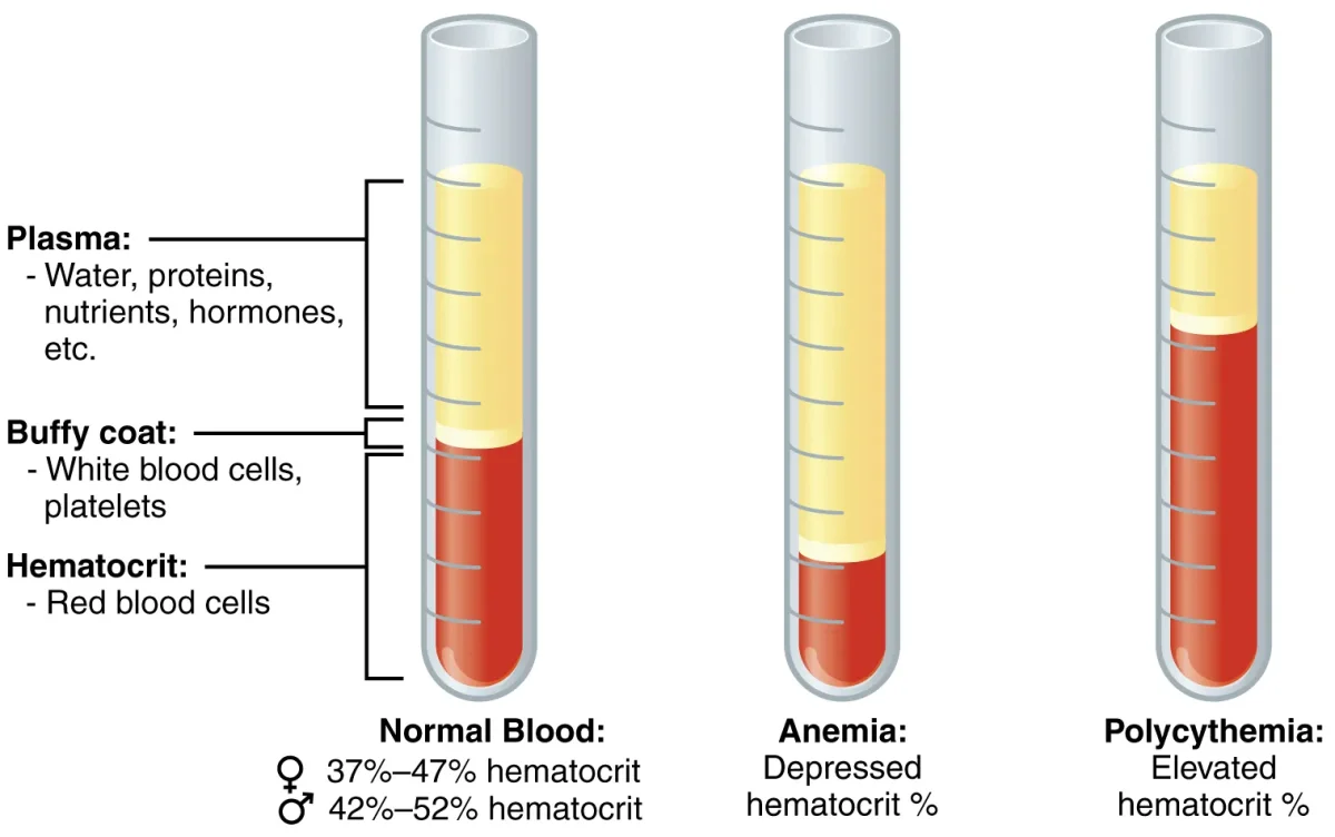
Plasma (~55% of Blood Volume)
Plasma is the liquid matrix of blood. It is about 90% water and 10% dissolved solutes:
- Plasma proteins (~7%) - albumin (maintains oncotic pressure), globulins (antibodies and transport), fibrinogen (clotting precursor)
- Electrolytes - Na+, K+, Ca2+, Cl-, HCO3-
- Nutrients - glucose, amino acids, lipids
- Waste products - urea, creatinine, bilirubin
- Dissolved gases - O2, CO2
- Hormones - transported to target organs
Serum is plasma minus the clotting factors (what you get after blood clots and the clot is removed).
Red Blood Cells (Erythrocytes)
RBCs are the most abundant cells in blood (~4.5-5.5 million per microliter). Their sole purpose is gas transport.
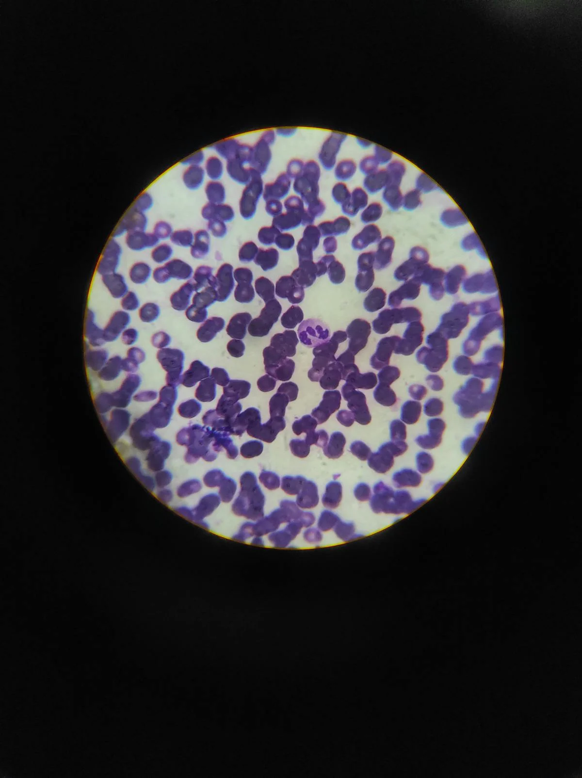
The proportion of blood volume occupied by RBCs is called the hematocrit (normally ~42-52% in males, ~37-47% in females). RBC production is stimulated by erythropoietin (EPO), a hormone released by the kidneys in response to low oxygen levels (hypoxia). This is why people living at high altitude develop higher hematocrit - their kidneys sense low O2 and ramp up RBC production.
Key features of RBCs:
- Biconcave disc shape - maximizes surface area for gas exchange and allows flexibility to squeeze through narrow capillaries
- No nucleus or organelles - mature RBCs eject their nucleus during development, leaving more room for hemoglobin
- Packed with hemoglobin - each RBC contains ~280 million hemoglobin molecules
- No mitochondria - RBCs rely entirely on anaerobic glycolysis for their own energy (they cannot consume the oxygen they carry)
- Lifespan: ~120 days - old RBCs are removed by macrophages in the spleen and liver
Hemoglobin is a tetrameric protein (two alpha and two beta subunits in adult hemoglobin, HbA), each containing a heme group with an iron (Fe2+) atom that binds one O2 molecule. Each hemoglobin molecule can carry 4 O2 molecules.
The Oxygen-Hemoglobin Dissociation Curve
Hemoglobin does not bind oxygen all-or-nothing. As each O2 binds, the hemoglobin subunits shift toward a higher-affinity state, making the next O2 easier to bind. This positive cooperativity produces the classic sigmoid (S-shaped) dissociation curve when % saturation is plotted against PO2.
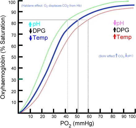
Two regions of the curve matter:
- Plateau (high PO2, ~100 mmHg in lungs): Hemoglobin stays ~97% saturated even if PO2 drops somewhat. Loading is protected.
- Steep portion (low PO2, ~40 mmHg in tissues): Small drops in PO2 cause large drops in saturation. Unloading is maximized where O2 is needed.
Right Shift = More Unloading
A rightward shift means hemoglobin releases O2 more readily at any given PO2 (lower affinity). This happens in metabolically active tissue, which is exactly where extra O2 is needed.
The Bohr effect is the specific case of ↑CO2 and ↑H+ lowering hemoglobin’s O2 affinity. Active muscle produces both, so the curve shifts right exactly where O2 needs to be delivered. A leftward shift (↓CO2, ↓H+, ↓temp, ↓2,3-BPG) means tighter O2 binding and less unloading.
Fetal Hemoglobin
Fetal hemoglobin (HbF) has two alpha and two gamma subunits instead of two alpha, two beta. The gamma subunits bind 2,3-BPG more weakly, so HbF has a higher O2 affinity than adult HbA. Its dissociation curve sits to the left of the adult curve, letting the fetus pull O2 off maternal hemoglobin at the placenta.
White Blood Cells (Leukocytes)
WBCs defend the body against infection. They are far less numerous than RBCs (~5,000-10,000 per microliter) but are larger and nucleated. The specific roles of neutrophils, macrophages, and other frontline defenders are covered in innate immune cells.
| Cell Type | Function | Key Facts |
|---|---|---|
| Neutrophils | First responders; phagocytose bacteria | Most abundant WBC; multilobed nucleus; short-lived; pus = dead neutrophils |
| Lymphocytes | Adaptive immunity (B cells, T cells, NK cells) | Second most abundant; small cells with large nucleus |
| Monocytes | Become macrophages in tissue; phagocytosis + antigen presentation | Largest WBC; kidney-shaped nucleus |
| Eosinophils | Combat parasites; modulate allergic response | Bilobed nucleus; red-staining granules |
| Basophils | Release histamine and heparin; involved in allergic reactions | Rarest WBC; large purple/blue granules; similar to mast cells |
Platelets (Thrombocytes)
Platelets are not true cells - they are small, anucleate cell fragments (~150,000-400,000 per microliter) produced by megakaryocytes in the bone marrow. Platelet production is stimulated by thrombopoietin, a hormone from the liver and kidneys.
Platelets are essential for hemostasis (stopping bleeding). When a blood vessel is damaged, platelets adhere to exposed collagen, activate, change shape, and aggregate to form a temporary platelet plug. They also release chemicals that initiate the coagulation cascade.
Blood Types: ABO System
Blood type is determined by antigens on the surface of RBCs and antibodies in the plasma.
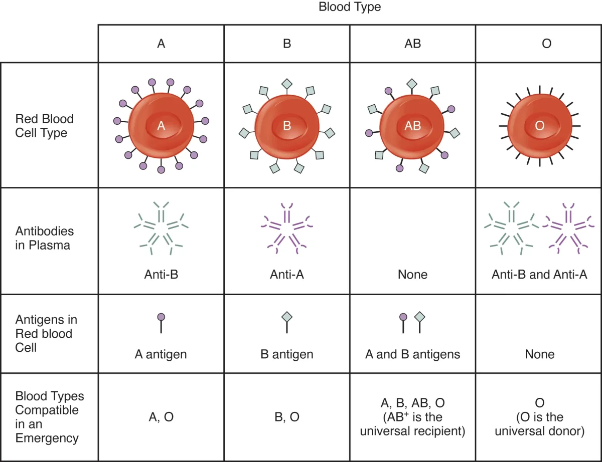
| Blood Type | Antigens on RBCs | Antibodies in Plasma | Can Donate To | Can Receive From |
|---|---|---|---|---|
| A | A antigen | Anti-B | A, AB | A, O |
| B | B antigen | Anti-A | B, AB | B, O |
| AB | Both A and B | Neither (universal recipient) | AB only | A, B, AB, O |
| O | Neither | Both anti-A and anti-B (universal donor) | A, B, AB, O | O only |
The Rh Factor
The Rh system adds a second layer of compatibility. If you have the Rh antigen (Rh D protein) on your RBCs, you are Rh-positive (+). If not, you are Rh-negative (-).
Rh incompatibility in pregnancy: An Rh-negative mother carrying an Rh-positive fetus can develop anti-Rh antibodies if fetal blood enters her circulation (usually during delivery). These antibodies are harmless to the first baby but can attack the RBCs of subsequent Rh-positive fetuses, causing hemolytic disease of the newborn. Prevention: the mother receives Rh immunoglobulin injections to destroy any fetal Rh+ cells before her immune system can mount a response.
True universal donor: O-negative (no A, B, or Rh antigens). True universal recipient: AB-positive (no antibodies against any common antigens).