
Oxygen Transport
Oxygen is not very soluble in blood. If you relied on dissolved oxygen alone, your blood could carry only about 3 mL of O2 per liter - nowhere near enough to meet your body’s demand of roughly 250 mL of O2 per minute. The solution? A molecular shuttle bus called hemoglobin, which increases oxygen-carrying capacity by nearly 70-fold.
Hemoglobin Structure
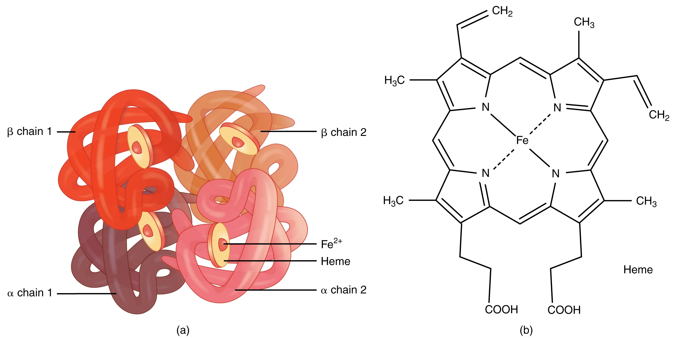
Hemoglobin is a quaternary protein consisting of 4 polypeptide subunits:
- Adult hemoglobin (HbA): 2 alpha chains + 2 beta chains (α2β2)
- Fetal hemoglobin (HbF): 2 alpha chains + 2 gamma chains (α2γ2)
Each subunit contains a heme group - a porphyrin ring with a central iron atom (Fe2+) that can bind one O2 molecule.
Cooperative Binding and the T/R State Model
Hemoglobin exists in two conformational states:
| State | Name | Oxygen Affinity | When? |
|---|---|---|---|
| T-state | Tense | Low affinity | Deoxyhemoglobin (no O2 bound) |
| R-state | Relaxed | High affinity | Oxyhemoglobin (O2 bound) |
When the first O2 molecule binds to a heme group, it triggers a conformational change that shifts the entire molecule from T-state to R-state, making it easier for the next O2 molecules to bind. This is positive cooperativity - each O2 binding event facilitates the next.
The reverse is also true: when the first O2 is released in the tissues, the remaining O2 molecules are released more easily. This creates a very efficient loading/unloading system.
The Oxygen-Hemoglobin Dissociation Curve
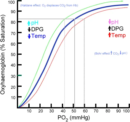
The O2-Hb dissociation curve plots hemoglobin saturation (%) against partial pressure of oxygen (). Because of cooperative binding, the curve is sigmoidal (S-shaped), not linear.
Key points on the curve:
| Saturation | Location | Meaning | |
|---|---|---|---|
| ~100 mmHg | ~98% | Lungs | Hemoglobin is nearly fully loaded |
| ~40 mmHg | ~75% | Resting tissues | About 25% of O2 has been unloaded |
| ~20 mmHg | ~35% | Exercising tissues | About 65% of O2 has been unloaded |
The steep middle portion of the curve ( ~20-60 mmHg) is where small changes in cause large changes in saturation. This is the tissue range, where hemoglobin efficiently unloads oxygen in response to small drops in .
The flat upper portion ( > 70 mmHg) means that hemoglobin stays nearly saturated even if drops somewhat. This is a safety buffer - even at altitude, saturation remains high until drops significantly.
Right Shift - Decreased Affinity (Releases O2 More Easily)
A right shift means hemoglobin releases oxygen more readily at any given . This is beneficial in the tissues where O2 is needed.
Factors causing a right shift:
- Increased CO2 (hypercapnia)
- Decreased pH / increased H+ (acidosis)
- Increased temperature
- Increased 2,3-BPG (2,3-bisphosphoglycerate)
All of these conditions are found in actively metabolizing tissues - muscles during exercise are hot, acidic, and producing CO2. The right shift ensures that hemoglobin dumps oxygen exactly where it is needed most.
Left Shift - Increased Affinity (Holds O2 Tighter)
A left shift means hemoglobin binds oxygen more tightly and is less willing to release it. This is beneficial in the lungs where O2 is being picked up.
Factors causing a left shift:
- Decreased CO2
- Increased pH (alkalosis)
- Decreased temperature
- Decreased 2,3-BPG
- Fetal hemoglobin (HbF)
- Carbon monoxide (CO) binding
The Bohr Effect
The Bohr effect specifically describes how CO2 and H+ concentration affect hemoglobin’s oxygen affinity:
- In the tissues: high CO2 and low pH cause a right shift, promoting O2 release
- In the lungs: low CO2 and high pH cause a left shift, promoting O2 binding
This creates an elegant delivery system: hemoglobin automatically releases more oxygen to tissues that are metabolically active (producing CO2 and acid) and picks up more oxygen in the lungs (where CO2 is being exhaled).
The Haldane Effect
The Haldane effect is the mirror image of the Bohr effect: it describes how oxygenation of hemoglobin affects its ability to carry CO2.
- In the tissues: deoxygenated hemoglobin binds CO2 more readily (forming carbaminohemoglobin)
- In the lungs: oxygenated hemoglobin releases CO2 more readily
The Bohr and Haldane effects work together: the Bohr effect promotes O2 delivery to tissues, and the Haldane effect promotes CO2 pickup from tissues and CO2 release in the lungs.
2,3-BPG (2,3-Bisphosphoglycerate)
2,3-BPG is a molecule produced by red blood cells during glycolysis. It binds to the central cavity of the deoxyhemoglobin tetramer, stabilizing the T-state and reducing oxygen affinity (right shift).
2,3-BPG levels increase in response to chronic hypoxia - for example, at high altitude or in anemia. This is an adaptive response that helps deliver more oxygen to tissues when overall oxygen availability is reduced.
Fetal Hemoglobin (HbF)
Fetal hemoglobin has gamma chains instead of beta chains. The gamma chains do not bind 2,3-BPG as well, so HbF has a higher oxygen affinity than adult HbA (left-shifted curve).
This is essential for fetal survival: at the placenta, fetal blood must “steal” oxygen from maternal hemoglobin. HbF’s higher affinity allows it to grab O2 even at the relatively low of the placenta.
Myoglobin
Myoglobin is a single-subunit oxygen-binding protein found in muscle tissue. Unlike hemoglobin’s 4 subunits, myoglobin has only one heme group and therefore shows no cooperativity.
Its dissociation curve is hyperbolic (not sigmoidal), and it has a much higher affinity for oxygen than hemoglobin. Myoglobin does not release oxygen until drops very low.
Function: myoglobin acts as an oxygen reservoir in muscle cells. During intense exercise, when capillary drops dramatically, myoglobin releases its stored O2 to sustain aerobic metabolism.
| Feature | Hemoglobin | Myoglobin |
|---|---|---|
| Subunits | 4 (quaternary) | 1 (tertiary) |
| Heme groups | 4 | 1 |
| Curve shape | Sigmoidal | Hyperbolic |
| Cooperativity | Yes | No |
| O2 affinity | Lower (releases easily) | Higher (stores O2) |
| Location | Red blood cells | Muscle cells |
| Function | O2 transport | O2 storage |
Carbon Monoxide Poisoning
Carbon monoxide (CO) binds hemoglobin with approximately 200-250 times greater affinity than oxygen. Even small amounts of CO in inhaled air can occupy a large fraction of heme binding sites, forming carboxyhemoglobin (COHb).
CO poisoning is devastating for two reasons:
- Reduced carrying capacity - CO occupies heme sites that would otherwise carry O2
- Left shift - CO binding causes the remaining heme groups to hold onto O2 more tightly (increased affinity), so even the O2 that is loaded cannot be delivered to tissues